. 2023 May;77(3):e22265.
doi: 10.1002/syn.22265. Epub 2023 Feb 23.

Abnormal [18 F]NIFENE binding in transgenic 5xFAD mouse model of Alzheimer's disease: In vivo PET/CT imaging studies of α4β2* nicotinic acetylcholinergic receptors and in vitro correlations with Aβ plaques

Affiliations

Abnormal [18 F]NIFENE binding in transgenic 5xFAD mouse model of Alzheimer's disease: In vivo PET/CT imaging studies of α4β2* nicotinic acetylcholinergic receptors and in vitro correlations with Aβ plaques

Christopher Liang et al. Synapse. 2023 May.

Abstract

Since cholinergic dysfunction has been implicated in Alzheimer's disease (AD), the effects of Aβ plaques on nicotinic acetylcholine receptors (nAChRs) α4β2* subtype were studied using the transgenic 5xFAD mouse model of AD. Using the PET radiotracer [18 F]nifene for α4β2* nAChRs, in vitro autoradiography and in vivo PET/CT studies in 5xFAD mice were carried out and compared with wild-type (C57BL/6) mice. Ratios of [18 F]nifene binding in brain regions versus cerebellum (CB) in 5xFAD mice brains were for thalamus (TH) = 17, hippocampus-subiculum = 7, frontal cortex (FC) = 5.5, and striatum = 4.7. [125 I]IBETA and immunohistochemistry (IHC) in 5xFAD brain slices confirmed Aβ plaques. Nicotine and acetylcholine displaced [18 F]nifene in 5xFAD mice (IC50 nicotine = 31-73 nM; ACh = 38-83 nM) and C57BL/6 (IC50 nicotine = 16-18 nM; ACh = 34-55 nM). Average [18 F]nifene SUVR (CB as reference) in 5xFAD mice was significantly higher in FC = 3.04 compared to C57BL/6 mice FC = 1.92 (p = .001), whereas TH difference between 5xFAD mice (SUVR = 2.58) and C57BL/6 mice (SUVR = 2.38) was not significant. Nicotine-induced dissociation half life (t1/2 ) of [18 F]nifene for TH were 37 min for 5xFAD mice and 26 min for C57BL/6 mice. Dissociation half life for FC in C57BL/6 mice was 77 min , while no dissociation of [18 F]nifene occurred in the medial prefrontal cortex (mFC) of 5xFAD mice. Coregistration of [18 F]nifene PET with MR suggested that the mPFC, and anterior cingulate (AC) regions exhibited high uptake in 5xFAD mice compared to C57BL/6 mice. Ex vivo [18 F]nifene and in vitro [125 I]IBETA Aβ plaque autoradiography after in vivo PET/CT scan of 5xFAD mouse brain were moderately correlated (r2 = 0.68). In conclusion, 5xFAD mice showed increased non-displaceable [18 F]nifene binding in mPFC.

Keywords: 5xFAD transgenic mice; Alzheimer's disease; PET/CT; [125I]IBETA; [18F]FEPPA; [18F]Nifene.


Conflict of interest statement

CONFLICT OF INTEREST STATEMENT

The authors declare no conflict of interest.

Figures

FIGURE 1
FIGURE 1
Schematic of α4β2* nAChRs in 5xFAD AD mice: (A) Schematic showing synaptic junction of α4β2* nAChR in normal mouse. (B) In 5xFAD transgenic mouse, Aβ plaques and microglia present in the synapses which may interact and/or interrupt [18F]nifene, acetylcholine, and nicotine binding. AChE, acetylcholinesterase; AChEI, acetylcholinesterase inhibitors; AD, Alzheimer’s disease; Ch, choline; nAChR, nicotinic acetylcholine receptor.
FIGURE 2
FIGURE 2
In vitro [18F]nifene in 5xFAD AD mice brain: (A) Anti-Aβ immunostain of 10 μm brain slice of 5xFAD mouse confirming the presence of Aβ plaques. (B) Total [18F]nifene binding in brain slices of 5xFAD mouse showing high levels in thalamus (TH) and other extrathalmic regions with little binding in the cerebellum (CB). (C and D) Close-up view of FC immunostain and [18F]nifene in 5xFAD mouse. (E and F) Close-up view of HP-SUB immunostain and [18F]nifene in 5xFAD mouse. (G) [18F]Nifene binding in brain slice of 5xFAD mouse in the presence of 0.1 μM nicotine. (H) [18F]Nifene binding in brain slice of 5xFAD mouse in the presence of 10 μM nicotine where most of the [18F]nifene is displaced (>95%); inset shows scan of brain slice. (I) Nicotine concentration effects on the specific binding of [18F]nifene in 5xFAD and control mice brain slices in TH, FC and SUB. ACC, anterior cingulate cortex; FC, frontal cortex; HP, hippocampus; HP-SUB, hippocampus-subiculum ; ST, striatum; SUB, subiculum; TH, thalamus.
FIGURE 3
FIGURE 3
Acetylcholine effects on [18F]nifene: (A) Total binding of [18F]nifene in control mouse brain 10 μm thick slice. (B) Acetylcholine (1 μM) displaces [18F]nifene binding in control mouse brain slice. (C) [18F]Nifene binding in brain 10 μm thick slice of 5xFAD mouse. (D) Acetylcholine (1 μM) displaces [18F]nifene in brain slice of 5xFAD mouse (inset shows scan of brain slice). (E) Acetylcholine concentration effects in 5xFAD and control mice brain slices in vitro. CB, cerebellum; FC, frontal cortex; HP, hippocampus; ST, striatum; SUB, subiculum; TH, thalamus.
FIGURE 4
FIGURE 4
In vitro [125I]IBETA for Aβ plaques in 5xFAD Mice: (A) [125I]IBETA binding to Aβ plaques in 10 μm brain slice of 5xFAD transgenic mice. (B) Adjacent slices immunostained with anti-Aβ showing corresponding location of Aβ plaques. (C) Correlation plot of [125I]IBETA and [18F]nifene (from Figure 2B) showing agreement in most regions except the hippocampus (HP) and lateral septal nuclei (LSN). CB, cerebellum; CER, cerebellum; FC, frontal cortex; HP, hippocampus; LSN, lateral septal nuclei; ST, striatum; SUB, subiculum; TH, thalamus.
FIGURE 5
FIGURE 5
In vivo [18F]nifene in 5xFAD AD mice brain: (A) PET/CT of intraperitoneal (IP) [18F]nifene (11 MBq in 50 μL saline) administered in control mouse (C57BL/6, male 42 g). Coronal, transaxial, and sagittal images of control mouse head at 60 min after injection showing [18F]nifene in the brain. (B) Time-activity curve of thalamus, frontal cortex, and cerebellum in control mouse brain. (C) Ratio of thalamus and frontal cortex to cerebellum of time-activity curve in control mouse. (D) PET/CT of [18F]nifene (6.25 MBq in 50 μL saline, IP) administered in AD mouse (5xFAD, male 30 g). Coronal, transaxial, and sagittal images of 5xFAD female mouse head at 60 min after injection showing [18F]nifene in the brain. (E) Time-activity curve of thalamus, frontal cortex, and cerebellum in 5xFAD female mouse brain. (F) Ratio of thalamus and frontal cortex to cerebellum of time-activity curve in 5xFAD female mouse. CB, cerebellum; FC, frontal cortex; HG, harderian gland; SUV, standard uptake value; TH, thalamus.
FIGURE 6
FIGURE 6
5xFAD mice [18F]nifene PET/CT: (A) [18F]Nifene in control group mice (male, 36 g, 2.1 MBq, intraperitoneal [IP]) showing binding in TH, FC, and low binding in CB with [18F]nifene greater in TH compared to FC. (B) [18F]Nifene in 5xFAD group mice (male, 20 g, 2.85 MBq, IP) showing binding in TH, FC, and low binding in CB with [18F]nifene greater in TH lower or similar compared to FC. (C) Bar graph comparing averages of control (n = 6) and 5xFAD (n = 6) in TH and FC SUVR (CB as reference). Differences in [18F]nifene TH binding between the two groups were not significant, but the [18F]nifene FC in 5xFAD was higher and very significant (p = .001) when compared to controls. CB, cerebellum; FC, frontal cortex; HG, harderian gland; TH, thalamus.
FIGURE 7
FIGURE 7
PET-MR of [18F]nifene 5xFAD mice: (A) Coregistered [18F]nifene PET-MR control mouse C57BL/6, sagittal and coronal brain sections. (B) Coregistered [18F]nifene PET-MR mouse 5xFAD, sagittal and coronal brain sections. (C–E) Three superior to inferior planes of coregistered [18F]nifene PET-MR 5xFAD mouse with cross hairs placed on the medial prefrontal cortex/anterior cingulate in sagittal and transaxial brain sections. AC, anterior cingulate; CB, cerebellum; HG, harderian gland; mPFC, medial prefrontal cortex; TH, thalamus.
FIGURE 8
FIGURE 8
Nicotine effects on [18F]nifene in C57BL/6 mice: (A) PET/CT of [18F]nifene (7.4 MBq, intraperitoneal [IP]) in control C57BL/6 mouse male (46 g) showing coronal, transaxial, and sagittal images at 30 min before nicotine administration. (B) Coronal, transaxial, and sagittal images at 30 min after nicotine (1 mg/kg, IP). (C) Time-activity curve of thalamus, frontal cortex, and cerebellum showing nicotine intervention (arrow). (D) Dissociation rate (ln X/X0) plot for thalamus (koff = 0.027 min−1), frontal cortex (koff = 0.009 min−1), and cerebellum (koff = 0.007 min−1).
FIGURE 9
FIGURE 9
Nicotine effects on [18F]nifene in 5xFAD mice: (A) PET/CT of [18F]nifene (5 MBq, intraperitoneal [IP]) in 5xFAD mouse female (20 g) showing coronal, transaxial, and sagittal images at 30 min before nicotine administration. (B) Coronal, transaxial, and sagittal images 30 min after nicotine (1 mg/kg, IP). (C) Time-activity curve of thalamus, frontal cortex, and cerebellum showing nicotine intervention (arrow). (D) Dissociation rate (ln X/X0) plot for thalamus (koff = 0.035 min−1) and cerebellum (koff = 0.006 min−1). Frontal cortex shows no dissociation in the 5xFAD mouse but continues to increase at a rate of 0.0005 min−1.
FIGURE 10
FIGURE 10
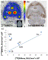
Ex vivo [18F]Nifene and [125I]IBETA in 5xFAD mice: (A) Sagittal in vivo PET/CT [18F]nifene image in 5xFAD mouse brain at approximately 70 min post-injection (male 26 g; 7.4 MBq intraperitoneal [IP]). (B) Ex vivo [18F]nifene autoradiograph (10 μm thick slice) of same mouse after PET/CT experiment. (C) In vitro autoradiograph of adjacent brain slice of same mouse (after fluorine-18 decay) labeled with [125I]IBETA for Aβ amyloid plaques. (D) In vitro immunostain of adjacent brain slice (after fluorine-18 decay) labeled with anti-Aβ for Aβ amyloid plaques. (E) Correlation of [125I]IBETA labeled Aβ plaques with [18F]nifene binding (r2 = 0.68). The outlier, mPFC was not used in the correlation. AC, anterior cingulate; BS, brain stem; BSHS, brain stem hotspot; CB, cerebellum; COL, colliculi; HP, hippocampus; mPFC, frontal cortex; PC, piriform cortex; TH, thalamus.
FIGURE 11
FIGURE 11
In vitro [18F]FEPPA in 5xFAD mice: (A) Control brain slice 10 μm scan showing various regions. (B) [18F]FEPPA binding in the 10 μm brain slice of control mice. (C) 5xFAD brain slice 10 μm scan showing various regions. (D) [18F]FEPPA binding in 10 μm brain slice of 5xFAD transgenic mice. (E) Comparison of [18F]FEPPA binding between control and 5xFAD mice. Significantly higher binding of [18F]FEPPA was seen in FC and HP-SUB. AC, anterior cingulate; CB, cerebellum; FC, frontal cortex; HP, hippocampus; SUB, subiculum; WM and WM2, white matter.

References

    1. Aggleton JP, Pralus A, Nelson AJD, & Hornberger M (2016). Thalamic pathology and memory loss in early Alzheimer’s disease: Moving the focus from the medial temporal lobe to Papez circuit. Brain, 139, 1877–1890. - PMC - PubMed
    1. Bankhead P, Loughrey MB, Fernandez JA, Dombrowski Y, McArt DG, Dunne PD, McQuaid S, Gray RT, Murray LJ, Coleman HG, James JA, Salto-Tellez M, & Hamilton PW (2017).QuPath: Open source software for digital pathology image analysis. Scientific Reports, 7, 16878. - PMC - PubMed
    1. Bieszczad KM, Kant R, Constantinescu CC, Pandey SK, Kawai HD, Metherate R, Weinberger NM, & Mukherjee J (2012). Nicotinic acetylcholine receptors in rat forebrain that bind ź8F-nifene: Relating PET imaging, autoradiography, and behavior. Synapse, 66, 418–434. - PMC - PubMed
    1. Boulter C, Irwin C, Frankle TN, Beindorff N, & Boulter Y (2021). Quantitative brain positron emission tomography in female 5xFAD Alzheimer mice: Pathological features and sex-specific alterations. Frontiers Medicine, 8, 745064. 10.3389/fmed.2021.745064 - DOI - PMC - PubMed
    1. Braak H, Thal DR, Ghebremedhin E, & Del Tredici K (2011). Stages of the pathologic process in Alzheimer disease: Age categories from 1 to 100 years. Journal of Neuropath & Experimental Neurology, 70(11), 960–969. - PubMed

Publication types