. 2023 Feb 16;186(4):864-876.e21.
doi: 10.1016/j.cell.2022.12.041. Epub 2023 Feb 6.

Bacteriophages inhibit and evade cGAS-like immune function in bacteria

Affiliations

Bacteriophages inhibit and evade cGAS-like immune function in bacteria

Erin Huiting et al. Cell. .

Erratum in

Abstract

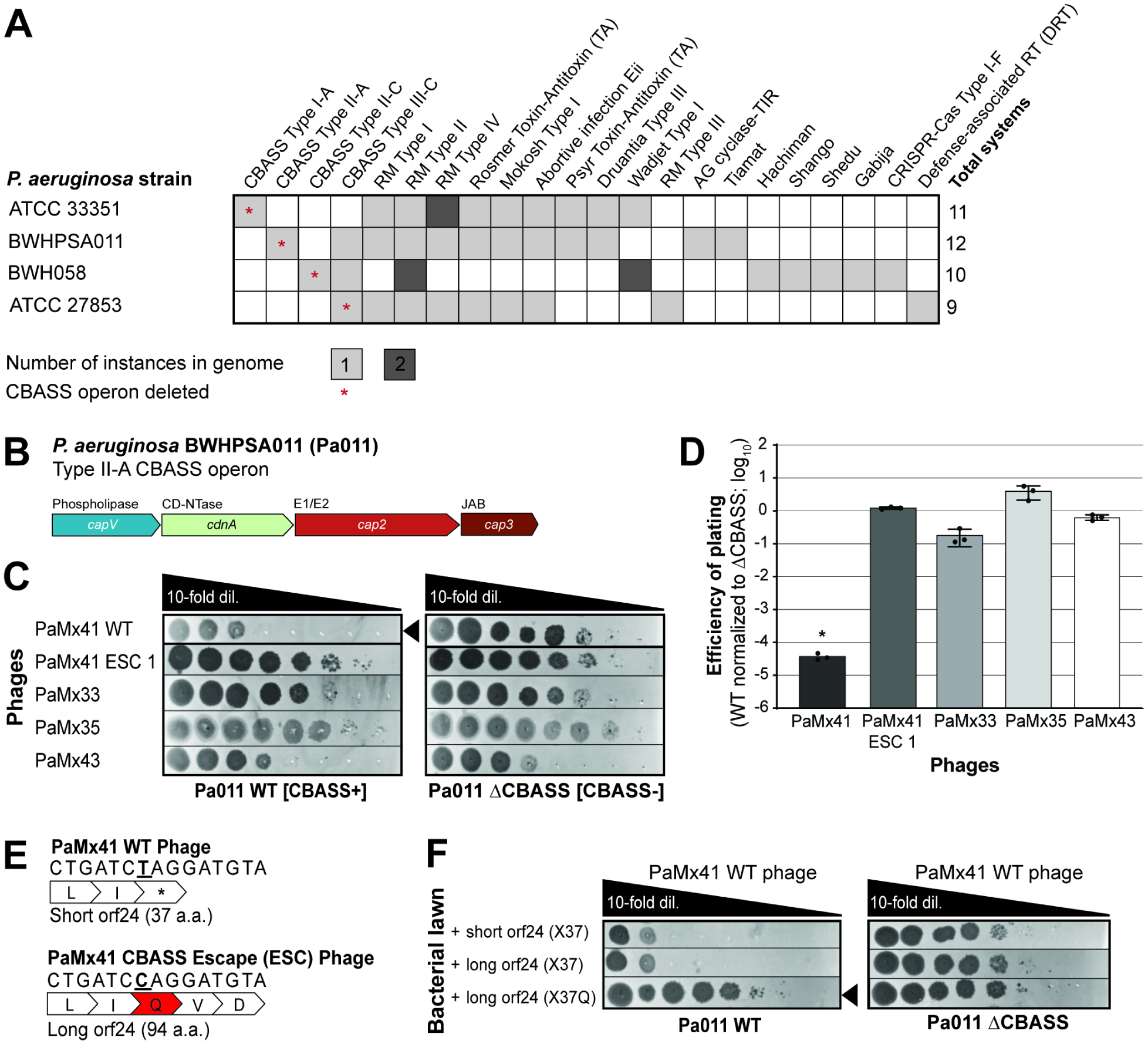
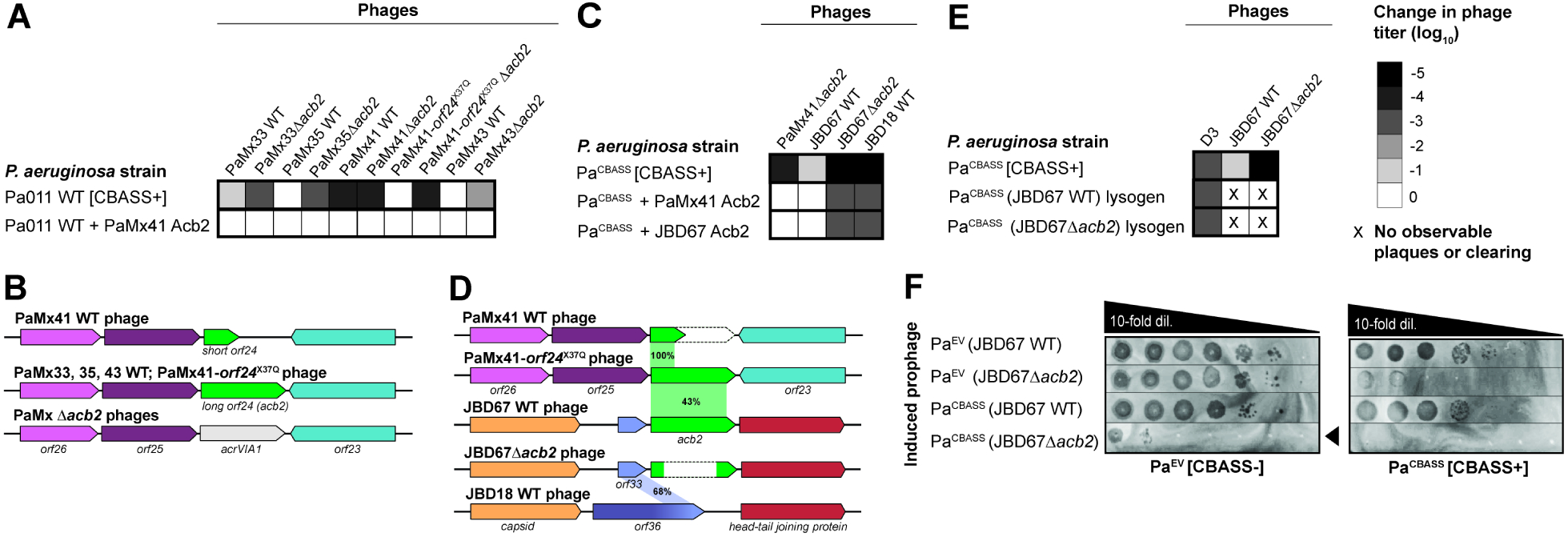
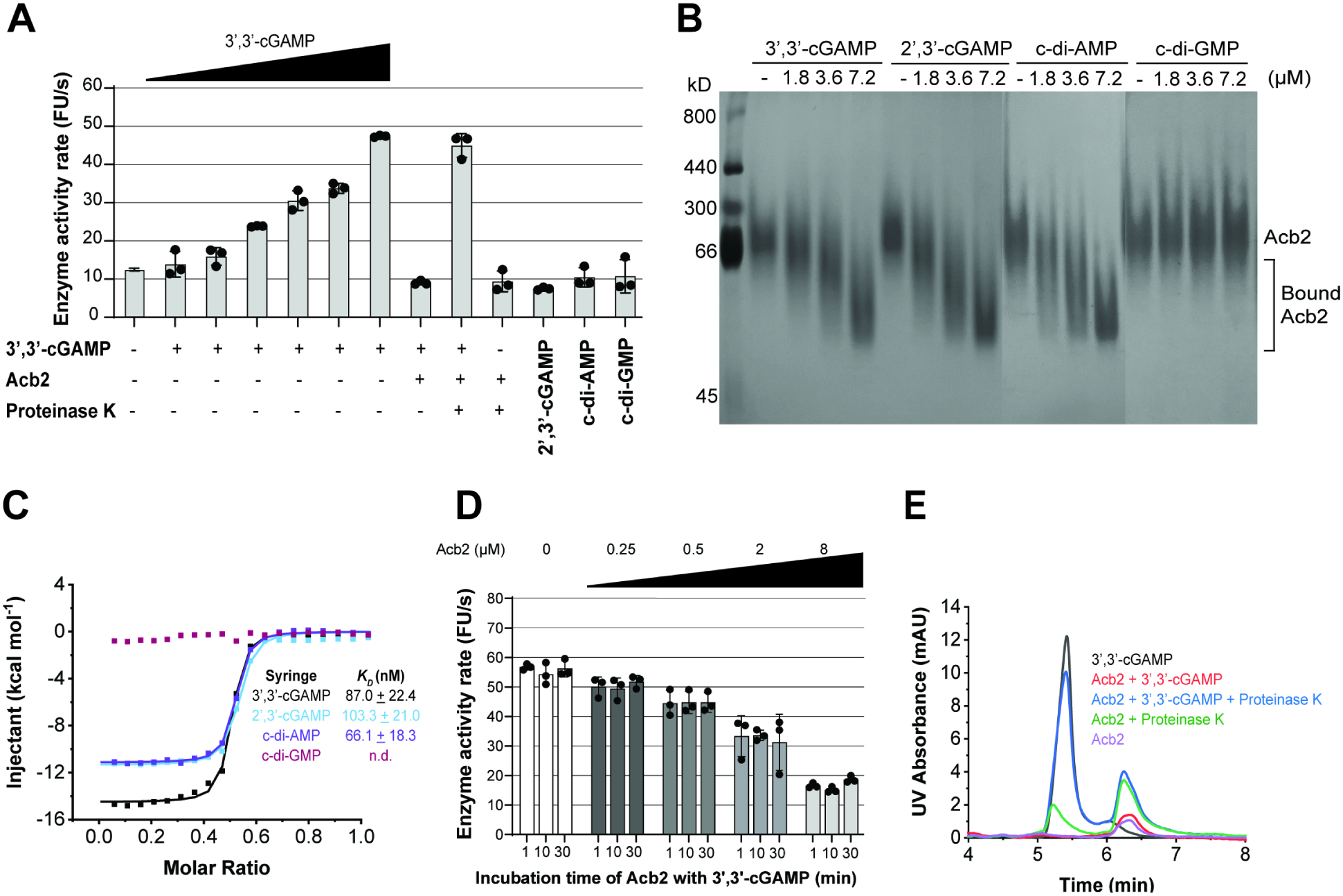
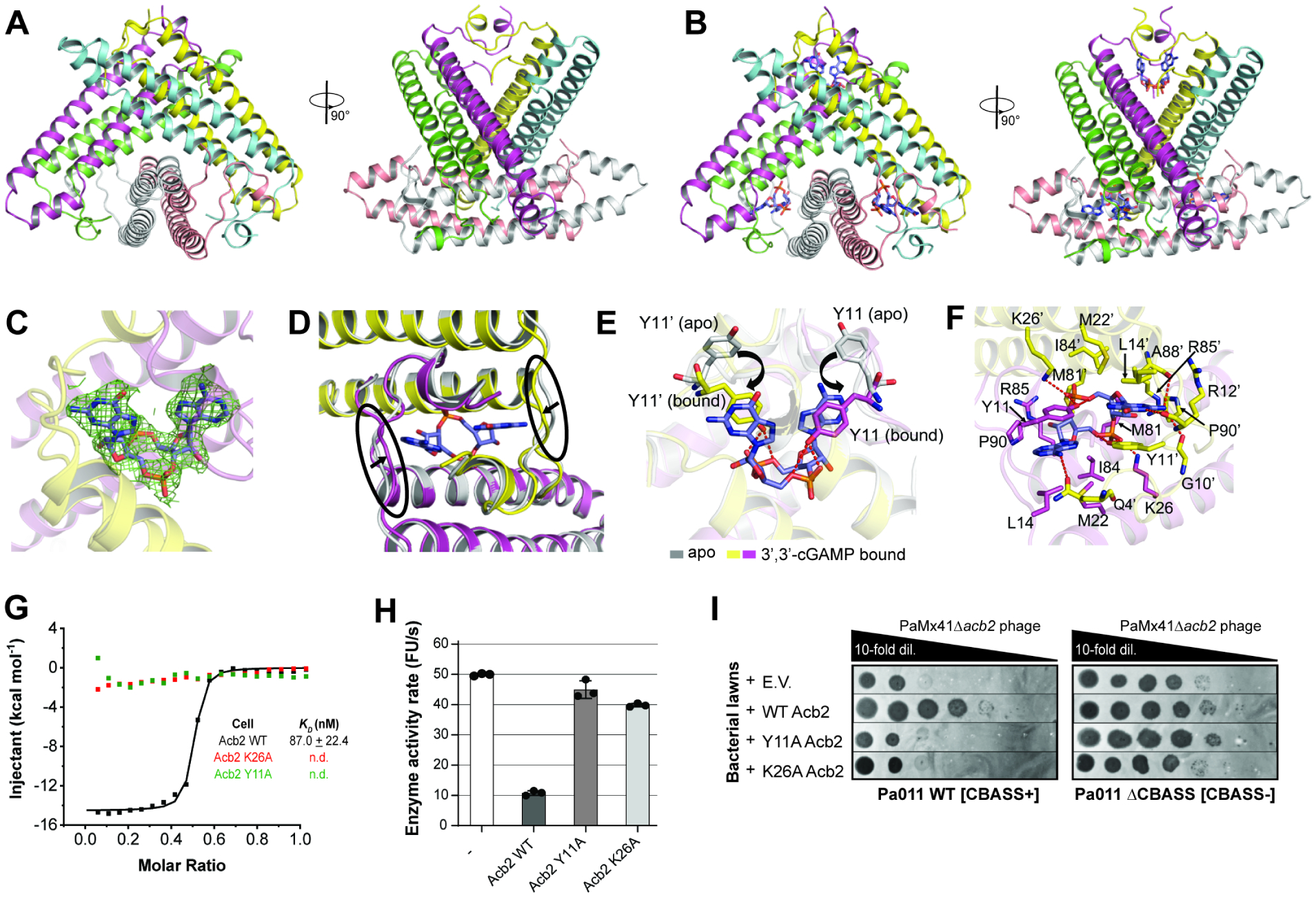
A fundamental strategy of eukaryotic antiviral immunity involves the cGAS enzyme, which synthesizes 2',3'-cGAMP and activates the effector STING. Diverse bacteria contain cGAS-like enzymes that produce cyclic oligonucleotides and induce anti-phage activity, known as CBASS. However, this activity has only been demonstrated through heterologous expression. Whether bacteria harboring CBASS antagonize and co-evolve with phages is unknown. Here, we identified an endogenous cGAS-like enzyme in Pseudomonas aeruginosa that generates 3',3'-cGAMP during phage infection, signals to a phospholipase effector, and limits phage replication. In response, phages express an anti-CBASS protein ("Acb2") that forms a hexamer with three 3',3'-cGAMP molecules and reduces phospholipase activity. Acb2 also binds to molecules produced by other bacterial cGAS-like enzymes (3',3'-cUU/UA/UG/AA) and mammalian cGAS (2',3'-cGAMP), suggesting broad inhibition of cGAS-based immunity. Upon Acb2 deletion, CBASS blocks lytic phage replication and lysogenic induction, but rare phages evade CBASS through major capsid gene mutations. Altogether, we demonstrate endogenous CBASS anti-phage function and strategies of CBASS inhibition and evasion.

Keywords: CBASS; Pseudomonas aeruginosa; acb; anti-CBASS; anti-phage immunity; bacteriophage; cGAS; capsid; escapers; innate immunity.


Conflict of interest statement

Declaration of interests J.B.-D. is a scientific advisory board member of SNIPR Biome, Excision Biotherapeutics, and LeapFrog Bio, and a scientific advisory board member and co-founder of Acrigen Biosciences. The Bondy-Denomy lab receives research support from Felix Biotechnology.

Figures

Figure 1
Figure 1. P. aeruginosa BWHPSA011 (Pa011) CBASS-based immunity protects against PaMx41 infection.
(A) The presence of different anti-phage immune systems in P. aeruginosa strains that were used in this study. Some systems are present in the genome twice as indicated by the darker shade of gray and number (2). An asterisk (*) indicates the CBASS operon was effectively deleted from the bacterial genome. (B) Pa011 CBASS operon. (C) Plaque assays with PaMx41-like phages and an evolved PaMx41 CBASS Escaper (ESC) phage spotted in 10-fold serial dilutions on a lawn of Pa011 WT [CBASS+] or ΔCBASS [CBASS-]; clearings represent phage replication. Black arrowhead highlights the reduction in PaMx41 WT phage titer. See also Figure S1. (D) Efficiency of plating was quantified as plaque-forming units (PFU) per ml on Pa011 WT divided by PFU/ml on the ΔCBASS (n=3). Data are mean ± s.d. Non-parametric ANOVA test yielded a P value of <0.0001. (E) Schematic of PaMx41 WT and CBASS escape phage genomes with the no-stop extension mutation. The bold underline indicates mutation of thymine (T) to cytosine (C), resulting in a stop codon (TAG, *) to glutamine (CAG, Q) substitution (in red). (F) Plaque assays with PaMx41 WT phage on a lawn of Pa011 WT or ΔCBASS over-expressing the indicated orf24 variants. See also Figure S2.
Figure 2.
Figure 2.. Phage-encoded acb2 is necessary for replication in the presence of CBASS.
(A, C, E) Heat maps representing the order of magnitude change in phage titer, where phage titer is quantified by comparing the number of spots (with plaques, or clearing if plaques were not visible) on the CBASS+ strain divided by the CBASS- strain. Plaque assays used for these quantifications can been seen in Figure S2 (n=3). (B, D) Comparison of the acb2 locus across phage genomes. PaMx41-like Δacb2 phages have the acb2 gene substituted with the type VI-A anti-CRISPR gene (acrVIA1) as part of the knockout procedure, and JBD67Δacb2 phages have the acb2 gene removed from its genome. Genes with known protein functions are indicated with names, and genes with hypothetical proteins are indicated with “orf”. Acb2 percent amino acid identity is shown in (D). (F) Plaque assays assessing the titer induced prophages spotted on a lawn of PaEV or PaCBASS. Black arrowhead highlights reduction in JBD67Δacb2 phage titer. See also Figure S2.
Figure 3.
Figure 3.. Acb2 antagonizes CBASS activity by sequestering the 3’,3’-cGAMP signaling molecule.
(A) CapV enzyme activity in the presence of the indicated cyclic dinucleotides and resorufin butyrate, which is a phospholipase substrate that emits fluorescence when hydrolyzed. The enzyme activity rate was measured by the accumulation rate of fluorescence units (FU) per second. The concentration of 3’,3’-cGAMP ranged from 0.025 to 0.8 μM (0.025, 0.05, 0.1, 0.2, 0.4, 0.8 μM), and the other cyclic dinucleotides were added at 0.8 μM. To test the effects of Acb2 to bind and release 3’,3’-cGAMP, Acb2 (32 μM) was incubated with 3’,3’-cGAMP (0.8 μM) for 10 minutes and then Proteinase K (0.065 mg/mL) was added to extract the nucleotides from the Acb2 protein. Filtered nucleotides products were used for the CapV activity assay. Data are mean ± s.d. (n=3). (B) Native PAGE showed the binding of Acb2 to cyclic dinucleotides. (C) Isothermal titration calorimetry (ITC) assays to test binding of cyclic dinucleotides to Acb2. Representative binding curves and binding affinities are shown. The KD values are mean ± s.d. (n=3). Raw data for these curves are shown in Figure S4. (D) CapV activity assay to test the effects of Acb2 on 3’,3’-cGAMP. The concentration of 3’,3’-cGAMP was 0.8 μM and Acb2 ranged from 0.25 to 8 μM (0.25, 0.5, 2, 8 μM). The 3’,3’-cGAMP was pre-incubated with Acb2 for 1, 10, 30 minutes, respectively. Data are mean ± s.d. (n=3). (E) The ability of Acb2 to bind and release 3’,3’-cGAMP when treated with proteinase K was analyzed by HPLC. 3’,3’-cGAMP standard was used as a control. The remaining 3’,3’-cGAMP after incubation with Acb2 were tested.
Figure 4.
Figure 4.. Structure of Acb2 reveals hexamer bound to three molecules of 3’,3’-cGAMP.
(A) Overall structure of the Acb2 hexamer. Two views are shown. (B) Overall structure of the Acb2 hexamer bound to three molecules of 3’,3’-cGAMP. 3’,3’-cGAMP molecules are colored in slate. (C) 2Fo-Fc electron density of 3’,3’-cGAMP in the binding pocket contoured at 1 σ. (D) Structural comparison between an Acb2 dimer in its apo (colored in gray) and 3’,3’-cGAMP-bound form (colored in yellow and light magenta). The loops which move upon 3’,3’-cGAMP binding are marked with black circles. (E) Structural alignment between apo and 3’,3’-cGAMP-bound Acb2, which are colored as in (B). Y11 from the two structures are highlighted as sticks. Red dashed lines represent poplar interactions. (F) Binding between Acb2 or Acb2 mutants and 3’,3’-cGAMP. Red dashed lines represent polar interactions. (G) ITC assays to test the binding of 3’,3’-cGAMP to Acb2 mutants. Representative binding curves and binding affinities are shown. The KD values are mean ± s.d. (n=3). Raw data for these curves are shown in Figure S4. (H) CapV activity assay to test the effects of Acb2 mutations. The concentration of 3’,3’-cGAMP was 0.8 μM and Acb2 or its mutants was 32 μM. The 3’,3’-cGAMP was pre-incubated with Acb2 or its mutants for 10 minutes. Bar graph represents average of three technical replicates. Data are mean ± s.d. (n=3). (I) Plaque assays on a lawn of Pa011 WT or ΔCBASS overexpressing empty vector (E.V.) or the indicated PaMx41 Acb2 mutants; clearings represent phage replication.
Figure 5.
Figure 5.. CBASS escape phages have mutations in the major capsid gene.
(A) Plaque assays were performed with the indicated control/WT and escape phages spotted in 10-fold serial dilutions on lawns of bacteria expressing CBASS+ (left) or lacking CBASS- (right); clearings represent phage replication. (B) Schematic of major capsid genes with corresponding missense mutations and associated CBASS Escape (ESC) phages. (C) Schematic of in vivo homologous recombination of parental phages with homology-directed repair (HDR) template 1 (encoding I121S or I121T capsid mutations) or template 2 (S330P capsid mutation) and resultant engineered/recombinant phages. (D) Plaque assays with recombinant phages possessing major capsid mutations, or WT capsid controls, spotted on lawns of Pa011 WT or ΔCBASS.

References

    1. Barbalat R, Ewald SE, Mouchess ML, and Barton GM (2011). Nucleic acid recognition by the innate immune system. Annu. Rev. Immunol 29, 185–214. 10.1146/annurev-immunol031210-101340. - DOI - PubMed
    1. Margolis SR, Wilson SC, and Vance RE (2017). Evolutionary Origins of cGAS-STING Signaling. Trends Immunol. 38, 733–743. 10.1016/j.it.2017.03.004. - DOI - PubMed
    1. Li X-D, Wu J, Gao D, Wang H, Sun L, and Chen ZJ (2013). Pivotal roles of cGAScGAMP signaling in antiviral defense and immune adjuvant effects. Science 341, 1390–1394. 10.1126/science.1244040. - DOI - PMC - PubMed
    1. Wu J, Sun L, Chen X, Du F, Shi H, Chen C, and Chen ZJ (2013). Cyclic GMP-AMP is an endogenous second messenger in innate immune signaling by cytosolic DNA. Science 339, 826–830. 10.1126/science.1229963. - DOI - PMC - PubMed
    1. Ishikawa H, and Barber GN (2008). STING is an endoplasmic reticulum adaptor that facilitates innate immune signalling. Nature 455, 674–678. 10.1038/nature07317. - DOI - PMC - PubMed

Publication types

Substances