. 2023 Feb 16;186(4):821-836.e13.
doi: 10.1016/j.cell.2023.01.016. Epub 2023 Feb 6.

Structures of LRP2 reveal a molecular machine for endocytosis

Affiliations

Structures of LRP2 reveal a molecular machine for endocytosis

Andrew Beenken et al. Cell. .

Abstract

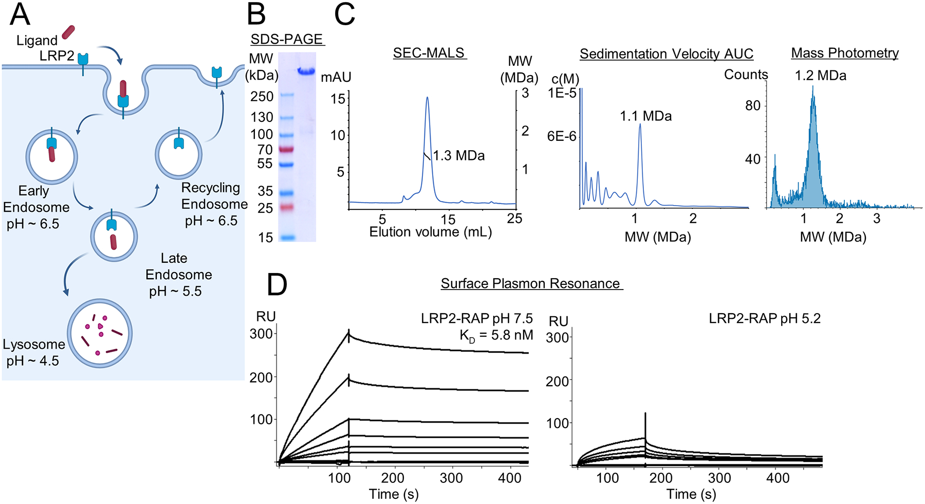
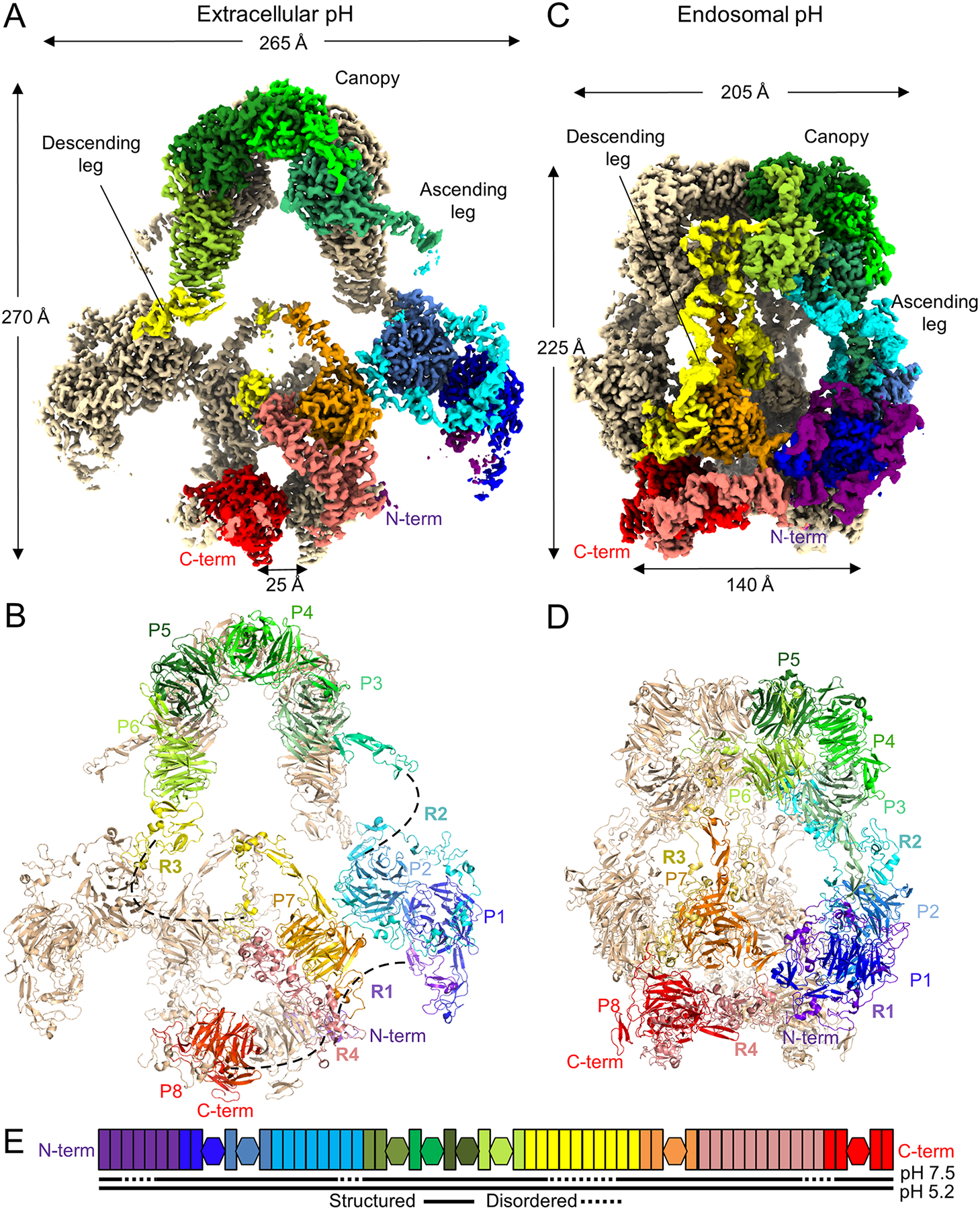
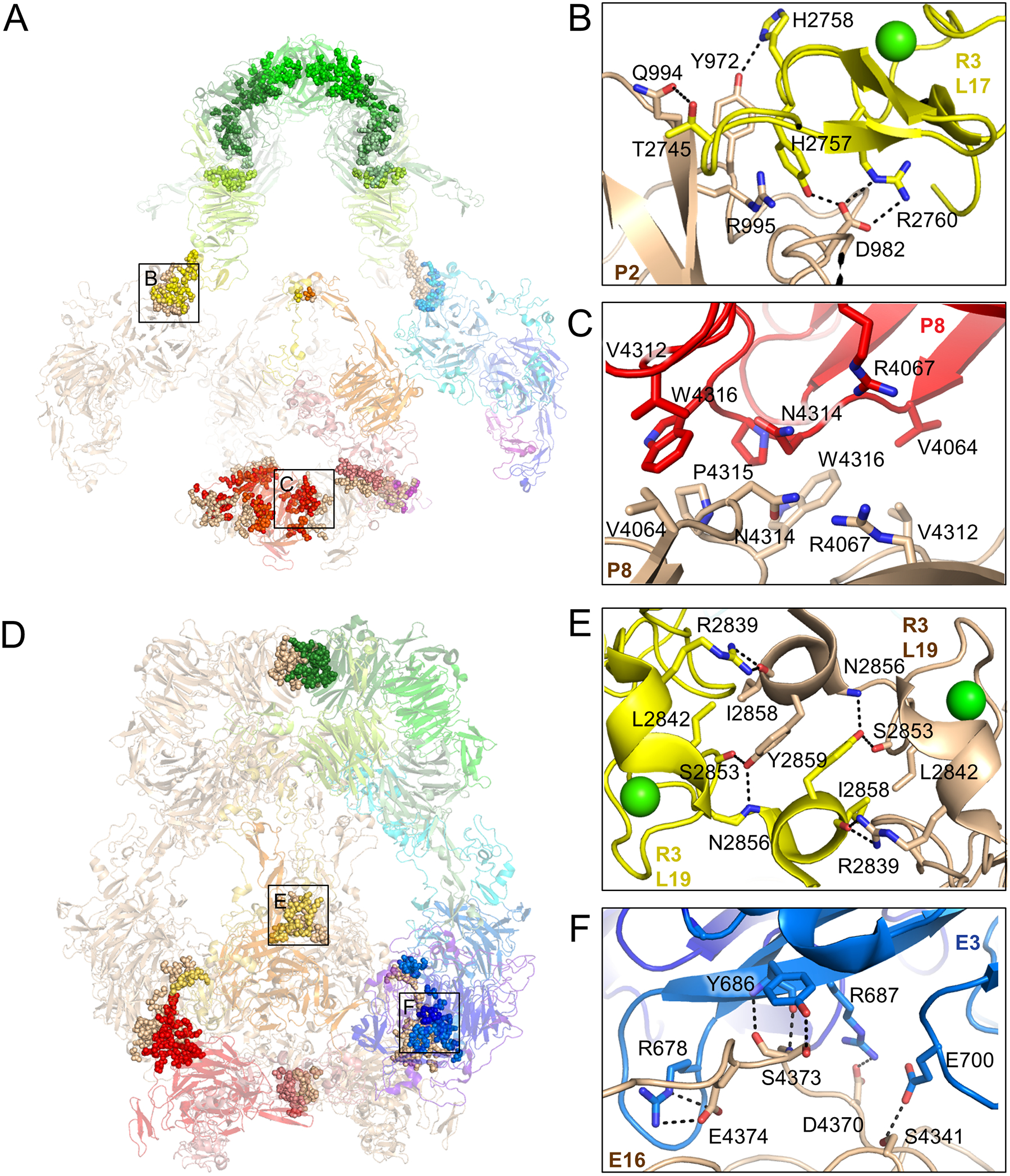
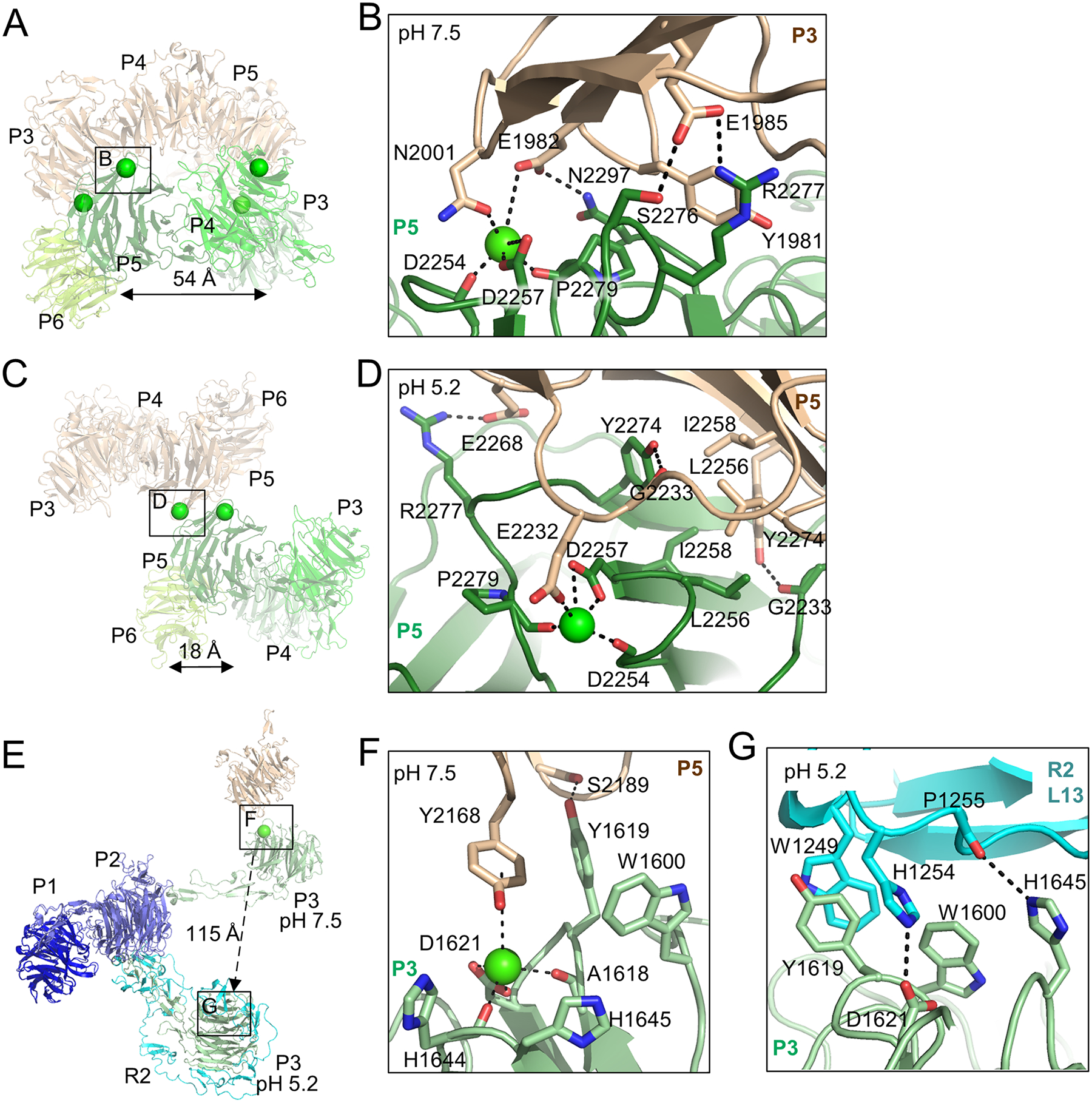
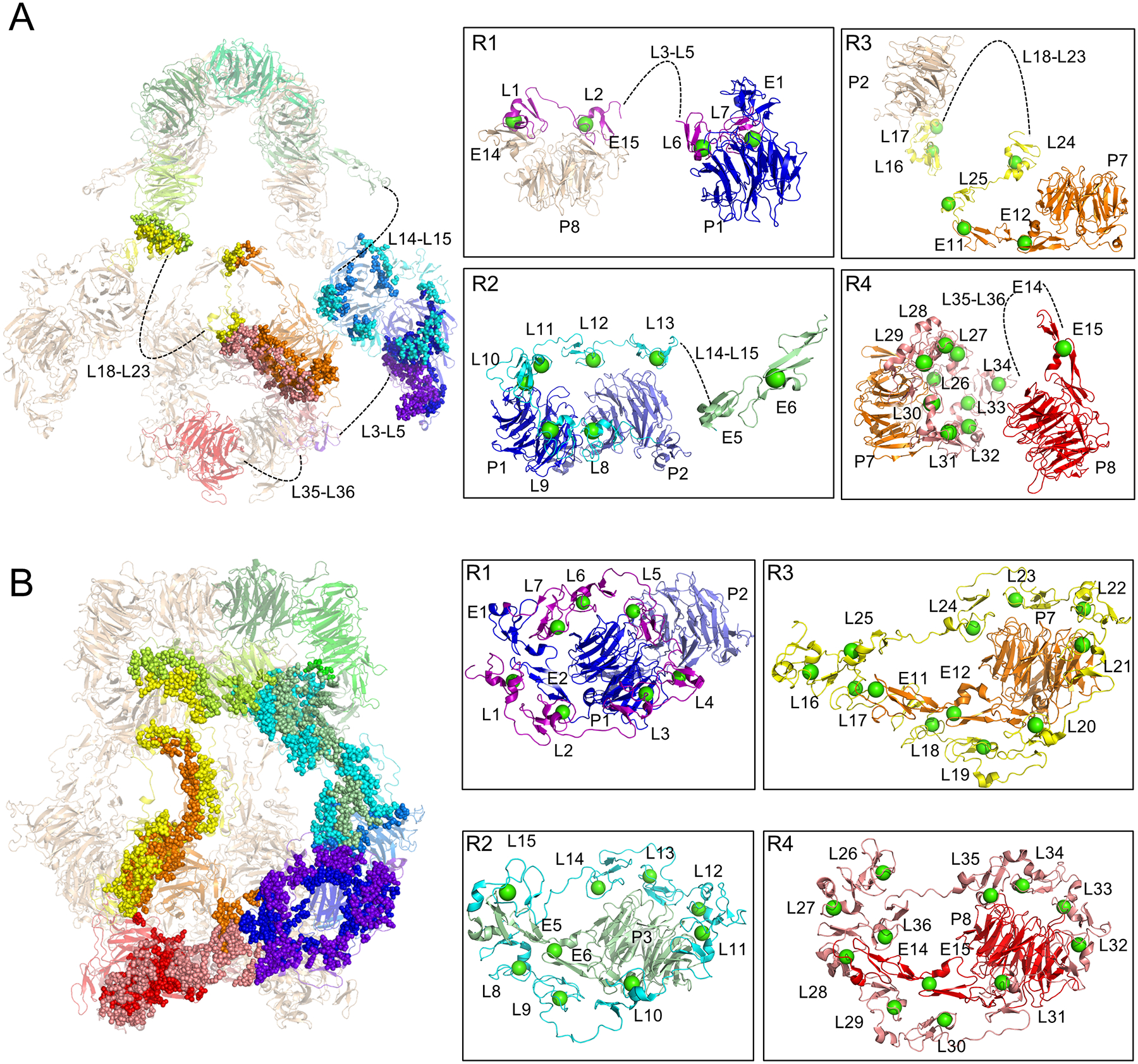
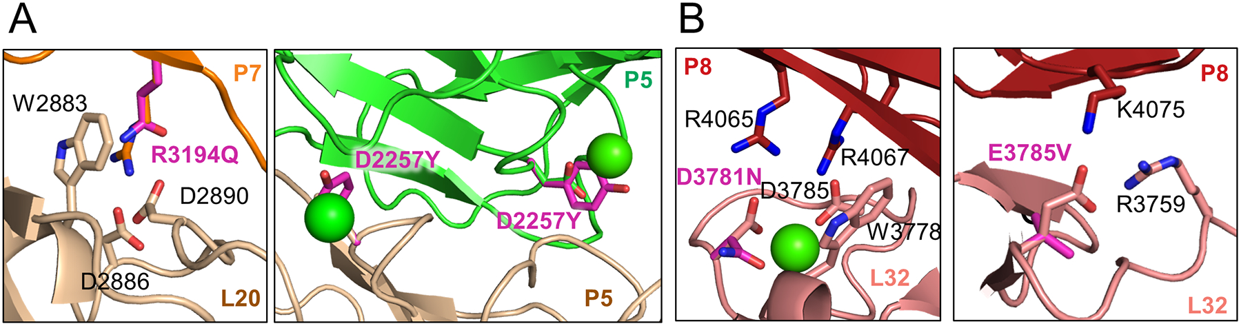
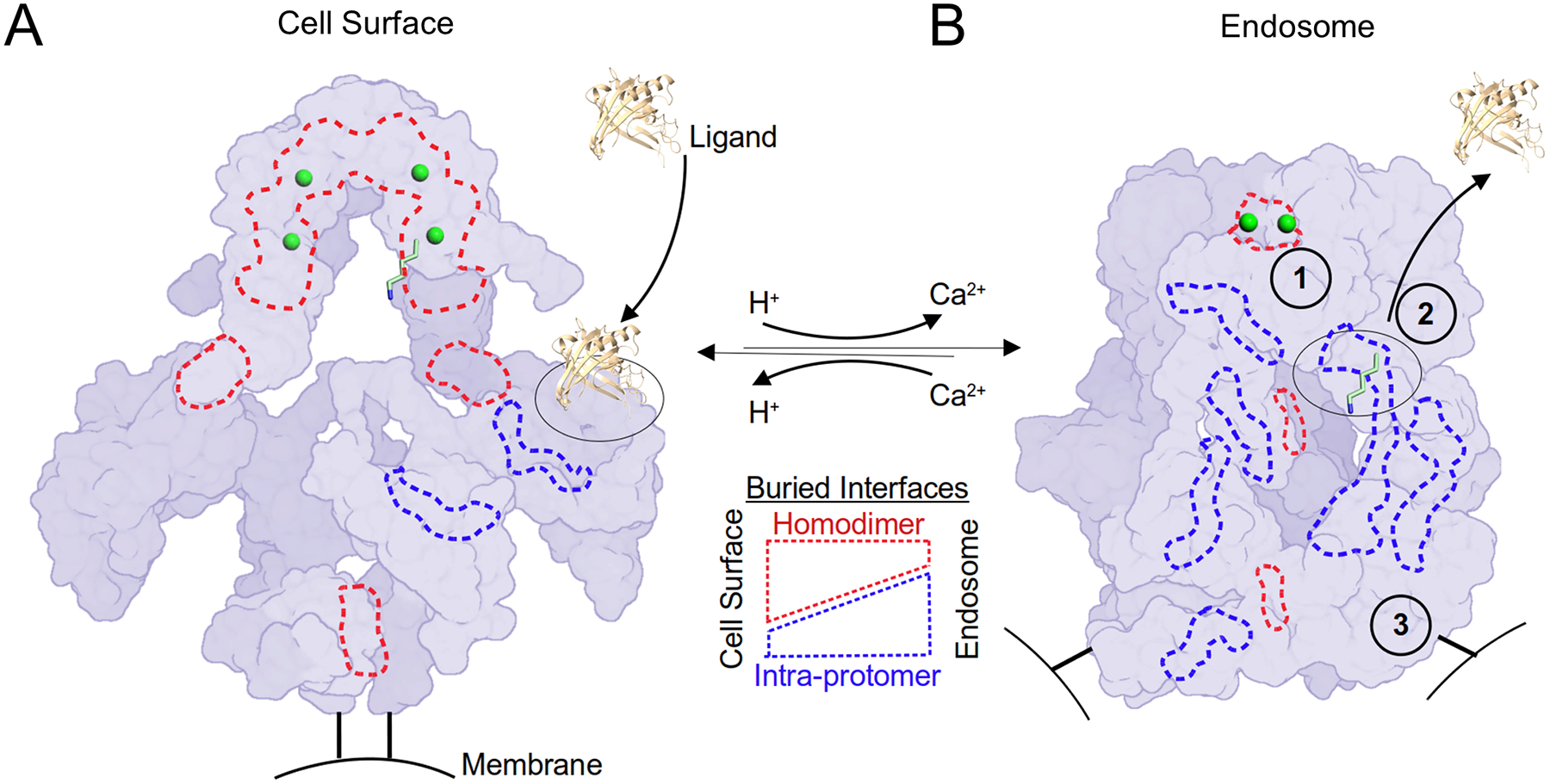
The low-density lipoprotein (LDL) receptor-related protein 2 (LRP2 or megalin) is representative of the phylogenetically conserved subfamily of giant LDL receptor-related proteins, which function in endocytosis and are implicated in diseases of the kidney and brain. Here, we report high-resolution cryoelectron microscopy structures of LRP2 isolated from mouse kidney, at extracellular and endosomal pH. The structures reveal LRP2 to be a molecular machine that adopts a conformation for ligand binding at the cell surface and for ligand shedding in the endosome. LRP2 forms a homodimer, the conformational transformation of which is governed by pH-sensitive sites at both homodimer and intra-protomer interfaces. A subset of LRP2 deleterious missense variants in humans appears to impair homodimer assembly. These observations lay the foundation for further understanding the function and mechanism of LDL receptors and implicate homodimerization as a conserved feature of the LRP receptor subfamily.

Keywords: CD91; LDL; LRP1; LRP2; cryo-EM; endocytosis; megalin; pH-sensitive; proteinuria; recycling.


Conflict of interest statement

Declaration of interests The authors declare no competing interests.

Figures

Figure 1.
Figure 1.. Biochemical characterization of endogenous LRP2 reveals a stable dimer
(A) Schematic representation of ligand binding by LRP2 and of LRP2 receptor recycling. (B) SDS-PAGE reducing, denaturing gel of the purified LRP2 sample following SEC showing a single major species at ~600kD (C) Biophysical characterization of LRP2 mass by SEC-MALS (left), Sedimentation velocity AUC (middle), and Mass Photometry (right). The three experiments demonstrate a dimeric form of purified LRP2 with a MW ~1.1 to 1.3 MDa. The fit of the MALS data is indicated as a black line on the SEC chromatogram. (D) SPR characterization of RAP binding to purified LRP2 at pH 7.5 (left) and pH 5.2 (right) using injections of LRP2 varying from 0.82–200 nM over a surface of immobilized RAP. SPR sensorgrams for pH 7.5 could be fit to determine KD of 5.8 nM, but sensorgrams for pH 5.2 could not be fit. Qualitatively, binding is substantially reduced at pH 5.2. See also Figure S1 and Tables S1, S2, S3
Figure 2.
Figure 2.. Architecture of the LRP2 homodimer at extracellular and endosomal pH.
(A) Cryo-EM map of LRP2 at pH 7.5. One protomer is colored in rainbow, from the N-terminus (purple) to the C-terminus (red); the other is in wheat. Distinctive regions are labelled, and dimensions are shown. Coloring and labeling is consistent throughout the figure. Distance between the transmembrane insertions is labeled. (B) Cryo-EM model of LRP2 at pH 7.5. β-propellers (P) and groups of L repeats (R) are labeled. Unmodeled portions of flexible ligand-binding regions in the structure are dashed lines. (C) Cryo-EM map of LRP2 at pH 5.2. (D) Cryo-EM model of LRP2 at pH 5.2. Labeling is as for panel B (E) Domain diagram as in Figure S1, now colored in rainbow from N-terminus to C-terminus. Below the diagram, structured disordered domains are indicated by solid and dotted lines for pH 7.5 and pH 5.2 respectively. See also Figures S2, S3, and Tables S1, S2, S4, and Movies S1, S2, S3
Figure 3.
Figure 3.. pH-specific homodimer interfaces regulate ligand-binding
(A) Residues involved in homodimer contacts between protomers at pH 7.5 are rendered as spheres superimposed on a semi-transparent cartoon depiction of the LRP2 structure (left panel). Residue level interactions are shown at right for the corresponding labeled boxed regions. Identities of interacting domains are labeled. (B) L17 of R3 is stabilized with its ligand-binding face facing solvent by virtue of polar contacts with P2 of the second protomer. (C) R4067, which functions as an intramolecular ligand at pH 5.2, is buried in a hydrophobic interface between the P8 domains of the two protomers at pH 7.5, preventing it from competing with ligand at ligand-binding repeats. (D) Depiction and labeling of LRP2 at pH 5.2 according to panel A. (E) L19 from R3 of the two protomers engage in symmetric contacts that tether the protomers together at the center of the assembly, excluding solvent and making ligand-binding surfaces inaccessible. (F) E3 of the first protomer packs against E16 of the second protomer in the homodimer, tethering the protomers together near the membrane insertion points. See also Figure S4
Figure 4.
Figure 4.. pH-sensitive interfaces define the morphology of the canopy in the homodimer assembly
(A) Depiction of the canopy as a semi-transparent cartoon at pH 7.5. The two pairs of symmetrically related putative Ca2+ ions coordinated in the canopy are depicted by spheres. The putative Ca2+ ion coordinated by conserved D2257 is boxed. The distance between the two putative Ca2+ ions coordinated by D2257 is indicated. (B) Residue-level interactions observed around the D2257-coordinated putative Ca2+ ion at pH 7.5. (C) Depiction of the canopy at pH 5.2. One pair of putative Ca2+ ions has dissociated due to the reduced pH, but the putative Ca2+ ions coordinated by D2257 persist, one of which is boxed. (D) Residue-level interactions observed around the D2257-coordinated putative Ca2+ ion at pH 5.2. (E) P1 from the LRP2 structures at pH 7.5 and pH 5.2 have been superimposed with RMSD 1.3 Å. The P1-P3 regions from both structures are depicted based on this superimposition as cartoons. Relative to the fixed position of P1, P3 translates 115Å following pH reduction from 7.5 to 5.2, and the putative Ca2+ ion coordinated by conserved D1621 dissociates, enabling interaction of P3 with ligand-binding repeats from R2. The regions that include conserved D1621 are boxed on the P3 domains at both pH’s. (F) Residue-level interactions observed around the D1621-coordinated putative Ca2+ ion at pH 7.5. Y2168 engages in a π-cation interaction with the putative Ca2+ ion. (G) Residue-level interactions observed around D1621 at pH 5.2 following dissociation of the putative Ca2+ ion. D1621 now forms an intra-protomer salt bridge with H1254. See also Figure S5
Figure 5.
Figure 5.. pH-dependent intra-protomer contacts govern ligand binding site availability
(A) Residues involved in intra-protomer contacts at pH 7.5 are rendered as spheres superimposed on a semi-transparent cartoon depiction of the LRP2 structure (left panel). Flexible ligand-binding repeats that could not be modeled are indicated with dashed lines and labeled. Residue level interactions are shown at right in boxes for each of the four ligand-binding domains. Ca2+ ions in the L repeats are rendered as green spheres. Individual ligand-binding repeats within the larger domains are labeled. Domains from one protomer are colored in rainbow, and from the other protomer in wheat. (B) Residues involved in intra-protomer contacts at pH 5.2 are rendered as spheres superimposed on a transparent cartoon depiction of the structure (left panel). See also Figure S4
Figure 6.
Figure 6.. pH-sensitive interfaces are essential for LRP2 function
(A) Representation of the residues surrounding human mutations in LRP2 at pH 5.2 at pH-sensitive sites associated with syndromic phenotypes. The mutations disturb both intra-protomer (left panel) and homodimer (right panel) interfaces. The p.R3194Q mutation ablates a π-cation interaction with W2883 as well as salt bridges with D2886 and D2890. The p.D2257Y mutation eliminates the ability to coordinate a putative Ca2+ ion at a pH-sensitive interface within the canopy of the homodimer. (B) Representation of the residues surrounding human mutations in LRP2 at pH 5.2 at pH-insensitive sites associated with non-syndromic phenotypes. The p.D3781N mutation weakens coordination of the Ca2+ ion and breaks a salt-bridge with R4065. The p.E3785V mutation breaks a salt bridge with R3759.
Figure 7.
Figure 7.. A homodimer coordinates the pH-sensitive LRP2 molecular machine
LRP2 is rendered as a schematic surface at extracellular and endosomal pH in the left and right panels respectively. Protomers are distinguished by two shades of purple. The C-termini of the extracellular domains that are directed towards the membrane insertions are rendered as black bars. Putative Ca2+ ions within the canopy are rendered as green spheres. K1430 is rendered in sticks with 6-fold magnification as one example of the 28 intramolecular ligands, and its binding partner, L8, is outlined with a dashed black oval. An extracellular ligand of ~20kDa rendered to scale is represented as a cartoon (PDB ID: 5JR8). Selected examples of buried surface area at the homodimer interfaces are outlined in dashed red lines, and intra-protomer interfaces are outlined in dashed blue lines. (A) At the cell surface, homodimer interfaces predominate. Hypothetical ligand binding to L8 is depicted. At the cell surface, the homodimer acts as a scaffold to keep L repeats open for ligand interactions and maintain distance between intramolecular ligands and their cognate L repeats. (B) In the endosomal compartment, 1) a pair of Ca2+ ions is released from the canopy and the homodimer interface unlocks to enable intramolecular ligands to interact with their cognate L repeats. 2) An intramolecular ligand displaces the extracellular ligand. As a result of these structural transitions, intra-protomer interfaces now predominate at endosomal pH. 3) The homodimer reorganization separates the membrane insertion points by >140 Å, which may serve as a compartment-specific trafficking signal. See also Movie S3

Comment in

References

    1. Nielsen R, Christensen EI, and Birn H (2016). Megalin and cubilin in proximal tubule protein reabsorption: from experimental models to human disease. Kidney Int 89, 58–67. 10.1016/j.kint.2015.11.007. - DOI - PubMed
    1. Christensen EI, and Nielsen R (2007). Role of megalin and cubilin in renal physiology and pathophysiology. Rev Physiol Biochem Pharmacol 158, 1–22. 10.1007/112_0604. - DOI - PubMed
    1. Gonias SL, and Campana WM (2014). LDL receptor-related protein-1: a regulator of inflammation in atherosclerosis, cancer, and injury to the nervous system. Am J Pathol 184, 18–27. 10.1016/j.ajpath.2013.08.029. - DOI - PMC - PubMed
    1. Consortium, C.e.S. (1998). Genome sequence of the nematode C. elegans: a platform for investigating biology. Science 282, 2012–2018. 10.1126/science.282.5396.2012. - DOI - PubMed
    1. Yochem J, and Greenwald I (1993). A gene for a low density lipoprotein receptor-related protein in the nematode Caenorhabditis elegans. Proc Natl Acad Sci U S A 90, 4572–4576. 10.1073/pnas.90.10.4572. - DOI - PMC - PubMed

Publication types

Substances

LinkOut - more resources