. 2023 Feb 7;14(1):660.
doi: 10.1038/s41467-023-36332-3.

Myristic acid as a checkpoint to regulate STING-dependent autophagy and interferon responses by promoting N-myristoylation

Affiliations

Myristic acid as a checkpoint to regulate STING-dependent autophagy and interferon responses by promoting N-myristoylation

Mutian Jia et al. Nat Commun. .

Abstract

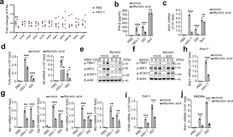
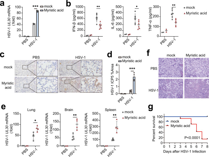
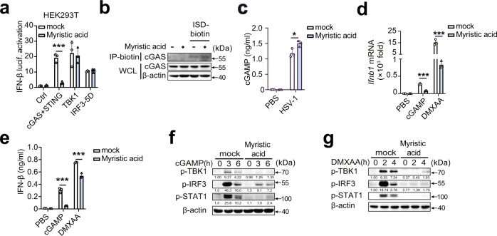
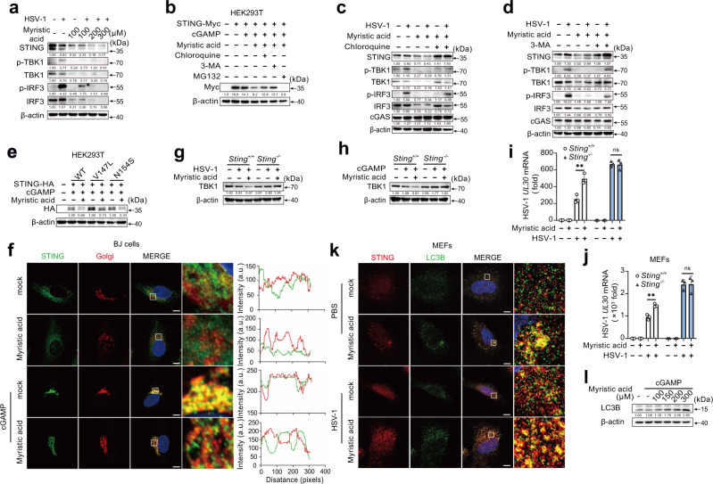
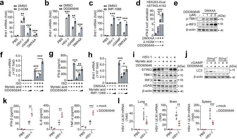
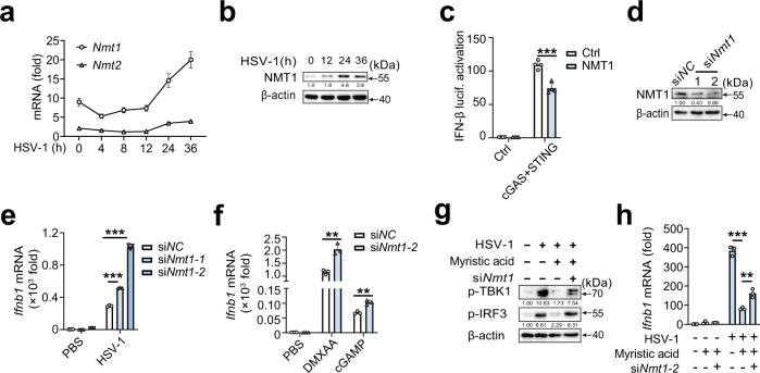
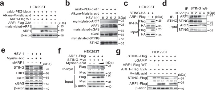
Stimulator of interferon gene (STING)-triggered autophagy is crucial for the host to eliminate invading pathogens and serves as a self-limiting mechanism of STING-induced interferon (IFN) responses. Thus, the mechanisms that ensure the beneficial effects of STING activation are of particular importance. Herein, we show that myristic acid, a type of long-chain saturated fatty acid (SFA), specifically attenuates cGAS-STING-induced IFN responses in macrophages, while enhancing STING-dependent autophagy. Myristic acid inhibits HSV-1 infection-induced innate antiviral immune responses and promotes HSV-1 replication in mice in vivo. Mechanistically, myristic acid enhances N-myristoylation of ARF1, a master regulator that controls STING membrane trafficking. Consequently, myristic acid facilitates STING activation-triggered autophagy degradation of the STING complex. Thus, our work identifies myristic acid as a metabolic checkpoint that contributes to immune homeostasis by balancing STING-dependent autophagy and IFN responses. This suggests that myristic acid and N-myristoylation are promising targets for the treatment of diseases caused by aberrant STING activation.


Conflict of interest statement

The authors declare no competing interests.

Figures

Fig. 1
Fig. 1. Myristic acid inhibits cGAS-dependent antiviral immune responses.
a Fold change of saturated fatty acids (SFAs), determined by gas chromatography in primary mouse peritoneal macrophages (PMs). Statistical significance was determined by paired two-sided multiple Student’s t-tests, n = 4 biologically independent samples. bd Enzyme-linked immunosorbent assay (ELISA) analysis of interferon (IFN)-β secretion (b) and quantitative polymerase chain reaction (qPCR) analysis of Ifnb1, Tnfa, or Il6 mRNA level (c and d) in mouse PMs pretreated with solvent (mock) or myristic acid, plus stimulation as indicated, n = 3 samples examined over 3 independent experiments. e, f Immunoblot assays of p-TBK1, p-IRF3, and p-STAT1 in PMs pretreated with solvent (mock) or myristic acid, and then infected with herpes simplex virus 1 (HSV-1) or Sendai virus (SeV). g qPCR analysis of Mx1, Isg15, Ifit1, and Ifit2 mRNA level in PMs pretreated with solvent (mock) or myristic acid, and then infected with HSV-1 or transfected with IFN-stimulating DNA (ISD). h qPCR analysis of Ifnb1 in Ifnar1-deficient PMs pretreated with solvent (mock) or myristic acid, and then infected with HSV-1, n = 3 samples examined over 3 independent experiments. i, j qPCR analysis of IFNB mRNA level in THP-1 (i) and bone marrow-derived macrophages (BMDMs) (j), n = 3 samples examined over 3 independent experiments. Statistical significance was determined by unpaired two-sided multiple Student’s t-tests in (bd) and (hj). Data are shown as mean ± standard deviation (SD) or typical photographs and are representative of three biological independent experiments with similar results. **P < 0.01, ***P < 0.001. Source data is provided in the Source data file.
Fig. 2
Fig. 2. Myristic acid inhibits immune responses against HSV-1 in vivo.
a Quantitative polymerase chain reaction (qPCR) analysis of HSV-1 UL30 mRNA level in peritoneal macrophages (PMs) pretreated with solvent (mock) or myristic acid, and then infected with HSV-1, n = 3 samples examined over 3 independent experiments. bg C57BL/6J mice were pretreated with myristic acid, and then infected with HSV-1 (2 × 107 p.f.u. per mouse) by intraperitoneal injection. Serum levels of IFN-β, IL-6, and TFN-α were analyzed by enzyme-linked immunosorbent assay (ELISA) (PBS group, n = 2; mock HSV-1 group, n = 6) (b). Immunohistochemistry (IHC) analysis of HSV-1 protein ICP5. Scale bar, 100 μm (c and d). qPCR analysis of HSV-1 UL30 mRNA level in lung, brain, and spleen (PBS group, n = 2; mock HSV-1 group, n = 4) (e). Haematoxylin and eosin staining of lung tissue sections. Scale bar, 100 μm (f), data are shown as typical photographs and are representative of biological samples (PBS group, n = 2; mock HSV-1 group, n = 4). Kaplan–Meier method was used to evaluate survival curves (n = 14 per group) (g). Statistical significance was determined by unpaired two-sided multiple Student’s t-tests in (a), (b), (d), and (e) or the log-rank Mantel–Cox test in (g). Data are shown as mean ± standard deviation (SD). *P < 0.05, ***P < 0.001. Source data is provided in the Source data file.
Fig. 3
Fig. 3. Myristic acid inhibits STING activation.
a Luciferase activity analysis of HEK293T cells transiently transfected with interferon (IFN)-β reporter plasmid and adapter plasmids as indicated, n = 3 samples examined over 3 independent experiments. b In vitro pull-down assay analysis of IFN-stimulating DNA (ISD)-biotin binding to cGAS. c Enzyme-linked immunosorbent assay (ELISA) analysis of cyclic adenosine monophosphate (AMP)–guanine monophosphate (GMP) (cGAMP) production in peritoneal macrophages (PMs) treated with solvent (mock) or myristic acid, followed by herpes simplex virus 1 (HSV-1) infection, n = 3 samples examined over 3 independent experiments. dg Quantitative polymerase chain reaction (qPCR) analysis of Ifnb1 mRNA level (d), ELISA analysis of IFN-β secretion (e), n = 3 samples examined over 3 independent experiments, and immunoblot assays of p-TBK1, p-IRF3, and p-STAT1 (f, g) in PMs pretreated with solvent (mock) or myristic acid, following stimulation with cGAMP or 5,6-dimethylxanthenone-4-acetic acid (DMXAA). Statistical significance was determined by unpaired two-sided multiple Student’s t-tests in (a) and (c–e). Data are shown as mean ± standard deviation (SD) or typical photographs and are representative of three biological independent experiments with similar results. *P < 0.05, ***P < 0.001. Source data is provided in the Source data file.
Fig. 4
Fig. 4. Myristic acid promotes STING-dependent autophagy.
a Immunoblot analysis of indicated proteins in peritoneal macrophages (PMs) pretreated with increasing concentrations of myristic acid, following herpes simplex virus 1 (HSV-1) infection. b Immunoblot analysis of STING expression in STING-Myc transfected HEK293T cells pretreated with chloroquine, 3-methyladenine (3-MA), or MG132, and then treated with myristic acid, following cyclic adenosine monophosphate (AMP)–guanine monophosphate (GMP) (cGAMP) stimulation. c, d Immunoblot analysis of indicated proteins in PMs pretreated with chloroquine (c) or 3-MA (d), and then treated with myristic acid, followed by HSV-1 infection. e Immunoblot analysis of STING expression in HEK293T cells transfected with empty vector (ctrl), wild-type STING (WT), or STING mutants (V147L or N154S), and then treated with myristic acid, following cGAMP stimulation. f Confocal analysis of STING and Golgi in BJ cells pretreated with solvent (mock) or myristic acid and then stimulated with cGAMP. Scale bar, 10 μm. Intensity profiles of each line were quantified by ImageJ software. g, h Immunoblot analysis of TBK1 expression in Sting or Sting PMs treated with myristic acid, following HSV-1 infection (g) or cGAMP stimulation (h). I, j Quantitative polymerase chain reaction (qPCR) analysis of HSV-1 UL30 mRNA level in peritoneal macrophages (PMs) (i) or mouse embryonic fibroblasts (MEFs) (j) from Sting or Sting mice, pretreated with solvent (mock) or myristic acid, and then infected with HSV-1, n = 3 samples examined over 3 independent experiments. k Confocal analysis of STING and LC3B-GFP in MEFs pretreated with solvent (mock) or myristic acid and then infected with HSV-1. Scale bar, 10 μm. l Immunoblot analysis of LC3 in PMs pretreated with chloroquine and increasing concentrations of myristic acid, followed by cGAMP stimulation. Statistical significance was determined by unpaired two-sided multiple Student’s t-tests in (i) and (j). Data are shown as mean ± standard deviation (SD) or typical photographs and are representative of three biological independent experiments with similar results. ***P < 0.001. Source data is provided in the Source data file.
Fig. 5
Fig. 5. Myristic acid inhibits cGAS-STING activation via facilitating N-myristoylation.
ac Quantitative polymerase chain reaction (qPCR) analysis of Ifnb1 mRNA in peritoneal macrophages (PMs) pretreated with DMSO, 2-HOM, DDD85646, or IMP-1088, following HSV-1 infection, cGAMP or 5,6-dimethylxanthenone-4-acetic acid (DMXAA) stimulation, n = 3 samples examined over 3 independent experiments. d Luciferase activity analysis of interferon (IFN)-β activation in HEK293-Dual hSTING-A162 cells pretreated with DMSO, 2-HOM, or DDD85646, followed by DMXAA stimulation, n = 3 samples examined over 3 independent experiments. e Immunoblot analysis of p-TBK1 and p-IRF3 in PMs pretreated with DMSO or DDD85646, and then stimulated with DMXAA. f, g qPCR analysis Ifnb1 mRNA expression (f) or enzyme-linked immunosorbent assay (ELISA) (h) analysis of IFN-β secretion in PMs pretreated with DDD85646, and then treated with myristic acid, followed by ISD stimulation, n = 3 samples examined over 3 independent experiments. h qPCR analysis of Ifnb1 mRNA in PMs pretreated with IMP-1088, and then treated with myristic acid, followed by DMXAA stimulation, n = 3 samples examined over 3 independent experiments. i Immunoblot analysis of indicated proteins in PMs pretreated with DDD85646, and then treated with myristic acid, followed by HSV-1 infection. j Immunoblot analysis of LC3 in PMs from Sting or Sting mice pretreated with DDD85646, followed by cGAMP stimulation. k, l C57BL/6J mice were orally administered with DDD85646 and then infected with HSV-1 (2 × 10+/+-/-7 p.f.u. per mouse) by intraperitoneal injection. Serum levels of IFN-β, IL-6, and TFN-α were analyzed by ELISA (PBS group, n = 2; mock HSV-1 group, n = 4) (k). qPCR analysis of HSV-1 UL30 mRNA level in lung, brain, and spleen (PBS group, n = 2; mock HSV-1 group, n = 6) (l). Statistical significance was determined by unpaired two-sided multiple Student’s t-tests in (ad), (fh), and (k, l). Data are shown as mean ± standard deviation (SD) or typical photographs and are representative of three biological independent experiments with similar results. *P < 0.05, **P < 0.01, ***P < 0.001. Source data is provided in the Source data file.
Fig. 6
Fig. 6. NMT1 attenuates cGAS-STING pathway.
a Quantitative polymerase chain reaction (qPCR) analysis of Nmt1 or Nmt2 mRNA in peritoneal macrophages (PMs) during HSV-1 infection, n = 3 samples examined over 3 independent experiments. b Immunoblot analysis of NMT1 in PMs during HSV-1 infection. c Luciferase activity analysis of interferon (IFN)-β activation in HEK293T cells transfected with empty vector or cGAS plus STING plasmids, together with N-myristoyltransferase 1 (NMT1) plasmid or empty vector plasmid, n = 4 samples examined over 3 independent experiments. d Immunoblot analysis of NMT1 expression in PMs transfected with negative control siRNA (siNC) or Nmt1 siRNA (siNmt1) for 48 h. e, f qPCR analysis of Ifnb1 mRNA expression in PMs transfected with siNC or siNmt1, followed by HSV-1 infection, DMXAA, or cGAMP stimulation, n = 3 samples examined over 3 independent experiments. g Immunoblot analysis of p-TBK1 and p-IRF3 in PMs transfected with siNC or siNmt1, and then stimulated with myristic acid, followed by HSV-1 infection. h qPCR analysis of Ifnb1 mRNA in PMs transfected with siNC or siNmt1, and then stimulated with myristic acid, followed by HSV-1 infection, n = 3 samples examined over 3 independent experiments. Statistical significance was determined by unpaired two-sided multiple Student’s t-tests in (c), (e), (f), and (h). Data are shown as mean ± standard deviation (SD) or typical photographs and are representative of three biological independent experiments with similar results. **P < 0.01, ***P < 0.001. Source data is provided in the Source data file.
Fig. 7
Fig. 7. Myristic acid promotes STING degradation by facilitating N-myristoylation of ARF1.
a Immunoblot analysis of N-myristoylation of ARF1 by selective labeling with alkyne-myristic acid in HEK293T cells transfected with wild-type ARF1 or its mutant ARF1 G2A. b Immunoblot analysis of N-myristoylation of ARF1, cyclic guanosine monophosphate (GMP)–adenosine monophosphate (AMP) synthase (cGAS), and STING by selective labeling with alkyne-myristic acid in HSV-1 infected peritoneal macrophages (PMs). c Co-immunoprecipitation analysis of the interaction between STING-HA with the ARF1-Flag in HEK293T cells. d Co-immunoprecipitation analysis of endogenous interaction between ARF1 and STING in herpes simplex virus 1 (HSV-1)-infected PMs. e Immunoblot analysis of indicated proteins in PMs transfected with siNC or siArf1 and then treated with solvent or myristic acid, followed by HSV-1 infection. f Co-immunoprecipitation analysis of the interaction between STING-Myc and ARF1-Flag in HEK293T cells, followed by myristic acid treatment. g Immunoblot analysis of STING expression in HEK293T cells co-transfected with STING-Flag, plus empty vector, wild type (WT) ARF1 or ARF1 G2A mutant, following myristic acid pretreatment and cGAMP stimulation. Data are shown as typical photographs and are representative of three biological independent experiments with similar results.

References

    1. Chen Q, Sun L, Chen ZJ. Regulation and function of the cGAS-STING pathway of cytosolic DNA sensing. Nat. Immunol. 2016;17:1142–1149. - PubMed
    1. Hopfner KP, Hornung V. Molecular mechanisms and cellular functions of cGAS-STING signalling. Nat. Rev. Mol. Cell Biol. 2020;21:501–521. - PubMed
    1. Decout A, Katz JD, Venkatraman S, Ablasser A. The cGAS-STING pathway as a therapeutic target in inflammatory diseases. Nat. Rev. Immunol. 2021;21:548–569. - PMC - PubMed
    1. Motwani M, Pesiridis S, Fitzgerald KA. DNA sensing by the cGAS-STING pathway in health and disease. Nat. Rev. Genet. 2019;20:657–674. - PubMed
    1. Wu JX, et al. Cyclic GMP-AMP is an endogenous second messenger in innate immune signaling by cytosolic DNA. Science. 2013;339:826–830. - PMC - PubMed

Publication types