

Beta 2 Adrenergic Receptor Selective Antagonist Enhances Mechanically Stimulated Bone Anabolism in Aged Mice
- PMID: 36751418
- PMCID: PMC9893264
- DOI: 10.1002/jbm4.10712
Beta 2 Adrenergic Receptor Selective Antagonist Enhances Mechanically Stimulated Bone Anabolism in Aged Mice
Abstract
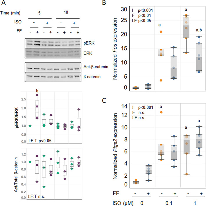
The anabolic response of aged bone to skeletal loading is typically poor. Efforts to improve mechanotransduction in aged bone have met with limited success. This study investigated whether the bone response to direct skeletal loading is improved by reducing sympathetic suppression of osteoblastic bone formation via β2AR. To test this possibility, we treated aged wild-type C57BL/6 mice with a selective β2AR antagonist, butaxamine (Butax), before each of nine bouts of cantilever bending of the right tibia. Midshaft periosteal bone formation was assessed by dynamic histomorphometry of loaded and contralateral tibias. Butax treatment did not alter osteoblast activity of contralateral tibias. Loading alone induced a modest but significant osteogenic response. However, when loading was combined with Butax pretreatment, the anabolic response was significantly elevated compared with loading preceded by saline injection. Subsequent studies in osteoblastic cultures revealed complex negative interactions between adrenergic and mechanically induced intracellular signaling. Activation of β2AR by treatment with the β1, β2-agonist isoproterenol (ISO) before fluid flow exposure diminished mechanically stimulated ERK1/2 phosphorylation in primary bone cell outgrowth cultures and AKT phosphorylation in MC3T3-E1 pre-osteoblast cultures. Expression of mechanosensitive Fos and Ptgs2 genes was enhanced with ISO treatment and reduced with flow in both MC3T3-E1 and primary cultures. Finally, co-treatment of MC3T3-E1 cells with Butax reversed these ISO effects, confirming a critical role for β2AR in these responses. In combination, these results demonstrate that selective inhibition of β2AR is sufficient to enhance the anabolic response of the aged skeleton to loading, potentially via direct effects upon osteoblasts. © 2022 The Authors. JBMR Plus published by Wiley Periodicals LLC on behalf of American Society for Bone and Mineral Research.
Keywords: BONE‐BRAIN‐NERVOUS SYSTEM INTERACTIONS; EXERCISE; OSTEOBLASTS; PRECLINICAL STUDIES; TRANSCRIPTION FACTORS.
© 2022 The Authors. JBMR Plus published by Wiley Periodicals LLC on behalf of American Society for Bone and Mineral Research.
Conflict of interest statement
The authors have no potential or real conflicts of interest to disclose.
Figures


Similar articles
-
Static Preload Inhibits Loading-Induced Bone Formation.JBMR Plus. 2018 Oct 11;3(5):e10087. doi: 10.1002/jbm4.10087. eCollection 2019 May. JBMR Plus. 2018. PMID: 31131340 Free PMC article.
-
Proliferation and Activation of Osterix-Lineage Cells Contribute to Loading-Induced Periosteal Bone Formation in Mice.JBMR Plus. 2019 Sep 11;3(11):e10227. doi: 10.1002/jbm4.10227. eCollection 2019 Nov. JBMR Plus. 2019. PMID: 31768488 Free PMC article.
-
β-adrenergic receptor signaling regulates Ptgs2 by driving circadian gene expression in osteoblasts.J Cell Sci. 2014 Sep 1;127(Pt 17):3711-9. doi: 10.1242/jcs.148148. Epub 2014 Jul 2. J Cell Sci. 2014. PMID: 24994935
-
Formoterol and isoproterenol induce c-fos gene expression in osteoblast-like cells by activating beta2-adrenergic receptors.Bone. 1998 May;22(5):471-8. doi: 10.1016/s8756-3282(98)00026-x. Bone. 1998. PMID: 9600780
-
Liraglutide, a glucagon-like peptide-1 receptor agonist, facilitates osteogenic proliferation and differentiation in MC3T3-E1 cells through phosphoinositide 3-kinase (PI3K)/protein kinase B (AKT), extracellular signal-related kinase (ERK)1/2, and cAMP/protein kinase A (PKA) signaling pathways involving β-catenin.Exp Cell Res. 2017 Nov 15;360(2):281-291. doi: 10.1016/j.yexcr.2017.09.018. Epub 2017 Sep 15. Exp Cell Res. 2017. PMID: 28919123
Cited by
-
Effects of Selective and Nonselective Beta Blockers on Bone Mineral Density in Mexican Patients with Breast Cancer.Cancers (Basel). 2024 Aug 20;16(16):2891. doi: 10.3390/cancers16162891. Cancers (Basel). 2024. PMID: 39199661 Free PMC article.
-
The OSR9 Regimen: A New Augmentation Strategy for Osteosarcoma Treatment Using Nine Older Drugs from General Medicine to Inhibit Growth Drive.Int J Mol Sci. 2023 Oct 23;24(20):15474. doi: 10.3390/ijms242015474. Int J Mol Sci. 2023. PMID: 37895152 Free PMC article. Review.
References
-
- Dunstan CR, Somers NM, Evans RA. Osteocyte death and hip fracture. Calcif Tissue Int. 1993;53 Suppl 1:S113‐S116 discussion S6–71. - PubMed
-
- Bergman RJ, Gazit D, Kahn AJ, Gruber H, McDougall S, Hahn TJ. Age‐related changes in osteogenic stem cells in mice. J Bone Miner Res. 1996;11(5):568‐577. - PubMed
-
- Silbermann M, Weiss A, Reznick AZ, Eilam Y, Szydel N, Gershon D. Age‐related trend for osteopenia in femurs of female C57BL/6 mice. Compr Gerontol A. 1987;1(1):45‐51. - PubMed
LinkOut - more resources
Full Text Sources
Research Materials
Miscellaneous