Review
. 2021 Apr 28;1(2):82-91.
doi: 10.1159/000515461. eCollection 2021 Jun.

Infection-Related Glomerulonephritis

Affiliations
Review

Infection-Related Glomerulonephritis

Mazdak A Khalighi et al. Glomerular Dis. .

Abstract

Background: There has been a long, storied relationship between various bacterial infections and glomerular injury, which is now encompassed under the term of infection-related glomerulonephritis (GN). The clinical and pathologic manifestations vary depending on the duration, magnitude, and underlying pathogen associated with the inciting infectious process. A brief and acute episode may lead to a self-limiting glomerular manifestation while a chronic or repetitive infection can result in persistent and irreversible injury. In this review, we will discuss the clinical and pathologic findings associated with the infection-related glomerulonephritides.

Summary: An acute exudative GN with an influx of neutrophils is the most characteristic morphologic alteration associated with infection-related glomerular injury. The immunofluorescence staining pattern often reveals prominent complement component C3 deposition in both capillary walls and mesangial regions with or without accompanying immunoglobulin. Large subepithelial electron-dense deposits known as "humps" are the hallmark ultrastructural finding; however, these features can also be present in C3 glomerulopathies, which are often triggered by infections and may have similar underlying abnormalities in alternative pathway complement activation. In addition, other glomerular injuries can simultaneously be present along with infection-related GN, such as diabetic nephropathy, lupus nephritis, or immunoglobulin A nephropathy, constituting a true diagnostic challenge for the pathologist.

Key messages: Bacterial infection-related GN represents a spectrum of glomerular injury with variable clinical and pathologic presentations. The pathologic findings can show overlap with other glomerular diseases, and different forms of infection-related GN vary in terms of prognosis and treatment approach.

Keywords: Complement; Kidney biopsy; Renal pathology.


Conflict of interest statement

Anthony Chang has served as a consultant for Amicus Therapeutics, Alexion Pharmaceuticals, GlaxoSmithKline, and PathAI. He is a member of the speakers bureau for Alexion Pharmaceuticals. He receives royalties from Elsevier.

Figures

Fig. 1
Fig. 1
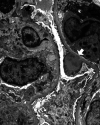
These glomeruli demonstrate global endocapillary hypercellularity primarily due to a prominent influx of neutrophils, which imparts an accentuation of the lobularity of the glomerular tuft and is descriptively also termed an acute exudative GN (hematoxylin and eosin, original magnification ×400). GN, glomerulonephritis.
Fig. 2
Fig. 2
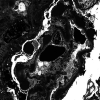
Aggregates of neutrophils in tubules with neutrophilic tubulitis can be numerous, which may be due to rupture of the glomerular basement membranes and glomerular crescents (not shown) as the possible source. Interstitial neutrophilic infiltration (not shown) may occur when tubular rupture occurs. These findings can mimic acute pyelonephritis (Jones methenamine silver, original magnification ×400).
Fig. 3
Fig. 3
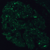
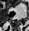
Diffuse granular C3 immunofluorescence staining along the glomerular capillaries can impart a “garland” pattern as it outlines the capillary walls and the larger globules may offer the first clue regarding the presence of subepithelial “humps” that can be subsequently visualized by electron microscopy (original magnification ×600).
Fig. 4
Fig. 4
Immunofluorescence microscopy for C3 can reveal a “starry-sky” pattern when there is both mesangial and capillary staining, but the degree of staining is not as prominent as the “garland” pattern seen in Figure 4 (original magnification ×600).
Fig. 5
Fig. 5
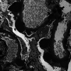
Numerous large subepithelial electron-dense deposits (arrowheads) with broad bases and an absence of basement membrane reaction to the deposit are located along the glomerular basement membrane. Some appear to be floating in the urinary space but simply represent tangential sectioning of the subepithelial hump-shaped deposits. Scale bar, 5 μm.
Fig. 6
Fig. 6
Many large subepithelial electron-dense deposits (arrowheads) often known as “humps” due to their shape as they protrude into the overlying podocyte cytoplasm. Additional aggregates of mesangial electron-dense deposits (arrows) are also noted. Scale bar, 2 μm.
Fig. 7
Fig. 7
This subepithelial “hump” (arrow) has a divot instead of a broad base, which can often be observed with infection-related GN. An adjacent neutrophil (arrowhead) may be responding to the subepithelial deposit, which may not be completely shielded from the circulation unlike the subepithelial deposits of membranous nephropathy that often do not elicit an inflammatory response. GN, glomerulonephritis.
Fig. 8
Fig. 8
Another subepithelial “hump” with variable electron density that has an unusually shaped divot at the base which appears to insert into the glomerular basement membrane (white arrow). A transmembranous electron-dense deposit (black arrow) and subendothelial (arrowhead) deposits are also present in adjacent glomerular capillaries.
Fig. 9
Fig. 9
An electron-dense deposit (arrow) is located in the mesangial notch while another large subepithelial “hump” (arrowhead) has a divot that traverses the entire width of the glomerular basement membrane as the base of the deposit appears to rest on the endothelial cell. Scale bar, 2 μm.
Fig. 10
Fig. 10
A large subepithelial “hump” (arrowhead) is located in the notch region where the glomerular basement membrane inserts in the mesangium. A few transmembranous electron-dense deposits (arrows) also accompany the large subepithelial “hump.” Scale bar, 1 μm.
Fig. 11
Fig. 11
Many large subepithelial electron-dense deposits (arrows) have a vague substructural appearance. Scale bar, 2 μm.
Fig. 12
Fig. 12
Several subepithelial electron-dense deposits (arrows) reveal variable electron lucency with a vague substructural organization of the deposits. A neutrophil is present in the capillary lumen. Scale bar, 2 μm.
Fig. 13
Fig. 13
Many small subepithelial electron-dense deposits (arrowheads) are scattered along the glomerular basement membrane in these glomerular capillaries and some deposits have a hint of basement membrane reaction, which can be very difficult to distinguish from the early stage of membranous nephropathy. Scale bar, 5 μm.
Fig. 14
Fig. 14
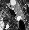
Massive accumulation of subendothelial electron-dense deposits (arrows) with focal duplication of the glomerular basement membrane correlates with the presence of “wire loops” in this unusual variant of IgA-dominant infection-related GN. GN, glomerulonephritis.
Fig. 15
Fig. 15
Numerous electron-dense deposits within several adjacent glomerular capillaries occupy the subendothelial region and extend into the lumina, which correlate with the presence of hyaline “thrombi” or pseudothrombi (arrows) in an unusual variant of IgA-dominant infection-related GN. Scale bar, 5 μm. GN, glomerulonephritis.

References

    1. Nasr SH, Markowitz GS, Stokes MB, Said SM, Valeri AM, D'Agati VD. Acute postinfectious glomerulonephritis in the modern era: experience with 86 adults and review of the literature. Medicine. 2008;87((1)):21–32. - PubMed
    1. Kambham N. Postinfectious glomerulonephritis. Adv Anat Pathol. 2012;19((5)):338–347. - PubMed
    1. Hunt EAK, Somers MJG. Infection-related glomerulonephritis. Pediatr Clin North Am. 2019;66((1)):59–72. - PubMed
    1. Courser WG, Johnson RJ. The etiology of glomerulonephritis: roles of infection and autoimmunity. Kidney Int. 2014;86:905–914. - PubMed
    1. Khalighi MA, Wang S, Henriksen KJ, Bock M, Keswani M, Meehan SM, et al. Revisiting post-infectious glomerulonephritis in the emerging era of C3 glomerulopathy. Clin Kidney J. 2016;9((3)):397–402. - PMC - PubMed