Review
. 2021 May 19;1(2):92-104.
doi: 10.1159/000516103. eCollection 2021 Jun.

Renal Disease in Cryoglobulinemia

Affiliations
Review

Renal Disease in Cryoglobulinemia

Thomas Menter et al. Glomerular Dis. .

Abstract

Background: Renal disease in cryoglobulinemia is difficult to grasp and diagnose because it is rare, serological testing is challenging and prone to artifacts, and its morphology is shared by other renal diseases resulting in a spectrum of differential diagnoses. On occasion, a definitive diagnosis cannot even be rendered after immunofluorescence and electron microscopic studies.

Summary: Based on kidney biopsies seen in our routine diagnostic and referral practice, we discuss and illustrate various morphological patterns of renal injury associated with cryoglobulins. We outline key pathophysiologic and clinical aspects associated with cryoglobulinemia induced renal disease and describe morphologic changes with a focus on electron microscopy. We present our practical, morphology-based approach to diagnostic decision-making with special consideration of differential diagnoses and disease mimickers. Since cryoglobulins are rarely tested for prior to kidney biopsy, pathologists and clinicians alike must have a high level of suspicion when interpreting renal biopsies and managing patients.

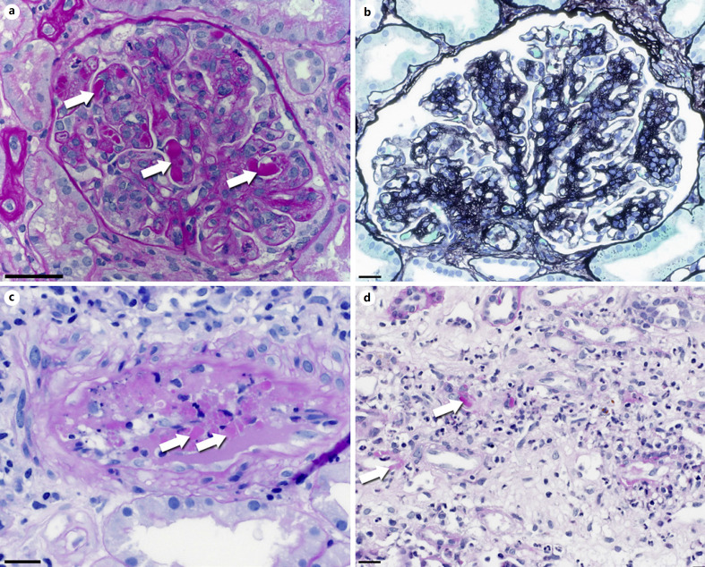
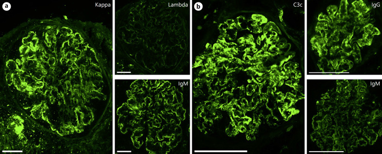
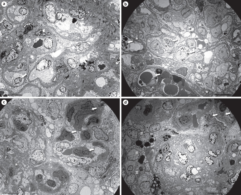
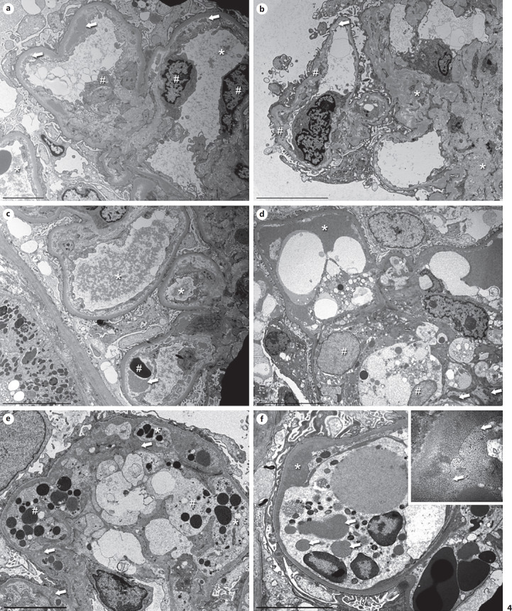
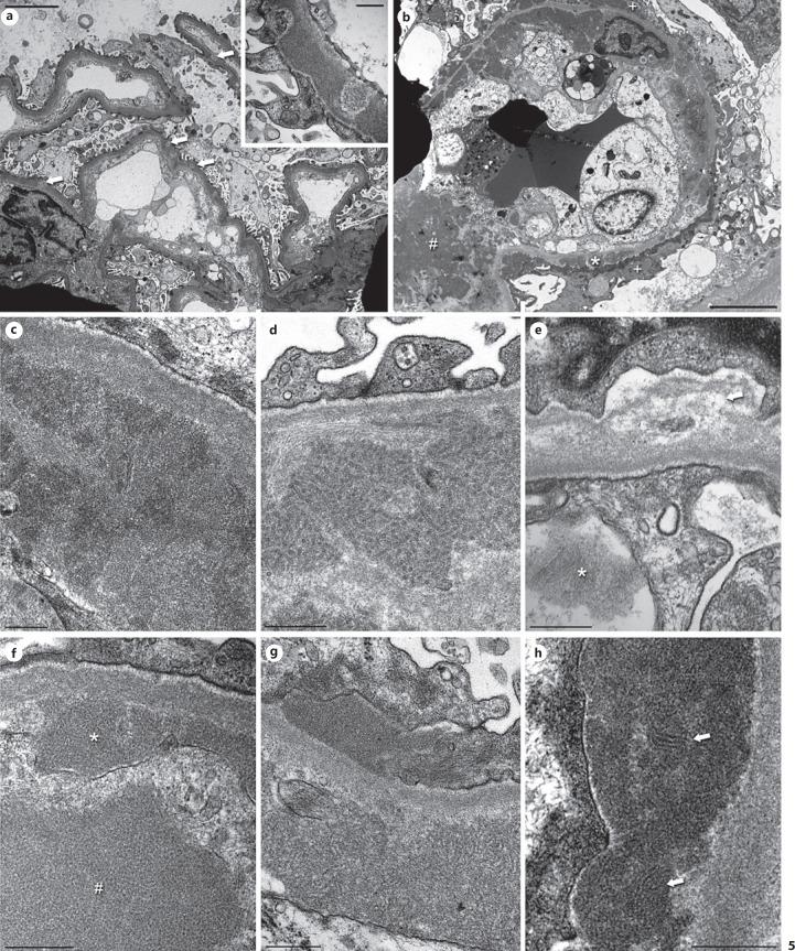
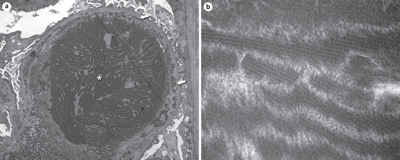
Key messages: Cryoglobulinemia-associated glomerulonephritis (GN) is a multifactorial disease which is important to recognize for clinical practice. Morphological features suggestive of cryoglobulinemia-associated GN include a pattern of membranoproliferative GN with abundance of monocytes and the presence of (pseudo)thrombi. By electron microscopy, the main diagnostic features are a prominent infiltration of monocytes/macrophages and the presence of mesangial and subendothelial deposits with frequently curved microtubular/cylindrical and annular substructures.

Keywords: Electron microscopy; Glomerulonephritis; Hepatitis C; Structured deposits.


Conflict of interest statement

The authors have no conflicts of interest to declare.

Figures

Fig. 1
Fig. 1
Light microscopy of cryoglobulinemic GN. a Early stage of MPGN in cryoglobulinemic GN with lobular architecture, prominent endocapillary thrombi (arrows), and hypercellularity. PAS stain. Original magnification 400-fold, bar = 50 µm. b Fully developed MPGN with expansion of the hypercellular mesangial matrix and double contours of the peripheral capillaries with cell interposition. The endocapillary cell proliferation is less pronounced. No thrombi. Methanamine stain. Original magnification 400-fold, bar = 20 µm. c Cryoglobulinemic vasculitis in an interlobular artery. Immune complexes are visible as PAS-positive material (arrows) in contrast to the PAS-negative fibrin precipitates. PAS stain. Original magnification 400-fold, bar = 20 µm. d Cryoglobulinemic capillaritis in the kidney medulla. A mixed, partly leukocytoclastic inflammatory infiltrate is visible within the interstitium. Some of the intact capillaries contain PAS-positive immune-complexes (arrows). PAS stain. Original magnification 400-fold, bar = 20 µm.
Fig. 2
Fig. 2
Immunofluorescence findings in cryoglobulinemic GN. a Type I cryoglobulins in a patient with splenic marginal zone ­lymphoma and viral hepatitis. The peripheral deposits consist of IgM-kappa. No lambda light chains are detected. Direct immunofluorescence. Original magnification 400-fold, bar = 50 µm. b Mixed cryoglobulins in a patient with Sjögren's syndrome. Mesangial and peripheral deposition of complement C3c, IgG, and to a lesser extent also IgM. Both kappa and lambda light chains were present (not shown). Direct immunofluorescence. Original magnification 400-fold, bar = 100 µm.
Fig. 3
Fig. 3
Electron microscopic findings in cryoglobulinemic GN at low power. a Mesangioproliferative cryoglobulinemic GN in a 61-year-old man with HCV-HIV-coinfection. The normal glomerular structures are preserved. The mesangial matrix is increased with an increase in the number of sectioned nuclei. The capillary lumina are open, and the endothelial cells are mildly swollen. In the lower left part, one endothelial cell nucleus is located in the periphery (arrow). Prominent monocyte/macrophage infiltration is not present. Original magnification 1,400-fold, bar = 10 µm. b Typical “nightmare” picture cryoglobulinemic MPGN in a 71-year-old woman with HCV-infection. Additional pictures of the same biopsy are shown in Figure 4d and f. Recognition of the lamina densa of the original basement membrane gives an overview. The image contains only little mesangial matrix (asterisks) infiltrated by macrophages. The capillary lumina are filled with swollen endothelial cells (hash signs) and many macrophages containing variable electron dense lysosomes (arrows). At this magnification, it is impossible to discriminate thrombi, deposits, and phagocytosed material. Even erythrocytes are difficult to differentiate. Original magnification 1,400-fold, bar = 10 µm. c A partly organized mesangiolysis (asterisk) on the left side of the picture in a 59-year-old man with HCV and a splenic marginal cell lymphoma. Within the mesangiolysis, it is impossible to differentiate the various cell types at this magnification. On the right side, a peripheral capillary is visible with massively swollen endothelial cells (hash signs) containing a more lucid cytoplasm compared to the monocytes in the lumen (arrows) as well as adjacent to the glomerular basement membrane. Original magnification 1,400-fold, bar = 10 µm. d A segmental sclerosis (asterisk) in the lower half of the picture in a 44-year-old woman with HCV-HIV-coinfection. In contrast to (c), the mesangiolysis shown here is older and contains much more mesangial matrix, and the macrophages have less cytoplasm. On the left side, a cellular crescent is visible (hash signs). The focus of the EM investigation should be the better-preserved capillaries seen in the upper right half. Some of the podocytes shown there contain myelin figures (arrows) raising the possibility of an additional Fabry's disease. Original magnification 1,400-fold, bar = 10 µm. HCV, hepatitis C virus.
Fig. 4
Fig. 4
Characteristic EM features of cryoglobulinemic GN at medium power: MPGN pattern, endothelial cell swelling, monocyte/macrophage infiltration, remodeling of the GBM, and electron-dense deposits. a Biopsy of a 70-year-old woman with HCV and M. Waldenström. Pictures of the same biopsy can be seen in c and Figure 5e. Two capillary loops with typical MPGN features. The flocculent material in the lumina probably resembles precipitated cryoglobulins, especially the larger aggregates (asterisk). Other loops showed typical thrombi (see c). The endothelial cells (hash signs) are mildly swollen with a segmental loss of the fenestration. Beneath them, an irregular newly formed basement membrane is visible. The lamina rara interna is widened and contains subendothelial deposits of variable size (arrows). Only little cell interposition is visible, most likely extensions of the endothelial cells. Podocytes show a partial loss of their foot processes. Original magnification 3,500-fold, bar = 5 µm. b Biopsy of a 61-year-old woman with HCV. The mesangium is markedly expanded due to an increased matrix (asterisks). The peripheral capillaries show prominent remodeling with very thin stretches of GBM and splitting of the lamina densa, probably due to resolved deposits. Segmentally, cellular interposition is present in the periphery (hash signs). The lamina rara interna is mildly widened with few small residual subendothelial deposits (arrow). Original magnification 2,800-fold, bar = 10 µm. c Same biopsy as in (a) and Figure 5e. The flocculent material within the lumina forms vague thrombi (asterisks), which extends below the detached endothelium (arrow) in the capillary loop on the lower right side surrounding an erythrocyte (hash sign). There is a prominent activation of the endothelium. Mesangial and subendothelial deposits are present. Original magnification 2,800-fold, bar = 10 µm. d Biopsy of a 71-year-old woman with HCV. Pictures of the same biopsy are shown in Figure 3b and (f). A compact thrombus (asterisk) is seen within the capillary lumen with an intact endothelial cell layer. In the lower half, the cytoplasm of 2 macrophages contains multiple lysosomes of variable electron density (hash signs). The mesangium to the right contains several small mesangial deposits (arrows). Original magnification 2,200-fold, bar = 10 µm. e 4th sequential biopsy of a 79-year-old woman with cryoglobulinemia in the context of a thymoma. A picture of the same biopsy can be seen in Figure 5d. This extended peripheral capillary shows macrophages (hash signs) extending below the swollen endothelium (asterisk). The macrophages have a more lucent cytoplasm and contain multiple lysosomes, mostly filled with an electron dense material. Adjacent to the macrophages, few poorly formed deposits are present (arrows). The cytoplasm of the endothelial cell appears darker. A partly detached endothelial cell is visible in the middle of the lower edge. Original magnification 4,400-fold, bar = 5 µm. f Same biopsy as in Figure 3b and (d). This tangentially sectioned capillary is filled by a macrophage with multiple lysosomes of variable sizes. The material within some of the vesicles (arrows) has an identical appearance to the adjacent electron-dense deposits (asterisk). Original magnification 5,600-fold, bar = 5 µm. Inset A high magnification picture taken at a different location reveals a blurred interface between the extracellular deposits and the macrophage. In some areas, it is impossible to delineate the cell membrane (arrows). Original magnification 36,000-fold, bar = 500 nm. HCV, hepatitis C virus.
Fig. 5
Fig. 5
Localization and different quantity and quality of deposits in cryoglobulinemic GN. a Biopsy of an 85-year-old woman with HCV and presence of cryoglobulins type II. This medium magnification shows an overall normal architecture of the capillary loops, yet the peripheral basement membranes are mildly thickened and contain several partly dissolved intramembranous deposits (arrows). The endothelium is activated. Original magnification 2,800-fold, bar = 5 µm. Inset Higher magnification of some deposits without substructures. Original magnification 36,000-fold, bar = 500 nm. b Biopsy of a 19-year-old woman with a known history of SLE, cryoglobulinemia, and increasing disease activity. In contrast to (a), this capillary loop contains abundant subendothelial (asterisk), subepithelial (plus signs), and mesangial deposits (hash sign), some of which are dissolving. The peripheral basement membranes show duplication with matrix and cellular interposition (arrow) as well as massively swollen endothelial cells, which lead to a markedly narrowed lumen. The podocytes show extensive foot process effacement. Original magnification 3,500-fold, bar = 5 µm. c Biopsy of a 55-year-old woman with essential cryoglobulinemia. This amorphous deposit shows some structures which are reminiscent of tubules and curvilinear structures, yet they are not well enough formed to consider them as structured (compare to d). Original magnification 28,000-fold, bar = 500 nm. d 4th sequential biopsy of a 79-year-old woman with cryoglobulinemia in the context of a thymoma. A picture of the same biopsy can be seen in Figure 4e. Prototypical annular and tubular curvilinear substructures of a deposit. The diameter of the structures was 22 nm. This is a very characteristic feature of nonlupus cryoglobulinemic GN. However, these types of deposits are only present in about 50% of the patients. Original magnification 36,000-fold, bar = 500 nm. e Biopsy of a 70-year-old woman with HCV and M. Waldenström. Pictures of the same biopsy can be seen in Figure 4a and c. High magnification of a small thrombus with flocculent material in the capillary lumen consisting of fibrils (asterisk). The endothelial cells are activated, adjacent to the mesangial cell in the upper part, there is focal resorption and subsequent edema of the mesangial matrix (arrows). Original magnification 36,000-fold, bar = 500 nm. f Biopsy of a 74-year-old man with lymphoplasmacytic lymphoma. High magnification of a deposit with annular substructure (asterisk), partly within the cytoplasm of an adjacent macrophage (hash sign). Original magnification 36,000-fold, bar = 500 nm. g Biopsy of a 9-year-old girl with type III cryoglobulinemia in the context of an IgA-dominant postinfectious glomerulonephritis. High magnification of a partly amorphous, partly structured subepithelial deposit and a structured mesangial deposit consisting of fibrils with a diameter of 15–25 nm. Original magnification 28,000-fold, bar 500 nm. h Same biopsy as in (b). At high magnification, fingerprint-like tubular substructures (arrows) can be visualized in this subendothelial deposit. This is a typical finding in patients with lupus nephritis and has been described both with and without cryoglobulinemia. Original magnification 28,000-fold, bar = 500 nm. HCV, hepatitis C virus.
Fig. 6
Fig. 6
Electron microscopic findings in cryocrystalglobulinemia. a Biopsy of a 64-year-old woman with type I cryoglobulinemia in the context of kappa light chain myeloma. A glomerular capillary is occluded by a thrombus consisting of mostly structured electron dense material arranged in thick parallel bundles (asterisk). The endothelial cells are intact but have lost their fenestration. There is a duplication of the glomerular basement membrane with frequent subendothelial deposits (arrows) and cell interposition. Original magnification 7,200-fold. b High magnification reveals a crystalline substructure with a regular periodicity of more and less electron dense areas within the bundles. Original magnification 140,000-fold.

Similar articles

Cited by

References

    1. Basile U, Torti E, Dell'Abate MT, Colacicco L, Gulli F, Zuppi C, et al. Pre-analytical phase in cryoglobulin detection: an alternative method for sample transport. Clin Chem Lab Med. 2016;54:e123–e126. - PubMed
    1. Brouet JC, Clauvel JP, Danon F, Klein M, Seligmann M. Biologic and clinical significance of cryoglobulins. A report of 86 cases. Am J Med. 1974;57((5)):775–788. - PubMed
    1. Kolopp-Sarda MN, Nombel A, Miossec P. Cryoglobulins today: detection and immunologic characteristics of 1,675 positive samples from 13,439 patients obtained over six years. Arthritis Rheumatol. 2019;71((11)):1904–1912. - PubMed
    1. Roccatello D, Saadoun D, Ramos-Casals M, Tzioufas AG, Fervenza FC, Cacoub P, et al. Cryoglobulinaemia. Nat Rev Dis Primers. 2018;4((1)):11. - PubMed
    1. Strait RT, Posgai MT, Mahler A, Barasa N, Jacob CO, Köhl J, et al. Erratum: IgG1 protects against renal disease in a mouse model of cryoglobulinaemia. Nature. 2015;526((7575)):728–724. - PubMed