

Paraspinal muscle abscesses in children - A case report and review of the literature
- PMID: 36751462
- PMCID: PMC9899453
- DOI: 10.25259/SNI_994_2022
Paraspinal muscle abscesses in children - A case report and review of the literature
Abstract
Background: Few pediatric cases with myositis and abscesses of the paraspinal muscles have been previously reported.
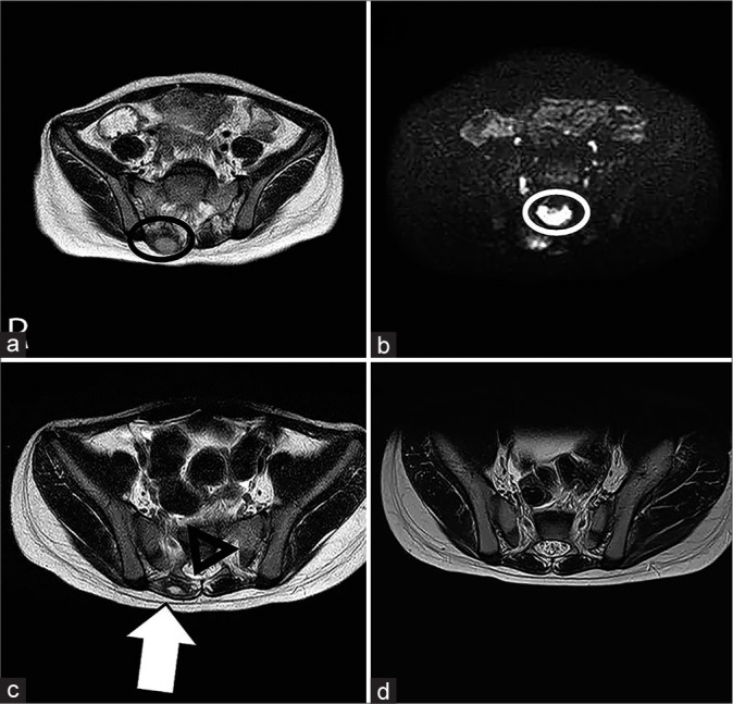
Case description: We herein report on a 3-year-old patient who developed an epidural abscess in a paraspinal muscle abscess, after a complication of infectious impetigo due to atopic dermatitis. The child improved through the administration of an antibacterial drug. The median age of seven patients with a history of paraspinal muscle inflammation and muscle abscess was 12 years old (3-15-years-old), few of which had underlying diseases, with methicillin-sensitive Staphylococcus aureus being the main causative agent. Although the prognosis was well in many cases following the administration of antibacterial agents, there were also cases in which extensive lesions were found and neurological sequelae remained.
Conclusion: The current case was the youngest case ever reported of a paraspinal muscle abscess. Although pediatric cases are rare, in the episode of a fever of unknown origin and difficulty walking, paraspinal muscle abscesses should be cited as a differential diagnosis and appropriate early diagnostic imaging and treatment should be performed.
Keywords: Abscess; Epidural abscess; Low back pain; Paraspinal muscles.
Copyright: © 2023 Surgical Neurology International.
Conflict of interest statement
There are no conflicts of interest.
Figures
Similar articles
-
Paraspinal abscess communicated with epidural abscess after extra-articular facet joint injection.Yonsei Med J. 2007 Aug 31;48(4):711-4. doi: 10.3349/ymj.2007.48.4.711. Yonsei Med J. 2007. PMID: 17722247 Free PMC article.
-
A Rare Case of the Ipsilateral Paraspinal Muscle Abscess Communicating with a Psoas Major Abscess: A Case Report.J Orthop Case Rep. 2025 Jan;15(1):150-154. doi: 10.13107/jocr.2025.v15.i01.5158. J Orthop Case Rep. 2025. PMID: 39801857 Free PMC article.
-
Pediatric spinal epidural abscess in an immunocompetent host without risk factors: Case report and review of the literature.IDCases. 2015 Oct 22;2(4):109-15. doi: 10.1016/j.idcr.2015.09.008. eCollection 2015. IDCases. 2015. PMID: 26793474 Free PMC article.
-
Review and case report demonstrate that spontaneous spinal epidural abscesses are rare but dangerous in childhood.Acta Paediatr. 2019 Jan;108(1):28-36. doi: 10.1111/apa.14579. Epub 2018 Oct 15. Acta Paediatr. 2019. PMID: 30222897 Review.
-
A Staphylococcus aureus paraspinal abscess associated with epidural analgesia in labour.Anaesthesia. 2001 Sep;56(9):873-8. doi: 10.1046/j.1365-2044.2001.02130-2.x. Anaesthesia. 2001. PMID: 11531675 Review.
References
-
- Bowen DK, Mitchell LA, Burnett MW, Rooks VJ, Martin JE. Spinal epidural abscess due to tropical pyomyositis in immunocompetent adolescents. J Neurosurg Pediatr. 2010;6:33–7. - PubMed
-
- Chiedozi LC. Pyomyositis. Review of 205 cases in 112 patients. Am J Surg. 1979;137:255–9. - PubMed
-
- Miller NJ, Duncan RD, Huntley JS. The conservative management of primary pyomyositis abscess in children: Case series and review of the literature. Scott Med J. 2011;56:181. - PubMed
Publication types
LinkOut - more resources
Full Text Sources