. 2023 Aug;18(8):1734-1742.
doi: 10.4103/1673-5374.360249.

Knockdown of NADPH oxidase 4 reduces mitochondrial oxidative stress and neuronal pyroptosis following intracerebral hemorrhage

Affiliations

Knockdown of NADPH oxidase 4 reduces mitochondrial oxidative stress and neuronal pyroptosis following intracerebral hemorrhage

Bo-Yun Ding et al. Neural Regen Res. 2023 Aug.

Abstract

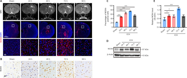
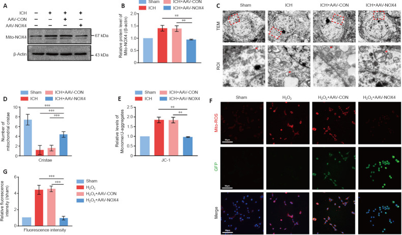
Intracerebral hemorrhage is often accompanied by oxidative stress induced by reactive oxygen species, which causes abnormal mitochondrial function and secondary reactive oxygen species generation. This creates a vicious cycle leading to reactive oxygen species accumulation, resulting in progression of the pathological process. Therefore, breaking the cycle to inhibit reactive oxygen species accumulation is critical for reducing neuronal death after intracerebral hemorrhage. Our previous study found that increased expression of nicotinamide adenine dinucleotide phosphate oxidase 4 (NADPH oxidase 4, NOX4) led to neuronal apoptosis and damage to the blood-brain barrier after intracerebral hemorrhage. The purpose of this study was to investigate the role of NOX4 in the circle involving the neuronal tolerance to oxidative stress, mitochondrial reactive oxygen species and modes of neuronal death other than apoptosis after intracerebral hemorrhage. We found that NOX4 knockdown by adeno-associated virus (AAV-NOX4) in rats enhanced neuronal tolerance to oxidative stress, enabling them to better resist the oxidative stress caused by intracerebral hemorrhage. Knockdown of NOX4 also reduced the production of reactive oxygen species in the mitochondria, relieved mitochondrial damage, prevented secondary reactive oxygen species accumulation, reduced neuronal pyroptosis and contributed to relieving secondary brain injury after intracerebral hemorrhage in rats. Finally, we used a mitochondria-targeted superoxide dismutase mimetic to explore the relationship between reactive oxygen species and NOX4. The mitochondria-targeted superoxide dismutase mimetic inhibited the expression of NOX4 and neuronal pyroptosis, which is similar to the effect of AAV-NOX4. This indicates that NOX4 is likely to be an important target for inhibiting mitochondrial reactive oxygen species production, and NOX4 inhibitors can be used to alleviate oxidative stress response induced by intracerebral hemorrhage.

Keywords: NADPH oxidase 4; caspase 1; caspase4/11; gasdermin D; intracerebral hemorrhage; mitochondria reactive oxygen species inhibitor; neuronal pyroptosis; neuronal tolerance; reactive oxygen species; secondary brain injury.


Conflict of interest statement

None

Figures

Figure 1
Figure 1
NOX4 expression reaches the peak at 72 hours in the brain after intracerebral hemorrhage (ICH) in rats. (A) MRI of rat brain at four time points (24, 48, 72, 96 hours) and the corresponding immunofluorescence expression of NOX4 (red) after ICH in rats. Images of selected regions (middle row, white squares) are shown at a higher magnification (bottom row). Compared with the other four groups, the 72 h group had the largest cerebral hemorrhage edema and the highest NOX4 expression. Scale bars: 1000 μm or 50 μm. (B) Representative immunohistochemical images of NOX4 expression at four time points (24, 48, 72, 96 hours) after ICH. NOX4 expression was highest in the 72 h group compared with the other four groups. Scale bars: 20 μm. (C) Percentage of NOX4 positive cells in rat brain from B. (D) Western blot analysis of NOX4 expression in rat brain after ICH in the perilesional basal ganglia at 24, 48, 72 and 96 hours following injury. (E) Quantitative analysis of NOX4 protein levels from D. Data are expressed as the mean ± SEM (n = 3 per group). *P < 0.05, ***P < 0.001 (one-way analysis of variance with Dunnett’s multiple comparison test). ICH: Intracerebral hemorrhage; MRI: magnetic resonance imaging; NOX4: nicotinamide adenine dinucleotide phosphate oxidase 4.
Figure 2
Figure 2
NOX4 knockdown relieves oxidative stress and enhances the neuronal tolerance to oxidative stress after intracerebral hemorrhage (ICH). (A) Schematic of AAV injection and ICH modeling in rats. (B) Fluorescence images of rat brain with AAV-NOX4 treatment (green). Selected regions (white squares) are shown at a higher magnification (a and b). Scale bars: 2000 µm or 20 µm. (C) Relative mRNA level of NOX4 (normalized by levels in the sham group) in rat brain with AAV-NOX4 or AAV-CON treatment before ICH. (D) Relative reactive oxygen species (ROS) levels (normalized by levels in the sham group) after NOX4 knockdown (n = 5 per group). (E) Western blot analysis of Nrf2 and Keap-1 expression of ICH rats in the indicated groups. (F, G) Quantitative analysis of Nrf2 (F) and Keap-1 (G) protein levels (normalized to levels in the sham group) as shown in E (n = 4 per group). (H) Representative immunofluorescence images of Nrf2 (pink) and NeuN (red) in ICH rats treated by AAV-CON/AAV-NOX4 (green, GFP) or as indicated. The expression of Nrf2 in neuron in the ICH and ICH + AAV-CON groups were decreased, while that in the ICH + AAV-NOX4 group was increased. Scale bars: 20 µm. (I) Relative fluorescence intensity of Nrf2 (normalized by levels in the sham group) after treatment as indicated (n = 5 per group). Data are expressed as the mean ± SEM. *P < 0.05, ***P < 0.001 (one-way analysis of variance with Dunnett’s multiple comparison test). AAV-CON: AAV9-U6-shRNA (Scramble)-CMV-GFP; AAV-NOX4: AAV9-U6-shRNA (NOX4)-CMV-GFP; ICH: intracerebral hemorrhage; Keap-1: Kelch like ECH associated protein 1; NOX4: nicotinamide adenine dinucleotide phosphate oxidase 4; Nrf2: nuclear factor erythroid2-related factor 2; ROS: reactive oxygen species.
Figure 3
Figure 3
Knockdown of NOX4 reduces cerebral edema and neuronal pyroptosis after intracerebral hemorrhage (ICH). (A) MRI of rat brains in the four groups (sham, ICH, ICH + AAV-CON, ICH + AAV-NOX4). The peribleeding edema was aggravated in the ICH and ICH+AAV-CON groups and alleviated in the ICH + AAV-NOX4 group. (B) The brain water content of rat brains evaluated using Evans blue staining (n = 6 per group). (C) Evans blue staining of AAV-treated rat brains in four groups (sham, ICH, ICH + AAV-CON, ICH + AAV-NOX4). Evans blue dye leakage was aggravated in the ICH and ICH + AAV-CON groups and was alleviated in the ICH + AAV-NOX4 group. Scale bars: 1000 µm. (D) Evans blue intensity in different groups (n = 5 per group). (E) Nissl staining of rat brains in the four groups (sham, ICH, ICH + AAV-CON, ICH + AAV-NOX4). Images of selected regions (black squares) are shown at a higher magnification. The number of Nissl bodies was reduced and the neurons were severely damaged in the ICH and ICH + AAV-CON groups; in the AAV-NOX4 group, the number of Nissl bodies and the morphology of neurons tended to be normal. Scale bars: 1000 µm, 50 µm, 20 µm. (F) Number of Nissl bodies (n = 5 per group). (G) mNSS in the four treatment groups (n = 6 per group). (H) TEM images of neurons in ICH rats in the four groups (sham, ICH, ICH + AAV-CON, ICH + AAV-NOX4). Images of selected regions (red squares) are shown at a higher magnification. Integrity of the neuronal membrane is shown by red lines. The membrane of neurons in ICH and ICH+AAV-CON groups was broken, while the membrane of neurons in ICH + AAV-NOX4 group was intact. Scale bars: 5 µm or 1 µm. (I) Images of primary neurons in ICH rats in the four groups under a light microscope. Images of selected regions (red squares) are shown at a higher magnification. Many vacuoles were found in the neurons of ICH and ICH + AAV-CON groups, and the cell membrane was damaged; no vacuoles were generated in the ICH + AAV-NOX4 group, and the cell membrane was smooth. Scale bars: 50 µm or 10 µm. Data are expressed as the mean ± SEM. *P < 0.05, **P < 0.01, ***P < 0.001 (one-way analysis of variance with Dunnett’s multiple comparison test). AAV-CON: AAV9-U6-shRNA (Scramble)-CMV-GFP; AAV-NOX4: AAV9-U6-shRNA (NOX4)-CMV-GFP; ICH: intracerebral hemorrhage; mNSS: modified neurological severity scores; MRI: magnetic resonance imaging; NOX4: nicotinamide adenine dinucleotide phosphate oxidase 4; ROI: region of interest; TEM: transmission electron microscopy.
Figure 4
Figure 4
Knockdown of NOX4 ameliorates neuronal pyroptosis through caspase 1/GSDMD-N and caspase4/11/GSDMD-C pathways after ICH. (A) Western blot analysis of Procaspase4/11, Cleaved caspase4/11 and Procaspase1 expression in the indicated groups. (B) Western blot analysis of GSDMD-FL, GSDMD-N and GSDMD-C expression in the indicated groups. (C–H) Quantitative analysis of Procaspase4/11 (C), Cleaved caspase4/11 (D), Procaspase1 (E), GSDMD-FL (F), GSDMD-N (G) and GSDMD-C (H) protein levels shown in A and B (n = 4 per group). (I) Representative immunofluorescence images showing the immunoreactivity of GSDMD (red) in neurons (NeuN-positive cells, yellow) in the indicated groups (AAV, GFP, green). The expression of GSDMD in the ICH and ICH + AAV-CON groups were increased, while that in the ICH + AAV-NOX4 group was decreased. Scale bars: 20 µm. (J) Representative immunohistochemical images illustrate the changes in the GSDMD protein expression in the indicated groups. The expression of GSDMD in the ICH and ICH + AAV-CON groups was increased, while that in the ICH + AAV-NOX4 group was decreased. Scale bars: 50 µm. (K, L) Percentage of GSDMD-positive cells in immunofluorescence (K) and immunohistochemical (L) images (n = 5 per group). Data are expressed as the mean ± SEM. ***P < 0.001 (one-way analysis of variance with Dunnett’s multiple comparison test). AAV-CON: AAV9-U6-shRNA (Scramble)-CMV-GFP; AAV-NOX4: AAV9-U6-shRNA (NOX4)-CMV-GFP; GSDMD: gasdermin D; ICH: intracerebral hemorrhage; MRI: magnetic resonance imaging; NOX4: nicotinamide adenine dinucleotide phosphate oxidase 4.
Figure 5
Figure 5
NOX4 knockdown reduces the NOX4 expression and reactive oxygen species (ROS) production in mitochondria, and improves neuronal mitochondrial function after intracerebral hemorrhage (ICH). (A) Western blot analysis of mitochondrial NOX 4 (Mito-NOX4) expression in the indicated groups. (B) Quantitative analysis of Mito-NOX4 protein levels (normalized by levels in the sham group) shown in A (n = 4 per group). (C) Images of neuron mitochondria in the indicated groups under the TEM. Images of selected regions (red squares) are shown at a higher magnification. Red arrows indicate the mitochondria of different treatment groups. The mitochondria in the ICH and ICH + AAV-CON groups were severely damaged and mitochondrial crest was reduced; these effects were not observed in the ICH + AAV-NOX4 group. Scale bars: 5 µm or 1 µm. (D) The number of mitochondrial cristae in different groups (n = 5 per group). (E) Comparison of mitochondrial membrane potential (JC-1) among different groups (n = 5 per group). (F) Representative immunofluorescence images showing the immunoreactivity of Mito-ROS (red) in PC-12 cells infected with AAV-CON or AAV-NOX4 (green, GFP) after treatment with H2O2. Mito-ROS in the H2O2 and H2O2 + AAV-CON groups was increased, while that in the H2O2 + AAV-NOX4 group was decreased. Scale bars: 50 µm. (G) The fluorescent intensity of mitochondrial ROS in different groups shown in (F) (n = 5 per group). Data are expressed as the mean ± SEM. **P < 0.01, ***P < 0.001 (one-way analysis of variance with Dunnett’s multiple comparison test). AAV-CON: AAV9-U6-shRNA (Scramble)-CMV-GFP; AAV-NOX4: AAV9-U6-shRNA (NOX4)-CMV-GFP; ICH: intracerebral hemorrhage; Mito-ROS: ROS in mitochondria; NOX4: nicotinamide adenine dinucleotide phosphate oxidase 4; TEM: transmission electron microscopy.
Figure 6
Figure 6
Mito-TEMPO reduces neuronal pyroptosis and oxidative stress response after intracerebral hemorrhage (ICH). (A) Evans blue staining following application of the mitochondrial ROS inhibitor Mito-TEMPO and treatment as shown in the indicated groups. Evans blue dye leakage was the strongest in the ICH group; leakage was not observed in the ICH + AAV-NOX4 and ICH + Mito-TEMPO groups, both under naked eye and fluorescence microscope. Scale bars: 100 µm. (B) Relative fluorescence intensity (normalized by levels in the sham group) of Evans blue (n = 5 per group). (C) Western blot analysis of Mito-NOX4 expression in the brain of ICH rats treated by AAV-NOX4 or Mito-TEMPO. (D) Relative protein levels (normalized by levels in the sham group) of Mito-NOX4 shown in C (n = 4 per group). (E) Nissl staining of rat brain in the indicated groups. The ICH group had the fewest Nissl bodies and the most serious neuron morphology damage; this was not observed in the ICH + AAV-NOX4 and ICH + Mito-TEMPO groups. Scale bars: 50 µm. (F) Quantitative analysis of the number of Nissl bodies shown in B (n = 5 per group). (G) Relative Mito-H2O2 levels (normalized by levels in the sham group) of mitochondria after treatment with AAV-NOX4 or Mito-TEMPO (n = 5 per group). (H) Representative immunofluorescence images illustrate the changes in GSDMD (red) expression of neurons (NeuN-positive cells, yellow) of ICH rats after treatment with AAV-NOX4 (green, GFP) or Mito-TEMPO. The expression of GSDMD in the ICH group was increased, while that in the ICH + AAV-NOX4 and ICH + Mito-TEMPO groups was decreased. Scale bars: 50 µm. (I) Percentage of GSDMD-positive cells among neurons after treatment with AAV-NOX4 or Mito-TEMPO (n = 5 per group). Data are expressed as means ± SEM. **P < 0.01, ***P < 0.001 (one-way analysis of variance with Dunnett’s multiple comparison test). AAV-NOX4: AAV9-U6-shRNA (NOX4)-CMV-GFP; GSDMD: gasdermin D; ICH: intracerebral hemorrhage; Mito-H2O2: H2O2 in mitochondria; Mito-NOX4: NOX4 in mitochondria; Mito-TEMPO: mitochondria-targeted superoxide dismutase mimetic; NOX4: nicotinamide adenine dinucleotide phosphate oxidase 4.

Similar articles

Cited by

References

    1. Bao Q, Hu P, Xu Y, Cheng T, Wei C, Pan L, Shi J. Simultaneous blood-brain barrier crossing and protection for stroke treatment based on edaravone-loaded ceria nanoparticles. ACS Nano. (2018);12:6794–6805. - PubMed
    1. Bao WD, Zhou XT, Zhou LT, Wang F, Yin X, Lu Y, Zhu LQ, Liu D. Targeting miR-124/Ferroportin signaling ameliorated neuronal cell death through inhibiting apoptosis and ferroptosis in aged intracerebral hemorrhage murine model. Aging Cell. (2020);19:e13235. - PMC - PubMed
    1. Bergsbaken T, Cookson BT. Macrophage activation redirects yersinia-infected host cell death from apoptosis to caspase-1-dependent pyroptosis. PLoS Pathog. (2007);3:e161. - PMC - PubMed
    1. Bergsbaken T, Fink SL, Cookson BT. Pyroptosis:host cell death and inflammation. Nat Rev Microbiol. (2009);7:99–109. - PMC - PubMed
    1. Brown GC, Borutaite V. There is no evidence that mitochondria are the main source of reactive oxygen species in mammalian cells. Mitochondrion. (2012);12:1–4. - PubMed