. 2023 Feb 8;8(3):e163150.
doi: 10.1172/jci.insight.163150.

Autoantibodies are highly prevalent in non-SARS-CoV-2 respiratory infections and critical illness

Affiliations

Autoantibodies are highly prevalent in non-SARS-CoV-2 respiratory infections and critical illness

Allan Feng et al. JCI Insight. .

Abstract

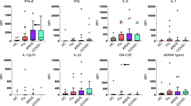
The widespread presence of autoantibodies in acute infection with SARS-CoV-2 is increasingly recognized, but the prevalence of autoantibodies in non-SARS-CoV-2 infections and critical illness has not yet been reported. We profiled IgG autoantibodies in 267 patients from 5 independent cohorts with non-SARS-CoV-2 viral, bacterial, and noninfectious critical illness. Serum samples were screened using Luminex arrays that included 58 cytokines and 55 autoantigens, many of which are associated with connective tissue diseases (CTDs). Samples positive for anti-cytokine antibodies were tested for receptor blocking activity using cell-based functional assays. Anti-cytokine antibodies were identified in > 50% of patients across all 5 acutely ill cohorts. In critically ill patients, anti-cytokine antibodies were far more common in infected versus uninfected patients. In cell-based functional assays, 11 of 39 samples positive for select anti-cytokine antibodies displayed receptor blocking activity against surface receptors for Type I IFN, GM-CSF, and IL-6. Autoantibodies against CTD-associated autoantigens were also commonly observed, including newly detected antibodies that emerged in longitudinal samples. These findings demonstrate that anti-cytokine and autoantibodies are common across different viral and nonviral infections and range in severity of illness.

Keywords: Adaptive immunity; Autoimmunity; Bacterial infections; Infectious disease; Influenza.


Figures

Figure 1
Figure 1. High prevalence of ACA in hospitalized ICU patients.
(A) Heatmap representing serum IgG ACA discovered using a 58-plex array of cytokines, chemokines, growth factors, and receptors. Stanford ICU patients who were infected with viruses, bacteria, fungi, or a combination of pathogens (n = 115), Stanford ICU patients with no evidence for infection (n = 52), and HC (n = 22) were analyzed for ACA. Cytokines are grouped on the y axis by category (IFNs, ILs, and other cytokines/growth factors/receptors). Colors indicate ACA whose MFI measurements are > 5 SD (red) or < 5 SD (black) above the average MFI for HC. MFIs < 3,000 were excluded. (B) Tukey box plots comparing MFI data from HC and Stanford ICU patients for the 7 antigens for which statistically significant differences (P < 0.05) were determined between patient groups using 2-tailed Wilcoxon rank-sum tests with Bonferroni correction. The middle line represents the median, while the lower and upper hinges correspond to the first and third quartiles. The upper whisker extends from the hinge to 1.5 times the interquartile range (IQR) above the 75th percentile MFI value, and the lower whisker extends from the hinge to 1.5 times the IQR below the 25th percentile MFI value.
Figure 2
Figure 2. IgG anti-cytokine autoantibodies in serum from patients with ARDS or patients acutely infected with influenza virus.
Tukey box plots comparing MFI data for 8 cytokines in patients with influenza (n = 25) and patients with ARDS (n = 17), both collected prior to the COVID-19 pandemic; patients with ARDS who were COVID-19 (n = 19); and HC (n = 11). One COVID-19 PCR-negative patient from the Marburg cohort had high levels of antibodies targeting SARS-CoV-2 proteins from our viral array and was excluded from this figure and other analyses (Supplemental Figure 6). The middle line represents the median, while the lower and upper hinges correspond to the first and third quartiles. The upper whisker extends from the hinge to 1.5 times the IQR above the 75th percentile MFI value, and the lower whisker extends from the hinge to 1.5 times the IQR below the 25th percentile MFI value. Black arrows indicate a serum sample with receptor-blocking activity (see Figure 4). Individual MFI values 1.5 times the IQR above the 75th percentile or 1.5 times the IQR below the 25th percentile are displayed as dots. MFI is shown on the y axis, which is hatched to reflect outlier samples with very high MFI. Cohorts are shown on the x axis.
Figure 3
Figure 3. Newly detectable autoantibodies in acutely infected patients with influenza.
(A) Longitudinal measurements of specific ACA over time in acutely infected patients (n = 40). Serum was collected at 3 time points for 18 influenza individuals, at 2 time points for 13 influenza individuals, and at only the first time point for 9 individuals. The first time point (T1) is from the day that the patient was admitted to the hospital and diagnosed with influenza. T2 and T3 refer to approximately 1 week and 1 month, respectively, following hospital admission. Black arrows indicate a serum sample with receptor blocking activity (see Figure 4). (B) Newly detectable IgG autoantibodies recognize CTD autoantigens. Line plots display MFI levels of antibodies targeting traditional autoantigens that are inducible (SRP54 in individual AA19; TPO in individual AA23), fluctuate (TPO in individual AA13), or do not change significantly over time (most individuals with TPO autoantibodies, and 2 individuals with anti–PDC-E2).
Figure 4
Figure 4. Cell-based cytokine receptor-blocking assays.
(A) FACS plots of IFN-α2, IL-6, and GM-CSF signaling assays. Cells were treated with media only; commercial blocking antibody or 10% positive control serum from a patient with atypical mycobacterial infection (AMI); 10% healthy control serum; or 10% test serum. Cells were treated with patient serum or a control in the unstimulated condition and with both cytokine and patient serum or a control in the stimulated condition. (B) Blocking activity of patient serum on cells in cytokine signaling assays, reported as percentage of pSTAT+ cells in the unstimulated and stimulated condition. Patient sera were from patients with influenza (nMarburg = 4, nAthens = 5), Stanford ICU (ninfected = 19, nnoninfected = 2), or ARDS (n = 8) . For IFN-α2 and IFN-α8, results shown represent 2 independent experiments (Supplemental Figure 7). HC and positive controls (PC; commercially available antibody or prototype patient serum with known blocking activity) are also included. (C) Neutralization activity to IFN-α2, IFN-γ, and IFN-λ3 in the serum samples of 2 patients. IFN-α2, IFN-γ, and IFN-λ3 were incubated with heat-inactivated serum from donor AMI (PC) and donor SU047 (infected Stanford ICU cohort) and added to HAP1 reporter cells. The serum samples were prepared and tested with a 5-fold serial dilution on HAP1 reporter cells. Final concentrations of IFN-α2, IFN-γ, and IFN-λ3 in the culture were 40 U/mL, 8 U/mL, and 1 ng/mL, respectively (Supplemental Figure 8). The percentages of GFP+ HAP1 reporter cells were evaluated 22–24 hours after the incubation with flow cytometry.

References

    1. Zuo Y, et al. Prothrombotic autoantibodies in serum from patients hospitalized with COVID-19. Sci Transl Med. 2020;12(570):eabd3876. doi: 10.1126/scitranslmed.abd3876. - DOI - PMC - PubMed
    1. Knight JS, et al. The intersection of COVID-19 and autoimmunity. J Clin Invest. 2021;131(24):154886. doi: 10.1172/JCI154886. - DOI - PMC - PubMed
    1. Bastard P, et al. Autoantibodies against type I IFNs in patients with life-threatening COVID-19. Science. 2020;370(6515):eabd4585. - PMC - PubMed
    1. Machhi J, et al. The natural history, pathobiology, and clinical manifestations of SARS-CoV-2 infections. J Neuroimmune Pharmacol. 2020;15(3):359–386. doi: 10.1007/s11481-020-09944-5. - DOI - PMC - PubMed
    1. Chang SE, et al. New-onset IgG autoantibodies in hospitalized patients with COVID-19. Nat Commun. 2021;12(1):5417. doi: 10.1038/s41467-021-25509-3. - DOI - PMC - PubMed

Publication types