. 2023 Apr 3;38(4):671-685.
doi: 10.1093/humrep/dead017.

Cortisol/glucocorticoid receptor: a critical mediator of the ovulatory process and luteinization in human periovulatory follicles

Affiliations

Cortisol/glucocorticoid receptor: a critical mediator of the ovulatory process and luteinization in human periovulatory follicles

H Jeon et al. Hum Reprod. .

Abstract

Study question: Do cortisol/glucocorticoid receptors play an active role in the human ovary during ovulation and early luteinization?

Summary answer: The ovulatory hCG stimulation-induced glucocorticoid receptor signaling plays a crucial role in regulating steroidogenesis and ovulatory cascade in human periovulatory follicles.

What is known already: Previous studies reported an increase in cortisol levels in the human follicular fluid after the LH surge or ovulatory hCG administration. However, little is known about the role of cortisol/glucocorticoid receptors in the ovulatory process and luteinization in humans.

Study design, size, duration: This study was an experimental prospective clinical and laboratory-based study. An in vivo experimental study was accomplished utilizing the dominant ovarian follicles from 38 premenopausal women undergoing laparoscopic sterilization. An in vitro experimental study was completed using the primary human granulosa/lutein cells (hGLC) from 26 premenopausal women undergoing IVF.

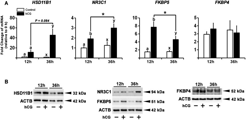
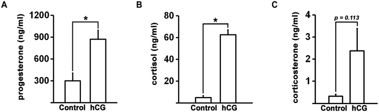
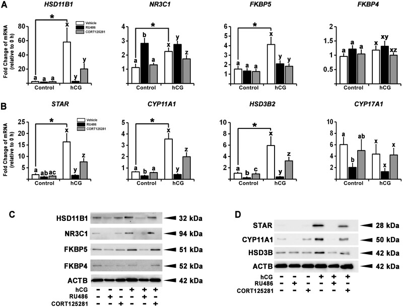
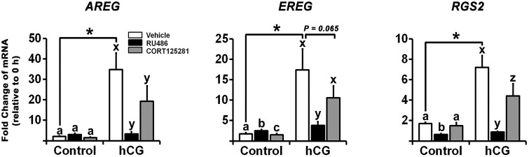
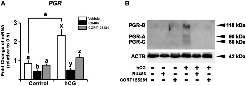
Participants/materials, setting, methods: This study was conducted in a private fertility clinic and academic medical centers. Dominant ovarian follicles were collected before the LH surge and at defined times after hCG administration from women undergoing laparoscopic sterilization. Primary hGLC were collected from women undergoing IVF. hGLC were treated without or with hCG in the absence or presence of RU486 (20 µM; dual antagonist for progesterone receptor and glucocorticoid receptor) or CORT125281 (50 µM; selective glucocorticoid receptor antagonist) for 12 or 36 h. The expression of genes involved in glucocorticoid receptor signaling, steroidogenesis, and ovulatory cascade was studied with RT-quantitative PCR and western blotting. The production of cortisol, corticosterone, and progesterone was assessed by hormone assay kits.

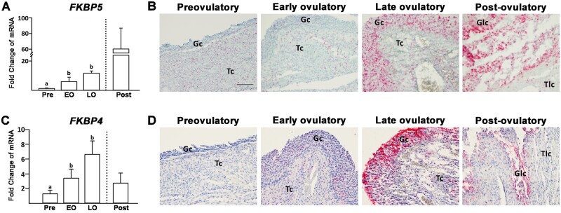
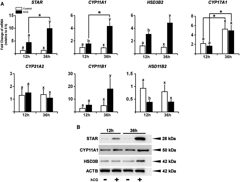
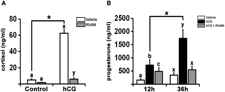
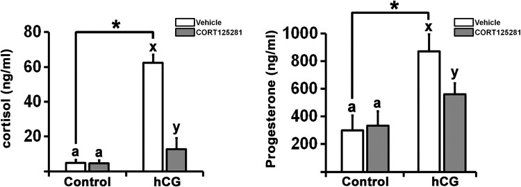
Main results and the role of chance: hCG administration upregulated the expression of hydroxysteroid 11-beta dehydrogenase 1 (HSD11B1), nuclear receptor subfamily 3 group C member 1 (NR3C1), FKBP prolyl isomerase 5 (FKBP5), and FKBP prolyl isomerase 4 (FKBP4) in human ovulatory follicles and in hGLC (P < 0.05). RU486 and CORT125281 reduced hCG-induced increases in progesterone and cortisol production in hGLC. The expression of genes involved in glucocorticoid receptor signaling, steroidogenesis, and the key ovulatory process was reduced by RU486 and/or CORT125281 in hGLC.

Large scale data: N/A.

Limitations, reasons for caution: The role of cortisol/glucocorticoid receptors demonstrated using the hGLC model may not fully reflect their physiological roles in vivo.

Wider implications of the findings: Successful ovulation and luteinization are essential for female fertility. Women with dysregulated cortisol levels often suffer from anovulatory infertility. Deciphering the functional role of glucocorticoid receptor signaling in human periovulatory follicles enhances our knowledge of basic ovarian physiology and may provide therapeutic insights into treating infertility in women.

Study funding/competing interest(s): This study was supported by P01HD71875 (to M.J., T.E.C., and M.B.) and R01HD096077 (to M.J.) from the Foundation for the National Institutes of Health and the BTPSRF of the University of Kentucky Markey Cancer Center (P30CA177558). The authors report no competing interests.

Trial registration number: N/A.

Keywords: cortisol; glucocorticoid receptor; granulosa cells; humans; luteinization; ovulation.


Conflict of interest statement

The authors declare no conflict of interest.

Figures

Figure 1.
Figure 1.
The expression of HSD11B1 and NR3C1 in periovulatory follicles after hCG administration. Dominant follicles were retrieved from the ovaries of women undergoing laparoscopic tubal sterilization before the LH surge or at defined h after recombinant hCG administration and divided into four phases: pre (Pre, n = 6), early (EO, n = 5), late (LO, n = 6), and post- (Post, n = 2) ovulatory phase. (A, C) The levels of mRNA for hydroxysteroid 11-beta dehydrogenase 1 (HSD11B1) and nuclear receptor subfamily 3 group C member 1 (NR3C1) were measured using quantitative PCR in granulosa cells isolated from a dominant follicle collected at Pre, EO, and LO and whole follicles retrieved at Post and normalized to the levels of glyceraldehyde-3-phosphate dehydrogenase (GAPDH) mRNA in each sample. The levels of each transcript were presented as fold change to Pre levels. Results are shown as the means ± SEM. Bars with no common superscripts are significantly different (P <0.05). The dotted line indicates that the post-ovulatory phase was not included in the statistical analysis due to the small sample size. (B, D) Paraffin-embedded sections of dominant follicles (n = 2/Pre, n = 4/EO, n = 4/LO, n = 1/Post) were subjected to immunohistochemical analyses to detect HSD11B1 and NR3C1. Pink/purple staining indicates positive signals of the targeted proteins. HSD11B1 shows staining in the cytoplasm, whereas NR3C1 shows staining in the nucleus. All sections were lightly stained with hematoxylin (blue) for nuclear counterstaining. Gc, granulosa cells; Tc, theca cells; Glc, granulosa-lutein cells; Tlc, theca-lutein cells. Scale bar, 500 µm for all the images.
Figure 2.
Figure 2.
The expression of FKBP5 and FKBP4 in periovulatory follicles after hCG administration. Dominant follicles were retrieved from the ovaries of women undergoing laparoscopic tubal sterilization before the LH surge or at defined h after recombinant hCG administration and divided into four phases: pre (Pre, n = 6), early (EO, n = 5), late (LO, n = 6), and post- (Post, n = 2) ovulatory phase. (A, C) The levels of mRNA for FKBP prolyl isomerase 5 (FKBP5) and FKBP prolyl isomerase 4 (FKBP4) were measured using quantitative PCR in granulosa cells isolated from a dominant follicle collected at Pre, EO, and LO and whole follicles retrieved at Post and normalized to the levels of glyceraldehyde-3-phosphate dehydrogenase (GAPDH) mRNA in each sample. The levels of each transcript were presented as fold change to Pre levels. Results are shown as the means ± SEM. Bars with no common superscripts are significantly different (P < 0.05). The dotted line indicates that the post-ovulatory phase was not included in the statistical analysis due to the small sample size. (B, D) Paraffin-embedded sections of dominant follicles (n = 2/Pre, n = 4/EO, n = 4/LO, n = 1/Post) were subjected to immunohistochemical analyses to detect FKBP5 and FKBP4. Pink/purple staining indicates positive signals of the targeted proteins. FKBP5 shows staining in the nucleus, and FKBP4 exhibits cytoplasmic staining. All sections were lightly stained with hematoxylin (blue) for nuclear counterstaining. Gc, granulosa cells; Tc, theca cells; Glc, granulosa-lutein cells; Tlc, theca-lutein cells. Scale bar, 100 µm for all the images.
Figure 3.
Figure 3.
The effect of hCG on the expression of HSD11B1, NR3C1, FKBP5, and FKBP4 in human granulosa/lutein cells (hGLC). Granulosa/lutein cells obtained from IVF patients were cultured for 6 days and then treated without or with hCG (1 IU/ml) for 12 or 36 h. (A) The levels of mRNA for hydroxysteroid 11-beta dehydrogenase 1 (HSD11B1), nuclear receptor subfamily 3 group C member 1 (NR3C1), FKBP prolyl isomerase 5 (FKBP5), and FKBP prolyl isomerase 4 (FKBP4) were measured by quantitative PCR and normalized to the levels of glucosamine-6-phosphate deaminase 2 (GNPDA2) mRNA in each sample (n = 5 individual patient samples). Results are shown as the means ± SEM. Different superscripts in each time point or bars with asterisk are significantly different (P < 0.05). (B) HSD11B1, NR3C1, FKBP5, and FKBP4 were detected by western blots using whole cell extracts. The membrane was reprobed with a monoclonal antibody against actin beta (ACTB) to assess the loading of protein in each lane. The experiments were repeated four times, each with individual patient samples.
Figure 4.
Figure 4.
The effect of hCG on the production of progesterone, cortisol, and corticosterone in human granulosa/lutein cells (hGLC). Granulosa/lutein cells obtained from IVF patients were cultured for 6 days and then treated without or with hCG (1 IU/ml) for 36 h. Concentrations of (A) progesterone, (B) cortisol, and (C) corticosterone in conditioned media were measured (n = 6 individual patient samples/progesterone, n = 8 individual patient samples/cortisol, n = 4 individual patient samples/corticosterone). Results are shown as the means ± SEM. Bars with asterisk are significantly different (P < 0.05).
Figure 5.
Figure 5.
Steroidogenesis pathway showing factors involved in synthesizing progesterone and cortisol from cholesterol. STAR, steroidogenic acute regulatory protein; CYP11A1, cytochrome P450 family 11 subfamily A member 1; CYP17A1, cytochrome P450 family 17 subfamily A member 1; HSD3B, hydroxy-delta-5-steroid dehydrogenase, 3 beta- and steroid delta-isomerase; CYP21A2, cytochrome P450 family 21 subfamily A member 2; CYP11B1, cytochrome P450 family 11 subfamily B member 1; HSD11B1, hydroxysteroid 11-beta dehydrogenase 1; HSD11B2, hydroxysteroid 11-beta dehydrogenase 2.
Figure 6.
Figure 6.
The effect of hCG on the expression of factors involved in cortisol synthesis in human granulosa/lutein cells (hGLC). Granulosa/lutein cells obtained from IVF patients were cultured for 6 days and then treated without or with hCG (1 IU/ml) for 12 or 36 h. (A) The levels of mRNA for steroidogenic acute regulatory protein (STAR), cytochrome P450 family 11 subfamily A member 1 (CYP11A1), hydroxy-delta-5-steroid dehydrogenase, 3 beta- and steroid delta-isomerase 2 (HSD3B2), cytochrome P450 family 17 subfamily A member 1 (CYP17A1), cytochrome P450 family 21 subfamily A member 2 (CYP21A2), cytochrome P450 family 11 subfamily B member 1 (CYP11B1), and hydroxysteroid 11-beta dehydrogenase 2 (HSD11B2) were measured by quantitative PCR and normalized to the levels of glucosamine-6-phosphate deaminase 2 (GNPDA2) mRNA in each sample (n = 5 individual patient samples). Results are shown as the means ± SEM. Different superscripts in each time point or bars with asterisk are significantly different (P < 0.05). (B) STAR, CYP11A1, and HSD3B were detected by western blots using whole cell extracts. The membrane was reprobed with a monoclonal antibody against actin beta (ACTB) to assess the loading of protein in each lane. The experiments were repeated four times, each with individual patient samples.
Figure 7.
Figure 7.
The effects of RU486 or CORT125281 on the expression of genes involved in glucocorticoid action and synthesis in human granulosa/lutein cells (hGLC). Granulosa/lutein cells obtained from IVF patients were cultured for 6 days and then treated without or with RU486 (mifepristone, 20 µM) or CORT125281 (50 µM) in the absence or presence of hCG (1 IU/ml) for 36 h. (A, B) The levels of mRNA for hydroxysteroid 11-beta dehydrogenase 1 (HSD11B1), nuclear receptor subfamily 3 group C member 1 (NR3C1), FKBP prolyl isomerase 5 (FKBP5), FKBP prolyl isomerase 4 (FKBP4), steroidogenic acute regulatory protein (STAR), cytochrome P450 family 11 subfamily A member 1 (CYP11A1), hydroxy-delta-5-steroid dehydrogenase, 3 beta- and steroid delta-isomerase 2 (HSD3B2), and cytochrome P450 family 17 subfamily A member 1 (CYP17A1) were measured by quantitative PCR and normalized to the levels of glucosamine-6-phosphate deaminase 2 (GNPDA2) mRNA (n = 5–6 individual patient samples). Results are shown as the means ± SEM. Bars with asterisk between treatments or with no common superscripts within each treatment are significantly different (P < 0.05). (C, D) HSD11B1, NR3C1, FKBP5, FKBP4, STAR, CYP11A1, and HSD3B were detected by western blots using whole cell extracts. The membrane was reprobed with a monoclonal antibody against actin beta (ACTB) to assess the loading of protein in each lane. The experiments were repeated three times, each with individual patient samples.
Figure 8.
Figure 8.
The effects of RU486 on the production of cortisol and progesterone in human granulosa/lutein cells (hGLC). Granulosa/lutein cells obtained from IVF patients were cultured for 6 days and then treated without or with RU486 (20 µM) in the absence or presence of hCG (1 IU/ml) for 12 and/or 36 h. (A) The concentrations of cortisol in 36 h conditioned media were measured (n = 8 individual patient samples). (B) The concentrations of progesterone in 12 and 36 h conditioned media were measured (n = 5 individual patient samples). Results are shown as the means ± SEM. Bars with asterisk or no common superscripts are significantly different (P < 0.05).
Figure 9.
Figure 9.
The effects of CORT125281 on the production of cortisol and progesterone in human granulosa/lutein cells (hGLC). Granulosa/lutein cells obtained from IVF patients were cultured for 6 days and then treated without or with CORT125281 (50 µM) in the absence or presence of hCG (1 IU/ml) for 36 h. The concentrations of cortisol and progesterone inconditioned media were measured (n = 8 individual patient samples/cortisol, n = 6 individual patient samples/progesterone). Results are shown as the means ± SEM. Bars with asterisk or no common superscripts are significantly different (P < 0.05).
Figure 10.
Figure 10.
The effects of CORT125281 on the expression of ovulatory genes in human granulosa/lutein cells (hGLC). Granulosa/lutein cells obtained from IVF patients were cultured for 6 days and then treated without or with RU486 (20 µM) or CORT125281 (50 µM) in the absence or presence of hCG (1 IU/ml) for 36 h. The levels of mRNA for amphiregulin (AREG), epiregulin (EREG), and regulator of G protein signaling 2 (RGS2) were measured by quantitative PCR and normalized to the levels of glucosamine-6-phosphate deaminase 2 (GNPDA2) mRNA (n = 6 individual patient samples). Results are shown as the means ± SEM. Bars with asterisk or no common superscripts are significantly different (P < 0.05).
Figure 11.
Figure 11.
The effects of RU486 or CORT125281 on the hCG-induced PGR expression in human granulosa/lutein cells (hGLC). Granulosa/lutein cells obtained from IVF patients were cultured for 6 days and then treated without or with RU486 (20 µM) or CORT125281 (50 µM) in the absence or presence of hCG (1 IU/ml) for 36 h. (A) The levels of mRNA for progesterone receptor (PGR) were measured by quantitative PCR and normalized to the levels of glucosamine-6-phosphate deaminase 2 (GNPDA2) mRNA (n = 6 individual patient samples). Results are shown as the means ± SEM. Bars with asterisk between treatments or no common superscripts within each treatment are significantly different (P < 0.05). (B) PGR was detected by western blots using whole cell extracts. The membrane was reprobed with a monoclonal antibody against actin beta (ACTB) to assess the loading of protein in each lane. The experiments were repeated three times, each with individual patient samples.

Similar articles

Cited by

References

    1. Akison LK, Robker RL.. The critical roles of progesterone receptor (PGR) in ovulation, oocyte developmental competence and oviductal transport in mammalian reproduction. Reprod Domest Anim 2012;47(Suppl 4):288–296. - PubMed
    1. Al-Alem L, Puttabyatappa M, Rosewell K, Brannstrom M, Akin J, Boldt J, Muse K, Curry TE Jr. Chemokine ligand 20: a signal for leukocyte recruitment during human ovulation? Endocrinology 2015;156:3358–3369. - PMC - PubMed
    1. Amin M, Simerman A, Cho M, Singh P, Briton-Jones C, Hill D, Grogan T, Elashoff D, Clarke NJ, Chazenbalk GD. et al. 21-Hydroxylase-derived steroids in follicles of nonobese women undergoing ovarian stimulation for in vitro fertilization (IVF) positively correlate with lipid content of luteinized granulosa cells (LGCs) as a source of cholesterol for steroid synthesis. J Clin Endocrinol Metab 2014;99:1299–1306. - PMC - PubMed
    1. Andersen CY, Morineau G, Fukuda M, Westergaard LG, Ingerslev HJ, Fiet J, Byskov AG.. Assessment of the follicular cortisol:cortisone ratio. Hum Reprod 1999;14:1563–1568. - PubMed
    1. Binder EB. The role of FKBP5, a co-chaperone of the glucocorticoid receptor in the pathogenesis and therapy of affective and anxiety disorders. Psychoneuroendocrinology 2009;34(Suppl 1):S186–S195. - PubMed

Publication types