. 2023 Feb 28;42(2):112048.
doi: 10.1016/j.celrep.2023.112048. Epub 2023 Feb 6.

Genetic determinants of host tropism in Klebsiella phages

Affiliations

Genetic determinants of host tropism in Klebsiella phages

Beatriz Beamud et al. Cell Rep. .

Abstract

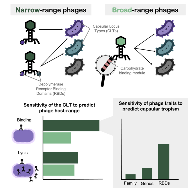
Bacteriophages play key roles in bacterial ecology and evolution and are potential antimicrobials. However, the determinants of phage-host specificity remain elusive. Here, we isolate 46 phages to challenge 138 representative clinical isolates of Klebsiella pneumoniae, a widespread opportunistic pathogen. Spot tests show a narrow host range for most phages, with <2% of 6,319 phage-host combinations tested yielding detectable interactions. Bacterial capsule diversity is the main factor restricting phage host range. Consequently, phage-encoded depolymerases are key determinants of host tropism, and depolymerase sequence types are associated with the ability to infect specific capsular types across phage families. However, all phages with a broader host range found do not encode canonical depolymerases, suggesting alternative modes of entry. These findings expand our knowledge of the complex interactions between bacteria and their viruses and point out the feasibility of predicting the first steps of phage infection using bacterial and phage genome sequences.

Keywords: CP: Microbiology; Klebsiella; bacterial capsule; bacteriophage; depolymerase; genomics; horizontal gene transfer; host range; microbial evolution.


Conflict of interest statement

Declaration of interests The authors declare no competing interests.

Figures

None
Graphical abstract
Figure 1
Figure 1
Diversity of isolated Klebsiella phages IGS values were used to define 13 similarity groups and to classify phages in the closest viral family, subfamily (if applicable), and genus after comparison with database sequences. Phages were named after the first letter of the associated viral family, the number of the phylogenetic group, and a letter to identify each species or strain. A, Autographiviridae; D, Drexlerviridae; M, Myoviridae; P, Podoviridae; S, Siphoviridae. The genome organization of each phage is shown. Arrows represent coding sequences (CDSs) and are colored based on functional categories of PHROGS. See also Figures S1–S4 and Table S1.
Figure 2
Figure 2
Host tropism of isolated Klebsiella phages Each row represents a phage (n = 46) and each column a bacterial strain (n = 138). For phages, the dendrogram constructed from IGS values is shown, whereas the maximum likelihood (ML) core phylogeny is shown for bacteria. Original host-phage pairs in which each phage was primarily isolated are indicated in purple. The black histogram and associated numbers indicate the host breadth of each phage in the 138 total bacterial strains analyzed, excluding isolation hosts. Three independent replicates were performed for all phage-bacteria pairs (n = 6,348). For a subset of interactions (n = 1,242), three additional replicates were performed using a different concentration of phage (Table S1). Finally, a subset of phage-bacteria of interest was reassayed twice by spotting several dilutions (n = 95). A given bacterial strain was considered to be susceptible to a given phage (green boxes) if at least two-thirds of the replicates of the spot assay were positive (clear spot, turbid spot, or single plaques). See also Figures S5 and S6 and Tables S1, S2, and S3.
Figure 3
Figure 3
Predictability of phage-host interactions based on bacterial capsular type (A) Sensitivity or true positive rate (TPR) of different host traits to predict positive spot tests for non-S8/S9 phages and phages from groups S8/S9. Three independent calculations were performed. Bars represent the standard error (n = 3). Asterisks represent the mean TPR values obtained with randomized data (null expectation). CLT, capsular locus type; OLT, O-antigen locus type; ST, sequence type. Other: presence of 45 secondary receptors (pooled). (B) Dpo activity of halo-producing non-S8/S9 phages and non-halo-producing S8/S9 phages relative to non-inoculated controls. Each point represents the mean activity of two biological replicates of a given phage-bacterium combination. Four technical replicates were performed, and the median obtained. Asterisks indicate statistical significance (t test: p < 0.001). (C) Predicted architecture of TSPs for non-S8/S9 phages with or without Dpos. The primary TSP is attached via an N-terminal anchor and the secondary TSP via a conserved short peptide. Top: podoviruses. Bottom: sipho- and myoviruses. Only one TSP copy is shown for simplicity. See also Table S1 and Figure S7A.
Figure 4
Figure 4
RBD-based prediction of phage capsular tropism (A) Left panel: sensitivity or TPR of different phage traits to predict infections in capsular types of bacteria. Three independent calculations were performed. For RBDs, the average of the 35 RBD clusters across calculations is shown. Asterisks represent the mean TPR values obtained with randomized data (null expectation). Right panel: TPR for each individual RBD cluster. Bars represent the standard error of independent calculations (n = 3). (B) Representation of the 35 RBD similarity clusters obtained. RBDs are named by the phage name followed by the phage protein in which they were detected. Clusters labeled in red are those for which a consensus capsular tropism could be obtained. Colors of nodes indicate phage taxonomic families. Sequences obtained in the present work are shown as triangles and previous published sequences as circles. Edge weight (width) represents amino acid identity between RBDs within the same cluster. Each RBD is annotated with the corresponding phage CLT tropism. White labels represent tropisms that did not match the consensus CLT of an RBD cluster. See also Table S4.
Figure 5
Figure 5
Validation of RBD-based capsular tropism prediction using prophage sequences (A) Ability of the consensus RBD clusters to predict the CLT tropism of prophages obtained from the RefSeq database as described in the main text. The percentage of total and matching predictions by bacterial species is shown. The n values indicate the total predictions for each RBD. “Other” refers to spp. within Klebsiella sp. and also from other genera. Ns denotes no significance by Fisher’s exact test (p > 0.05). (B) Distribution of RBD coverage and identity for prophage predictions. Colors indicate whether the CLT of the lysogenized host matched the consensus CLT of the RBD cluster used as a query. Hits with ≤40% RBD cover were not considered.
Figure 6
Figure 6
Non-depolymerase determinants of host tropism (A) Summary of host ranges for phages that showed both virulent and avirulent infections. Virulent infections were considered when the phage was able to produce a spot and to reduce bacterial density or produce progeny. Avirulence was considered when despite spot formation, no productive infection, and no significant effect on host density were observed. (B) Quantitation of Dpo activity and adsorption in 25 avirulent phage-host pairs. Dpo activity was quantified by comparing capsule polysaccharide levels in phage-treated versus untreated controls. Adsorption was quantified by measuring phage titer following inoculation relative to cell-free mock cultures (eclipse phase). Each point represents the mean of 3 independent replicates with 4 and 3 technical replicates for Dpo activity and adsorption, respectively. Bars represent the standard deviation between biological replicates. (C) Differential probability of resistance (dPR) for each phage-defense system. For avirulent host-phage combinations, only those in which adsorption was observed (B) were considered. Values of 1 indicate that the defense system was exclusively found in avirulent combinations. On the contrary, negative values indicate that the defense system was overrepresented in virulent combinations and thus probably did not contribute to the observed resistant phenotype. (D) Genomic comparison of the RBPs of S8/S9 phages. Domains found with InterProScan5 are shown. Pident shows the aa percentage identity after blastp comparison. See also Table S5. (E) Spotting of serial dilutions of S8/S9 phages (10–104 PFUs) as a function of the fraction of acapsular bacteria (Cap−) in the plate. Three independent replicates were performed, and a representative image was chosen. WT, wild type bacteria only; WT:Cap−, mix of WT-Cap− (1:1); Cap−, Cap only. Combinations CU630-S8c and NTUH-S9a were omitted as spots were not observed with these concentrations. See also Table S5 and Figure S7B.

References

    1. Abedon S.T. Cambridge University Press; 2008. Bacteriophage Ecology: Population Growth, Evolution, and Impact of Bacterial Viruses.
    1. Chevallereau A., Pons B.J., van Houte S., Westra E.R. Interactions between bacterial and phage communities in natural environments. Nat. Rev. Microbiol. 2022;20:49–62. doi: 10.1038/s41579-021-00602-y. - DOI - PubMed
    1. Lamy-Besnier Q., Brancotte B., Ménager H., Debarbieux L. Viral Host Range database, an online tool for recording, analyzing and disseminating virus-host interactions. Bioinformatics. 2021;37:2798–2801. doi: 10.1093/bioinformatics/btab070. - DOI - PMC - PubMed
    1. Edwards R.A., McNair K., Faust K., Raes J., Dutilh B.E. Computational approaches to predict bacteriophage-host relationships. FEMS Microbiol. Rev. 2016;40:258–272. doi: 10.1093/femsre/fuv048. - DOI - PMC - PubMed
    1. Roux S., Hallam S.J., Woyke T., Sullivan M.B. Viral dark matter and virus-host interactions resolved from publicly available microbial genomes. Elife. 2015;4:e08490. doi: 10.7554/eLife.08490. - DOI - PMC - PubMed

Publication types

LinkOut - more resources