. 2023 Feb 8;17(2):e0011055.
doi: 10.1371/journal.pntd.0011055. eCollection 2023 Feb.

Genomic and phenotypic analyses suggest moderate fitness differences among Zika virus lineages

Affiliations

Genomic and phenotypic analyses suggest moderate fitness differences among Zika virus lineages

Glenn Oliveira et al. PLoS Negl Trop Dis. .

Abstract

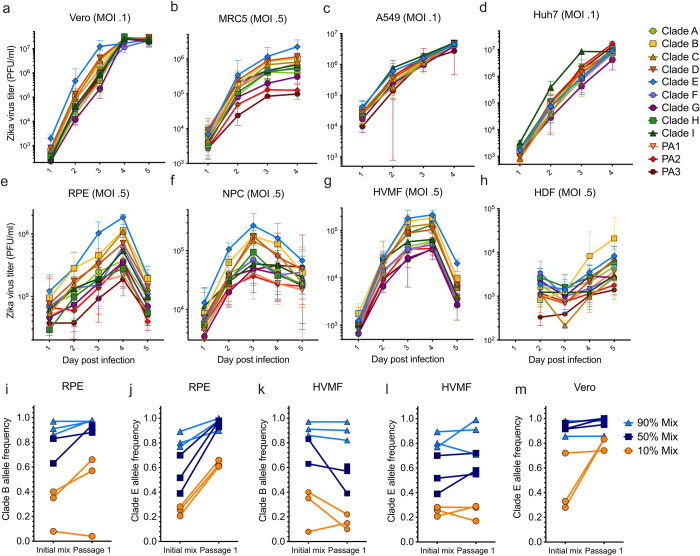
RNA viruses have short generation times and high mutation rates, allowing them to undergo rapid molecular evolution during epidemics. However, the extent of RNA virus phenotypic evolution within epidemics and the resulting effects on fitness and virulence remain mostly unknown. Here, we screened the 2015-2016 Zika epidemic in the Americas for lineage-specific fitness differences. We engineered a library of recombinant viruses representing twelve major Zika virus lineages and used them to measure replicative fitness within disease-relevant human primary cells and live mosquitoes. We found that two of these lineages conferred significant in vitro replicative fitness changes among human primary cells, but we did not find fitness changes in Aedes aegypti mosquitoes. Additionally, we found evidence for elevated levels of positive selection among five amino acid sites that define major Zika virus lineages. While our work suggests that Zika virus may have acquired several phenotypic changes during a short time scale, these changes were relatively moderate and do not appear to have enhanced transmission during the epidemic.


Conflict of interest statement

The authors have declared that no competing interests exist.

Figures

Fig 1
Fig 1. 13 major Zika virus lineages defined by nonsynonymous mutations.
(a) Zika phylogeny partitioned into 13 lineages defined by nonsynonymous mutations. Large red and white star: location of the initial infectious clone into which specific mutations were introduced. White boxes: nonsynonymous mutations introduced into the initial infectious clone to model each lineage. Red arrows: lineage-defining nonsynonymous mutations that were reverted to their ancestral states. (b) Proportions of clades circulating in countries across the Americas and Asia. Pie sizes represent the number of sequences. (Map made with the coastlines basemap from Natural Earth.) Temporal frequencies of sequenced ZIKV isolates across the Americas (c), North America (including the Caribbean and Central America) (d), South America (e) with 95% confidence intervals. Timepoint 2015- represents isolates collected during or before 2015. Timepoint 2017+ represents isolates collected during or after 2017.
Fig 2
Fig 2. Fitness assays of clade-defining infectious clones in continuous and human primary cells.
Replicative fitness of 12 clade-defining recombinant Zika viruses in (a) Vero cells, (b-d) human continuous cell lines, and (e-h) human primary cells. Growth curve points represent the average of the 5 biological replicates and error bars represent their range. Clade B and clade E viruses competed against clade A in (i, j) retinal pigment epithelial cells, (k-l) human villous mesenchymal cells, and (m) the clade E virus competed with clade G in Vero cells. All competitive fitness assays were conducted in triplicate at roughly 90%, 50%, and 10% clade-defining initial frequencies. Vero, African green monkey kidney cells; MRC-5, human lung fibroblasts; A549, human adenocarcinoma alveolar basal epithelial cells; Huh7, human hepatocyte-derived carcinoma cells; RPE, primary human retinal pigment epithelial cells; NPC, human neural progenitor cells; HVMF, primary human villous mesenchymal fibroblasts; HDF, human dermal fibroblasts.
Fig 3
Fig 3
Replicative and competitive fitness results of Zika virus isolates in human primary cells: Infections of (a) HVMF and (b) RPE cells with nine Zika virus isolates representing clades B, E, G, and I. Growth curve points represent the average of the 5 biological replicates and error bars represent their range. Zika virus isolates from clades with high fitness (clades E and B) competed against a low fitness clade (clade G) using (c-d) RPE cells and (f-h) HVMF cells. Competitive fitness assays conducted at 90%, 50%, and 10% mixtures and each in triplicate, with three separate isolates representing clade G (PA259359, PA259634, and PAN259249). RPE, human retinal pigment epithelial cells; HVMF, human villous mesenchymal fibroblasts.
Fig 4
Fig 4. Evaluation of Zika virus fitness in three mosquito cell lines and in Ae. aegypti mosquitoes.
Replicative fitness of 12 Zika virus clades among infected (a) Aag2 (Ae. aegypti), (b) U4.4 (Ae. albopictus), and (c) Hsu (Culex quinquefasciatus) cells for 3–5 replicates at a multiplicity of infection of 0.1. Shapes represent mean titers with error bars depicting the range. In vivo fitness of the 12 Zika virus clades by feeding Ae. aegypti mosquitoes (Poza Rica, Mexico) with infectious blood meals. (d) Infection (percent females with Zika virus-infected body out of the total number exposed females), (e) dissemination (percent females with Zika-virus infected legs and wings out of the total number of exposed females), and (f) transmission rates (percent females with Zika-virus infected saliva out of the total number of exposed females). Dots represent the rates expressed as percentages and error bars depict the 95% confidence intervals.
Fig 5
Fig 5. Site-specific dN/dS values across the ZIKV polyprotein.
(a) Zika virus site-specific dN/dS analysis measured using 517 Zika virus isolates. Box plots: posterior probability distributions of dN/dS values across the Zika virus genome. Red bars: clade-defining loci. (b) Posterior probability densities for the five clade-defining amino acid loci that have dN/dS values greater than one. (c) The number of synonymous, nonsynonymous, and clade-defining mutations across the Zika virus genome. Clade-specific amino acid sites have significantly higher dN/dS values (p = .0002; Mann-Whitney U test) when compared to the remaining amino acid sites with nonsynonymous mutations.

References

    1. Metsky HC, Matranga CB, Wohl S, Schaffner SF, Freije CA, Winnicki SM, et al.. Zika virus evolution and spread in the Americas. Nature. 2017;546: 411–415. doi: 10.1038/nature22402 - DOI - PMC - PubMed
    1. Grubaugh ND, Faria NR, Andersen KG, Pybus OG. Genomic Insights into Zika Virus Emergence and Spread. Cell. 2018;172: 1160–1162. doi: 10.1016/j.cell.2018.02.027 - DOI - PubMed
    1. Avelino-Silva VI, Alvarenga C, Abreu C, Tozetto-Mendoza TR, Canto CLM do, Manuli ER, et al.. Potential effect of Zika virus infection on human male fertility? Rev Inst Med Trop Sao Paulo. 2018;60: e64. doi: 10.1590/S1678-9946201860064 - DOI - PMC - PubMed
    1. Parra B, Lizarazo J, Jiménez-Arango JA, Zea-Vera AF, González-Manrique G, Vargas J, et al.. Guillain-Barré Syndrome Associated with Zika Virus Infection in Colombia. N Engl J Med. 2016;375: 1513–1523. - PubMed
    1. Manangeeswaran M, Kielczewski JL, Sen HN, Xu BC, Ireland DDC, McWilliams IL, et al.. ZIKA virus infection causes persistent chorioretinal lesions. Emerg Microbes Infect. 2018;7: 96. doi: 10.1038/s41426-018-0096-z - DOI - PMC - PubMed

Publication types

LinkOut - more resources