. 2023 Feb 8;145(7):3926-3936.
doi: 10.1021/jacs.2c08004. Online ahead of print.

Chemical Features of Polyanions Modulate Tau Aggregation and Conformational States

Affiliations

Chemical Features of Polyanions Modulate Tau Aggregation and Conformational States

Kelly M Montgomery et al. J Am Chem Soc. .

Abstract

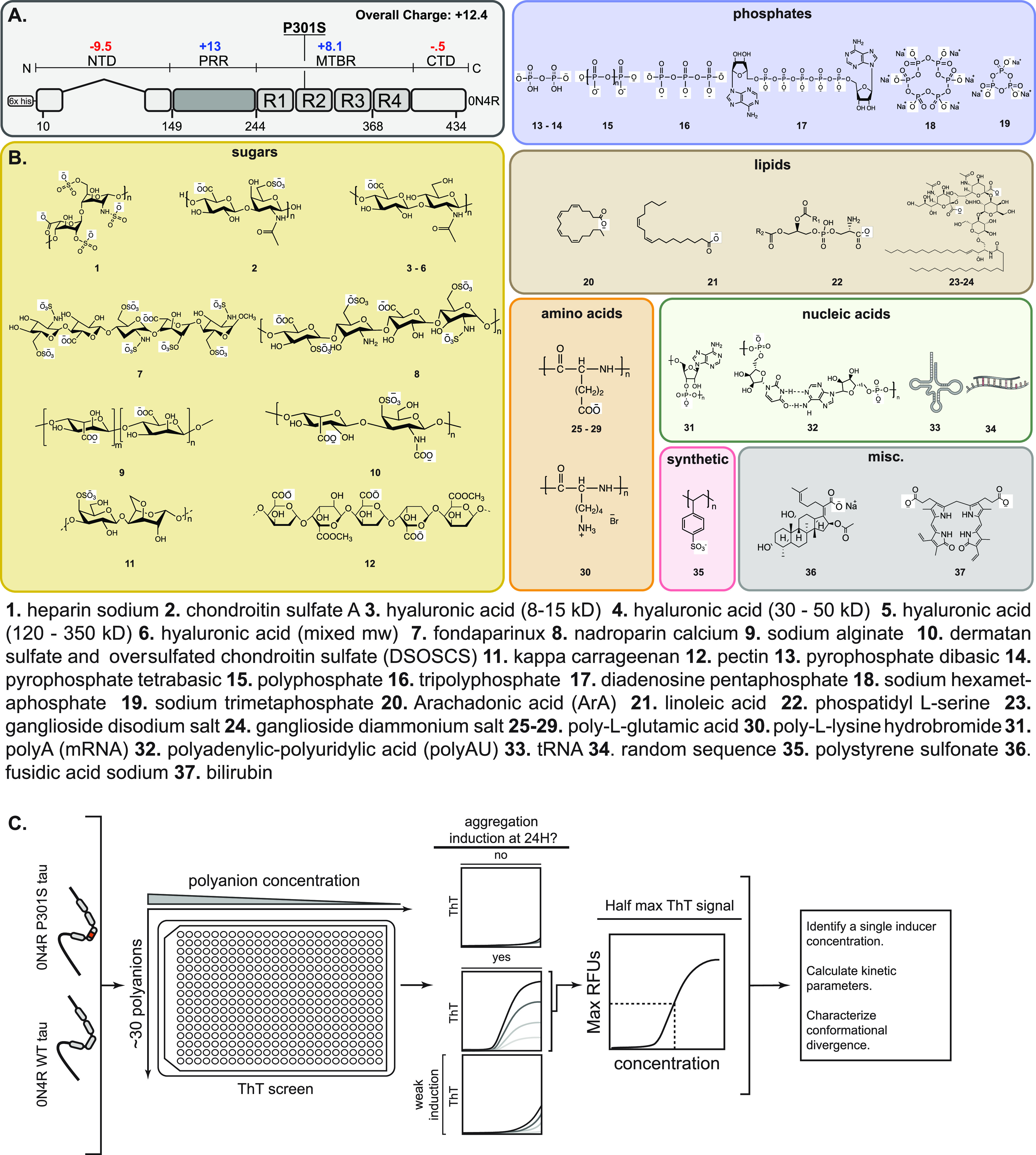
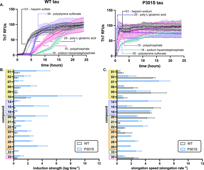
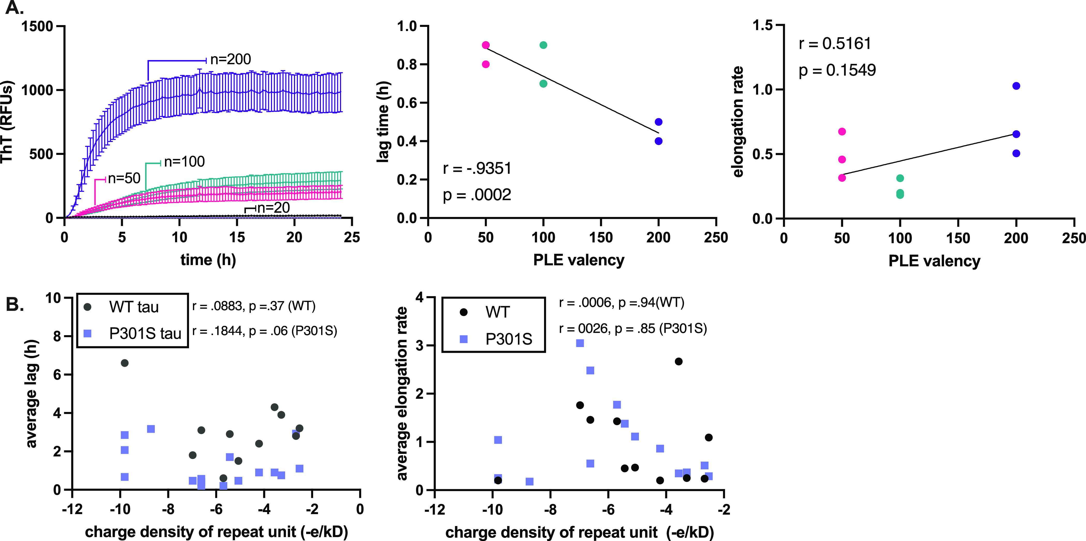
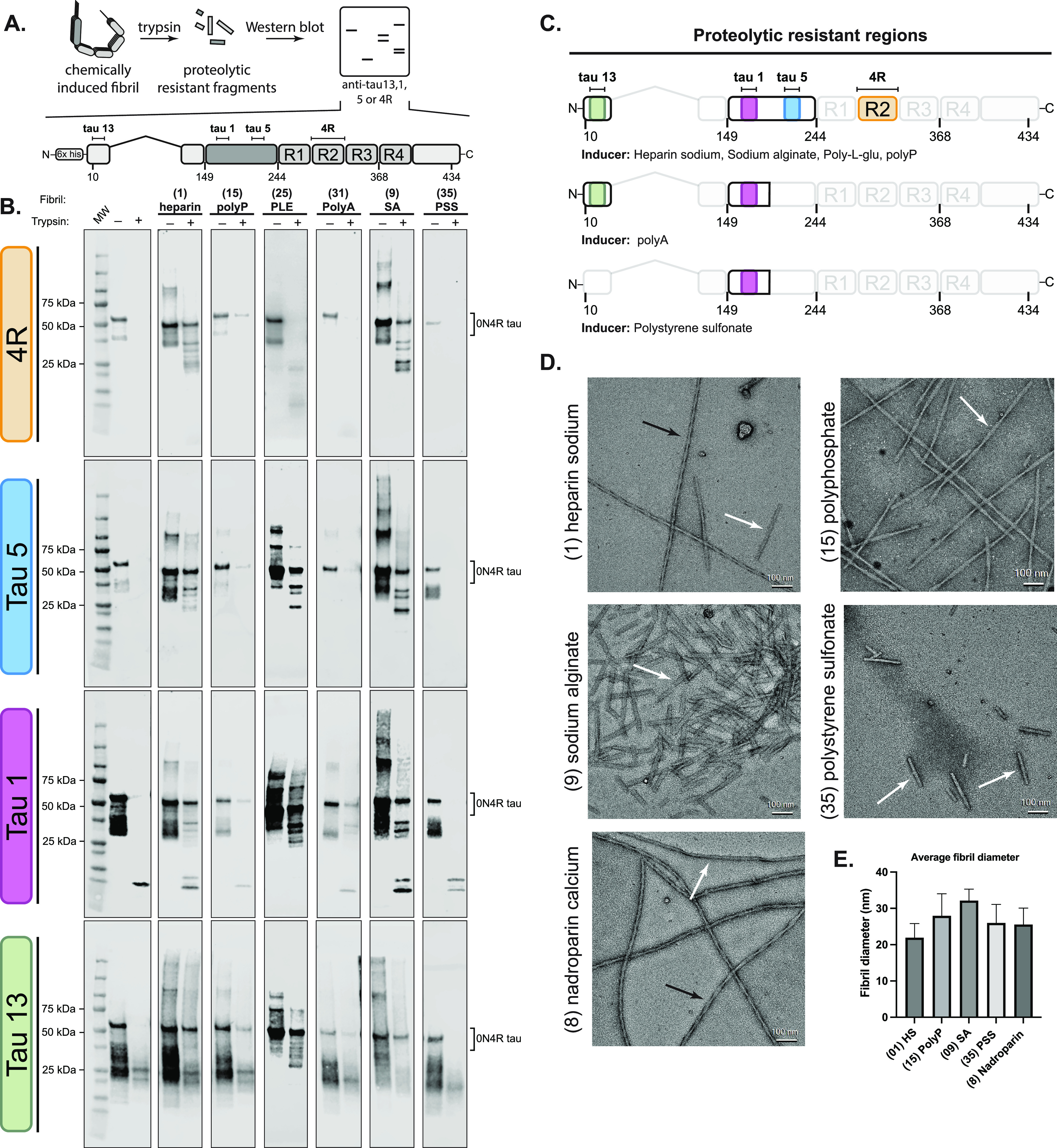
The aggregation of tau into insoluble fibrils is a defining feature of neurodegenerative tauopathies. However, tau has a positive overall charge and is highly soluble; so, polyanions, such as heparin, are typically required to promote its aggregation in vitro. There are dozens of polyanions in living systems, and it is not clear which ones might promote this process. Here, we systematically measure the ability of 37 diverse, anionic biomolecules to initiate tau aggregation using either wild-type (WT) tau or the disease-associated P301S mutant. We find that polyanions from many different structural classes can promote fibril formation and that P301S tau is sensitive to a greater number of polyanions (28/37) than WT tau (21/37). We also find that some polyanions preferentially reduce the lag time of the aggregation reactions, while others enhance the elongation rate, suggesting that they act on partially distinct steps. From the resulting structure-activity relationships, the valency of the polyanion seems to be an important chemical feature such that anions with low valency tend to be weaker aggregation inducers, even at the same overall charge. Finally, the identity of the polyanion influences fibril morphology based on electron microscopy and limited proteolysis. These results provide insights into the crucial role of polyanion-tau interactions in modulating tau conformational dynamics with implications for understanding the tau aggregation landscape in a complex cellular environment.


Conflict of interest statement

The authors declare no competing financial interest.

Figures

Figure 1
Figure 1
Workflow for testing the effects of a polyanion library on tau self-assembly. (A) Domain architecture of the common adult isoform of tau (0N4R). The overall charge of the domains is indicated, and the location of the his-tag and the P310S missense mutation are shown. N-terminal domain (NTD); proline-rich region (PRR); microtubule-binding repeats (MTBR); C-terminal domain (CTD). (B) Chemical structures of 37 anionic biomolecules, grouped by series. When appropriate, the minimal repeating unit is shown, and the average polymer length (n) is indicated in the Supplementary Tables 1 and 2. Compounds 33 and 34 are shown as cartoons because they do have repeating structures (see the Experimental Section). (C) Workflow for screening the anion library. Briefly, 0N4R tau (WT) and 0N4R P301S mutant tau (P301S) proteins were first tested against a range of concentrations of each library member in ThT assays. Anions were excluded from further analysis if they produced artifacts (Supplementary Figure 1) or if they failed to produce a ThT signal 40 RFUs above baseline fluorescence (ΔRFU ≤ 40) at 24 h. For the remaining molecules, the half-maximal effective concentration (EC50) was determined, and subsequent kinetic studies were performed at that anion concentration. From those studies, kinetic parameters, including lag time and elongation rate, were determined.
Figure 2
Figure 2
Anions have differential effects on lag time and elongation rate. (A) Representative ThT assay results, comparing heparin sodium (1), polyphosphate (15), sodium hexametaphosphate (18), poly-l-glutamic acid (25), and polystyrene sulfonate (35) on recombinant 0N4R WT tau (left) and 0N4R tau P301S (right) aggregation kinetics. The anions were used at their half-maximum concentration (see Supplementary Tables 1 and 2) and tau proteins at 10 μM. Results are the average of at least three independent experiments performed in triplicate, and the error bars represent SEM. For each result, the signal from control experiments using no tau was subtracted. (B) Anions have differential effects on lag time. Values are plotted as reciprocal (lag–1), termed the induction strength. Inactive inducers and those with weak signals were omitted from the analysis (see text). Results are the average of at least three independent experiments performed in triplicate, and the error bars represent SEM. (C) From the same aggregation reactions, the elongation rate was calculated and plotted as the reciprocal (elongation rate–1), termed the elongation speed. Results are the average of at least three independent experiments performed in triplicate, and the error bars represent SEM.
Figure 3
Figure 3
Polyanion valency is an important parameter in dictating tau fibril formation. (A) 2N solutions of poly-l-glutamic acid (PLE) of discrete molecular weights (n = 20, 50, 100, or 200) were used to induce WT and P301S tau (10 μM) for 24 h at 37 °C with constant shaking (left). From the resulting curves, lag time and elongation rate were extracted (right). A Pearson t-test was performed to measure the correlation between valency and kinetic parameters. Results are the average of at least three experiments performed in triplicate, and the error bars represent SEM (n = 9). Results are shown for P301S because it gives a more robust signal compared to WT tau. (B) A Pearson t-test was performed to determine the correlation between charge density (−e/kD) on the lag time (left) and elongation rate constants (right).
Figure 4
Figure 4
Identity of the polyanion impacts the tau fibril structure. (A) General workflow for tau fibril proteolysis. Fibrils were prepared using the corresponding inducer at its analysis concentration (see Supplementary Table 1), purified by ultracentrifugation, and subsequently proteolyzed using trypsin. Proteolysis products were separated by SDS-PAGE and probed using anti-tau antibodies (anti-tau 13, 1, 5, and 4R) (top). The domain architecture of 0N4R tau, showing the location of the epitopes for anti-tau antibodies (bottom). (B) Tau fibrils are generally resistant to proteolysis, but the digestion patterns depend on the identity of the inducer. The protease-resistant fragmentation of 0N4R WT tau filaments differentially induced using heparin (1), polyphosphate (15), poly-l-glutamic acid (25), polyA (31), sodium alginate (9), or polystyrene sulfonate (35). (C) Summary of the proteolytic resistant regions of each fibril sample. (D) Representative electron micrographs of negatively stained fibrils from recombinant 0N4R tau assembled in vitro at the end point of each reaction. Scale bar: 100 nm. Black arrows indicate twisted filaments, and white arrows indicate straight fibers. (E) Quantification of the average diameter of recombinant fibrils (n = 30).

References

    1. Orr M. E.; Sullivan A. C.; Frost B. A Brief Overview of Tauopathy: Causes, Consequences, and Therapeutic Strategies. Trends Pharmacol. Sci. 2017, 38, 637–648. 10.1016/j.tips.2017.03.011. - DOI - PMC - PubMed
    1. Holtzman D. M.; Carrillo M. C.; Hendrix J. A.; Bain L. J.; Catafau A. M.; Gault L. M.; Goedert M.; Mandelkow E.; Mandelkow E.-M.; Miller D. S.; Ostrowitzki S.; Polydoro M.; Smith S.; Wittmann M.; Hutton M. Tau: From Research to Clinical Development. Alzheimer’s Dementia 2016, 12, 1033–1039. 10.1016/j.jalz.2016.03.018. - DOI - PubMed
    1. Wang Y.; Mandelkow E. Tau in Physiology and Pathology. Nat. Rev. Neurosci. 2016, 17, 5–21. 10.1038/nrn.2015.1. - DOI - PubMed
    1. Goedert M.; Eisenberg D. S.; Crowther R. A. Propagation of Tau Aggregates and Neurodegeneration. Annu. Rev. Neurosci. 2017, 40, 189–210. 10.1146/annurev-neuro-072116-031153. - DOI - PubMed
    1. Jeganathan S.; Von Bergen M.; Mandelkow E. M.; Mandelkow E. The Natively Unfolded Character of Tau and Its Aggregation to Alzheimer-like Paired Helical Filaments. Biochemistry 2008, 47, 10526–10539. 10.1021/bi800783d. - DOI - PubMed