

Targeted Dephosphorylation of Tau by Phosphorylation Targeting Chimeras (PhosTACs) as a Therapeutic Modality
- PMID: 36753634
- PMCID: PMC11670127
- DOI: 10.1021/jacs.2c11706
Targeted Dephosphorylation of Tau by Phosphorylation Targeting Chimeras (PhosTACs) as a Therapeutic Modality
Abstract
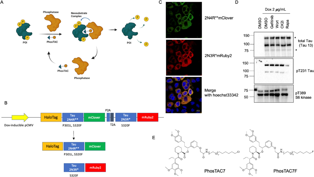
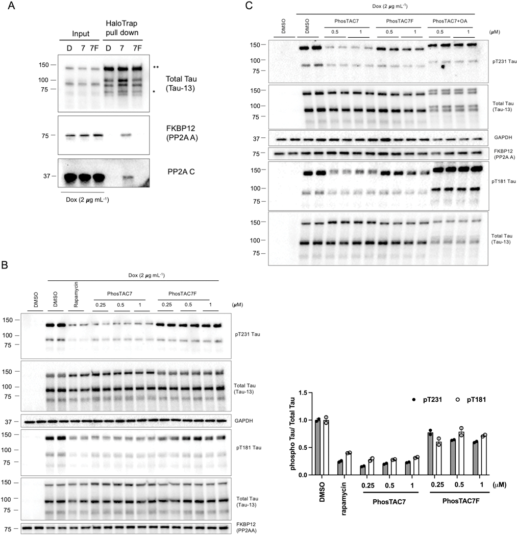
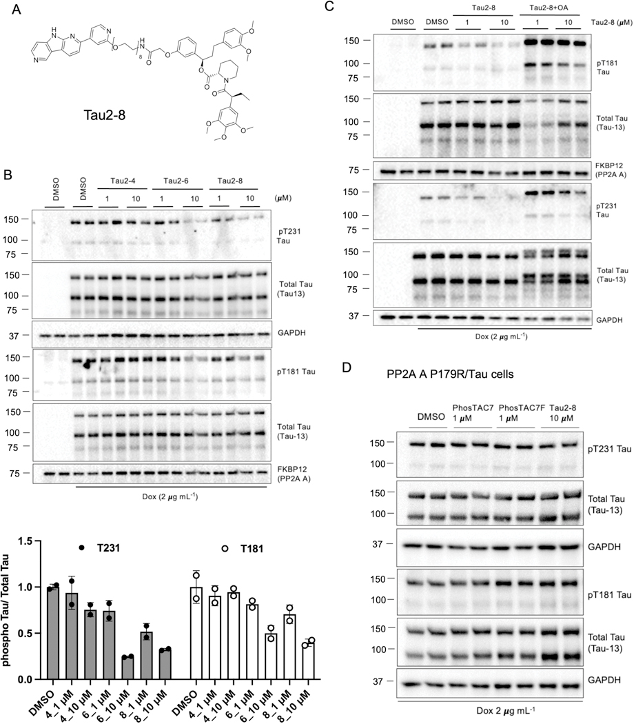
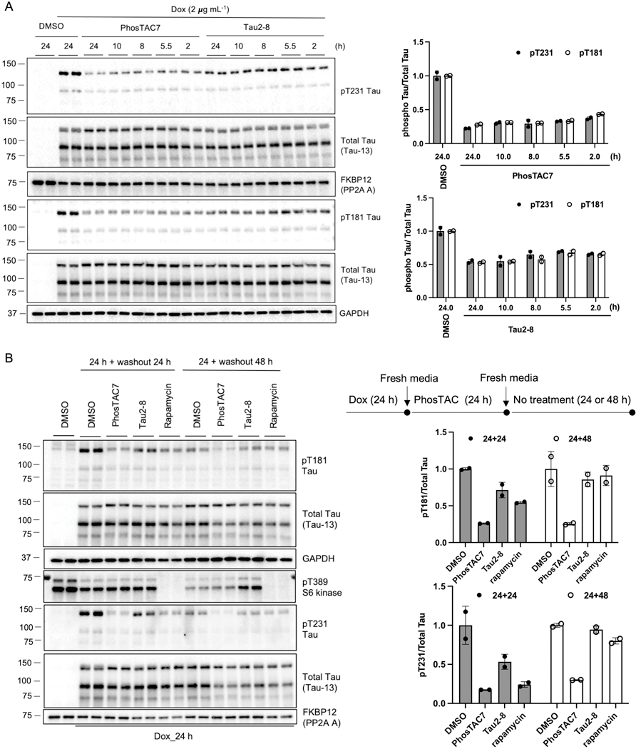
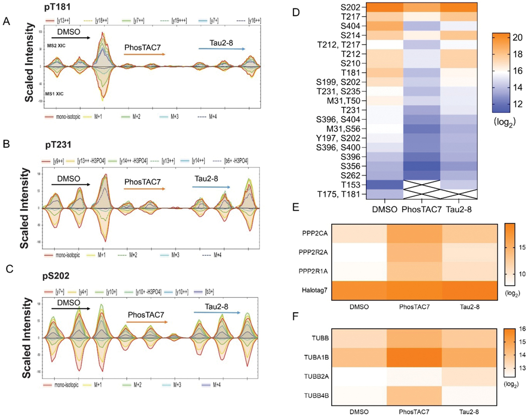
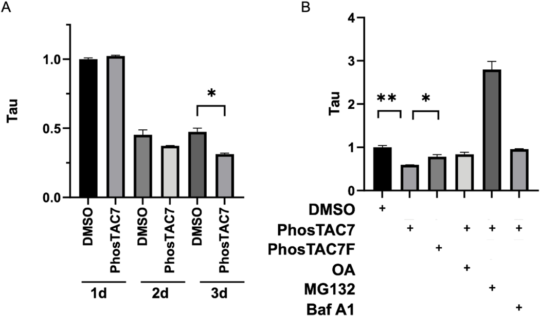
Microtubule-associated protein tau is essential for microtubule assembly and stabilization. Hyperphosphorylation of the microtubule-associated protein tau plays an important pathological role in the development of Alzheimer's disease and other tauopathies. In vivo studies using kinase inhibitors suggest that reducing tau phosphorylation levels has therapeutic potential; however, such approaches showed limited benefits. We sought to further develop our phosphorylation targeting chimera (PhosTAC) technology to specifically induce tau dephosphorylation. Herein, we use small molecule-based PhosTACs to recruit tau to PP2A, a native tau phosphatase. PhosTACs induced the formation of a stable ternary complex, leading to rapid, efficient, and sustained tau dephosphorylation, which also correlated with the enhanced downregulation of tau protein. Mass spectrometry data validated that PhosTACs downregulated multiple phosphorylation sites of tau. We believe that PhosTAC possesses several advantages over current strategies to modulate tau phosphorylation and represents a new avenue for disease-modifying therapies for tauopathies.
Conflict of interest statement
Conflict of interests
The authors declare no competing financial interest.
Figures
Similar articles
-
EGFR targeting PhosTACs as a dual inhibitory approach reveals differential downstream signaling.Sci Adv. 2024 Mar 29;10(13):eadj7251. doi: 10.1126/sciadv.adj7251. Epub 2024 Mar 27. Sci Adv. 2024. PMID: 38536914 Free PMC article.
-
Modulation of Phosphoprotein Activity by Phosphorylation Targeting Chimeras (PhosTACs).ACS Chem Biol. 2021 Dec 17;16(12):2808-2815. doi: 10.1021/acschembio.1c00693. Epub 2021 Nov 15. ACS Chem Biol. 2021. PMID: 34780684 Free PMC article.
-
Generation of tau dephosphorylation-targeting chimeras for the treatment of Alzheimer's disease and related tauopathies.Sci Bull (Beijing). 2024 Apr 30;69(8):1137-1152. doi: 10.1016/j.scib.2024.01.019. Epub 2024 Jan 19. Sci Bull (Beijing). 2024. PMID: 38341350
-
Protein phosphatase 2A and tau: an orchestrated 'Pas de Deux'.FEBS Lett. 2018 Apr;592(7):1079-1095. doi: 10.1002/1873-3468.12907. Epub 2017 Nov 19. FEBS Lett. 2018. PMID: 29121398 Review.
-
Phosphorylated tau targeted small-molecule PROTACs for the treatment of Alzheimer's disease and tauopathies.Biochim Biophys Acta Mol Basis Dis. 2021 Aug 1;1867(8):166162. doi: 10.1016/j.bbadis.2021.166162. Epub 2021 Apr 30. Biochim Biophys Acta Mol Basis Dis. 2021. PMID: 33940164 Free PMC article. Review.
Cited by
-
An artificial protein modulator reprogramming neuronal protein functions.Nat Commun. 2024 Mar 6;15(1):2039. doi: 10.1038/s41467-024-46308-6. Nat Commun. 2024. PMID: 38448420 Free PMC article.
-
Diverse perspectives on proteomic posttranslational modifications to address EGFR-TKI resistance in non-small cell lung cancer.Front Cell Dev Biol. 2024 Dec 24;12:1436033. doi: 10.3389/fcell.2024.1436033. eCollection 2024. Front Cell Dev Biol. 2024. PMID: 39777265 Free PMC article. Review.
-
EGFR targeting PhosTACs as a dual inhibitory approach reveals differential downstream signaling.Sci Adv. 2024 Mar 29;10(13):eadj7251. doi: 10.1126/sciadv.adj7251. Epub 2024 Mar 27. Sci Adv. 2024. PMID: 38536914 Free PMC article.
-
Targeted dephosphorylation of SMAD3 as an approach to impede TGF-β signaling.iScience. 2024 Jul 5;27(8):110423. doi: 10.1016/j.isci.2024.110423. eCollection 2024 Aug 16. iScience. 2024. PMID: 39104417 Free PMC article.
-
An Extensive Atlas of Proteome and Phosphoproteome Turnover Across Mouse Tissues and Brain Regions.bioRxiv [Preprint]. 2024 Oct 17:2024.10.15.618303. doi: 10.1101/2024.10.15.618303. bioRxiv. 2024. Update in: Cell. 2025 Apr 17;188(8):2267-2287.e21. doi: 10.1016/j.cell.2025.02.021. PMID: 39464138 Free PMC article. Updated. Preprint.
References
-
- Lee G; Cowan N; Kirschner M The primary structure and heterogeneity of tau protein from mouse brain. Science 1988, 239 (4837), 285–8. - PubMed
-
- Alzheimer A; Stelzmann RA; Schnitzlein HN; Murtagh FR An English translation of Alzheimer’s 1907 paper, “Uber eine eigenartige Erkankung der Hirnrinde”. Clin Anat 1995, 8 (6), 429–31. - PubMed
Grants and funding
LinkOut - more resources
Full Text Sources
Other Literature Sources
Molecular Biology Databases
