. 2023 Feb 8;14(1):686.
doi: 10.1038/s41467-023-36246-0.

Accelerating inhibitor discovery for deubiquitinating enzymes

Affiliations

Accelerating inhibitor discovery for deubiquitinating enzymes

Wai Cheung Chan et al. Nat Commun. .

Abstract

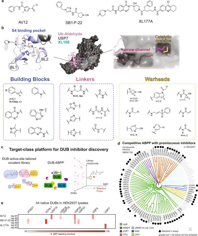
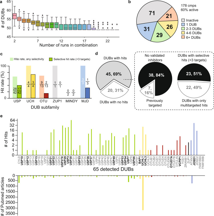
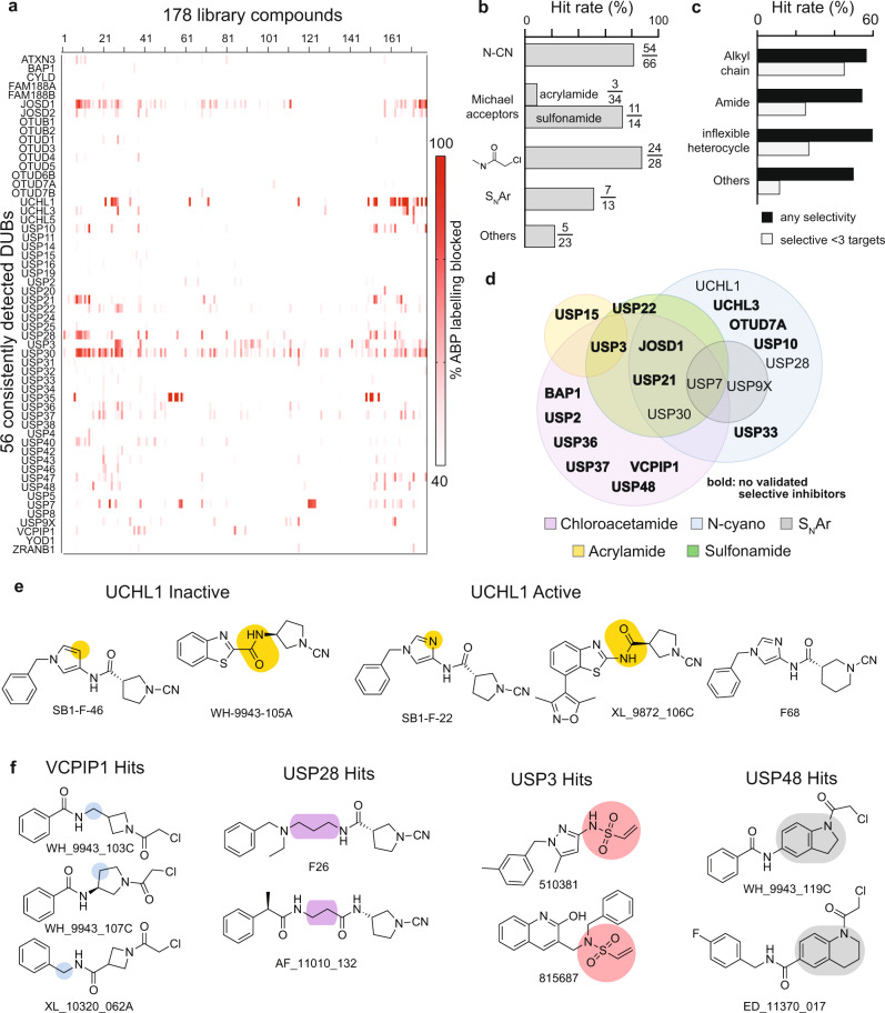
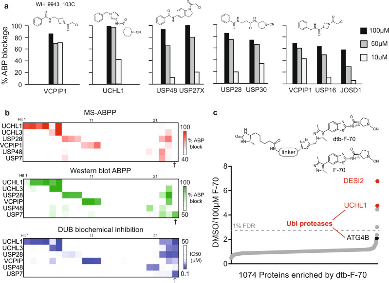
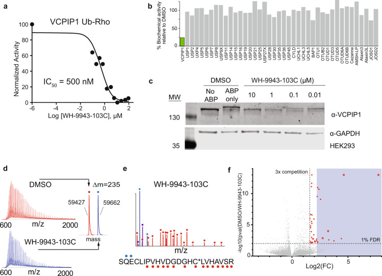
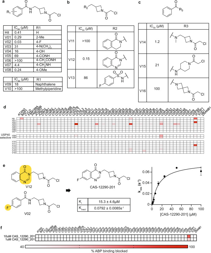
Deubiquitinating enzymes (DUBs) are an emerging drug target class of ~100 proteases that cleave ubiquitin from protein substrates to regulate many cellular processes. A lack of selective chemical probes impedes pharmacologic interrogation of this important gene family. DUBs engage their cognate ligands through a myriad of interactions. We embrace this structural complexity to tailor a chemical diversification strategy for a DUB-focused covalent library. Pairing our library with activity-based protein profiling as a high-density primary screen, we identify selective hits against 23 endogenous DUBs spanning four subfamilies. Optimization of an azetidine hit yields a probe for the understudied DUB VCPIP1 with nanomolar potency and in-family selectivity. Our success in identifying good chemical starting points as well as structure-activity relationships across the gene family from a modest but purpose-build library challenges current paradigms that emphasize ultrahigh throughput in vitro or virtual screens against an ever-increasing scope of chemical space.


Conflict of interest statement

J.A.M. is a founder, equity holder, and advisor to Entact Bio, serves on the SAB of 908 Devices, and receives sponsored research funding from Vertex, AstraZeneca, Taiho, Springworks and TUO Therapeutics. S.J.B. is a founder, equity holder, and advisor to Entact Bio and serves on the SAB of Adenoid Cystic Carcinoma Foundation. S.J.B. currently receives funding from AbbVie and Tuo Therapeutics, and has received in-kind services from AbbVie and Novartis Institutes for Biomedical Research during the past 24 months. The remaining authors declare no competing interests.

Figures

Fig. 1
Fig. 1. A DUB-tailored inhibitor discovery platform.
a Structures of DUB inhibitors AV12, SB1-F-22, and XL177A. b Library compounds followed a three-piece modular design, where noncovalent building blocks, linkers, and warhead moieties were diversified to target conserved and divergent aspects of the DUB catalytic domain and nearby regions. Blue: Noncovalent building blocks were designed to target the blocking loops (BL) 1 and 2, in addition to the P4 site. Red: Linkers were designed to traverse a long and narrow channel leading up to the catalytic cysteine residue, occupied by the C-terminus of the substrate ubiquitin in catalysis. In the top panel, an overlay of crystal structures for USP7 with Ub-aldehyde (PDB: 1NBF) and USP7 with XL188 (PDB: 5VS6) are shown, with USP7 in gray, Ub-aldehyde in pink, and XL188 in cyan. Yellow: electrophilic warheads were designed to target the invariant catalytic cysteine residue shown in yellow in the top panel. c The target-class platform yields high content data to drive DUB hit identification and SAR for compound optimization. d Analysis of multitargeted covalent DUB inhibitors (HBX41108 and PR619) confirmed that our DUB-ABPP platform spanned members from all subfamilies of cysteine protease DUBs,. The dendrogram is colored by DUB subfamily. e ABPP analysis using covalent inhibitors depicted in (a) in duplicate runs (each shown individually) validated the ability of our platform to read-out selective compound binding activity with deep coverage of the human DUBome.
Fig. 2
Fig. 2. DUB trends in primary screening.
a DUB ABPP assay performance The number of DUBs consistently detected across each unique combination of 2–24 assay runs (n = 25 biologically independent experiments) quickly stabilized to 49, with 56 DUBs detected in >80% of runs. Line at center of box shows median number of DUBs detected for the given number of assays considered, box encapsulates the middle two quartiles, while bars represent bounds for the rest of the distribution. “Outlier” points as calculated by an inter-quartile method are shown as diamonds. b Most compounds were active and spanned diverse selectivity profiles. c Hit rates were roughly consistent across the DUB subfamilies, with no hits identified for the MINDY subfamily. Insets above indicate number of DUBs targeted (numerator) with any selectivity (light color) or selectively with <3 targets (dark color) out of all DUBs in the subfamily (denominator). d left: Hits were successfully identified for 69% (45 out of 65) of the detected DUBs. Dashed box, left pie chart: 84% (38 out of 45) of hits did not have validated inhibitors reported. Dashed box, right pie chart: 51% (23 out of 45) of hits were targeted by selective compounds with <3 DUB targets. e DUBs targeted by library compounds. Individual DUBs showed a range of targetability by our library, within 1 to 65 library compounds hitting each targeted DUB, number of hits per DUB shown in the upper bar chart. Hit compounds were successfully identified against understudied members of the target class, including 11 DUBs with ≤ 20 publications, number of Pubmed articles for each DUB shown in lower bar chart. DUB from different subfamilies were grouped and shown in different colors: USP (green), UCH (yellow), OTU (maroon), ZUP1 (black), MJD (blue), MINDY (no hits, no colored bar). Selectively targeted DUBs are shown in bold.
Fig. 3
Fig. 3. Chemical trends in primary screening.
a Heatmap representing potency and selectivity data for all 178 library compounds against 56 DUBs detected consistently over 80% of runs. b Electrophiles displayed diverse DUB hit rates. Acrylamides exhibited poor activity towards DUB catalytic cysteines. Insets adjacent to each bar indicate the number of hits and the total number of compounds containing each electrophile. c Presence and type of linker influenced compound activity and selectivity. d Individual warheads showed partially overlapping DUB target profiles. Venn diagram is colored by warhead, legend in figure. Bolded DUBs are hitherto untargeted. e, f Selected representative examples for multiple chemically similar hits for the same DUB from the screen. In the case of UCHL1, stringent requirements surrounding hydrogen bond acceptor position and amide orientation on the linker are highlighted in yellow. For VCPIP1, compounds were structurally similar except for the methylene group which moved along the backbone of the molecule (in blue). Moieties common across hits are highlighted for USP28 (in purple), USP3 (in red), and USP48 (in gray).
Fig. 4
Fig. 4. Hit validation.
a Selected hits were confirmed by dose–response DUB-ABPP over 100, 50, 10 µM. b Overall, we observed high confirmation rates and excellent agreement for assays spanning compound binding in mass spectrometry (red), western blot (green), and enzyme inhibition (blue) for library hits targeting UCHL1, UCHL3, USP7, USP28, USP48 and VCPIP1. An outlier (XL-9872-111B) for which we observed poor agreement across assay types is shown on the rightmost column. Data for each DUB is shown in a row (6 DUBs total) and data for each compound is shown in a column (24 compounds total). For individual compound structures and primary screen activity, please refer to Supplementary Data 3. c In a competition experiment against a desthiobiotinylated analog, F-70 shows dose–response binding to only 4 proteins among 1074 proteins detected with a 1% FDR cutoff as measured by a two tailed Z-test. Top DUB hits are highlighted in red. Source data are provided as a source data file.
Fig. 5
Fig. 5. VCPIP1 hit validation.
a Hit molecule WH-9943-103C inhibited deubiquitination activity of VCPIP1 in Ub-Rho cleavage assay after 6 h incubation (n = 1). b WH-9943-103C inhibited VCPIP1 (highlighted in green) selectively out of a panel of 41 purified recombinant DUBs after 15 min incubation. c WH-9943-103C displayed in-cell target engagement as determined for DUB labeling by ABP then visualized on a Western blot. d WH-9943-103C labeled recombinant VCPIP1 with 1:1 stoichiometry as read out by intact protein mass spectrometry. Labeled ion envelope is shown in blue, unlabeled ion envelope from DMSO control is shown in red. e CE-MS/MS identified the catalytic cysteine of VCPIP1 to be covalently modified by WH-9943-103C, red and blue glyphs adjacent to the peptide sequence indicate y- and b-type fragment ions, respectively, detected in the MS/MS spectrum. The y-ion series confirms catalytic cysteine modification (C*). f A proteome-wide competitive binding survey of WH-9943-103C activity (50µM) indicated off-target modification of 39 (red dots, FDR <1%, >3-fold competition relative to DMSO) out of 24,579 unique cysteines detected (p-values for all detected ratios derived from maximum likelihood estimation and corrected by the Benjamini-Hochberg method). The blue box indicates >4.5-fold competition relative to DMSO, the observed fold competition for VCPIP1. This includes 4 cysteines that were previously characterized as ‘hyper-reactive’ (red stars). Source data are provided as a source data file.
Fig. 6
Fig. 6. Optimization of a potent and selective VCPIP1 inhibitor.
ac Structures and VCPIP1 biochemical inhibitory activity of WH-9943-103C analogs. Data for hit WH-9943-103C are duplicated from earlier figures for ease of comparison. d DUB-ABPP data for the WH-103C focused library. Compounds with VCPIP1 biochemical IC50 values below 250 nM (V02, V08, V12) were screened in ABPP at 10 μM, less potent compounds were screened at 50 μM. e, f SAR analysis suggested a combination of functionalities on V12 and V02 (yellow highlights) to yield CAS-12290-201. Binding kinetics and DUB-ABPP (10 and 1 µM) demonstrated that CAS-12290-201 to be a potent and selective inhibitor of the understudied DUB VCPIP1, error bars show the 95% confidence interval for parameters as fitted by GraphPad Prism 9.0.1.

References

    1. Komander D, Clague MJ, Urbé S. Breaking the chains: structure and function of the deubiquitinases. Nat. Rev. Mol. Cell Biol. 2009;10:550–563. - PubMed
    1. Harrigan JA, Jacq X, Martin NM, Jackson SP. Deubiquitylating enzymes and drug discovery: emerging opportunities. Nat. Rev. Drug Discov. 2018;17:57–78. - PMC - PubMed
    1. Ritorto MS, et al. Screening of DUB activity and specificity by MALDI-TOF mass spectrometry. Nat. Commun. 2014;5:4763. - PMC - PubMed
    1. Altun M, et al. Activity-based chemical proteomics accelerates inhibitor development for deubiquitylating enzymes. Chem. Biol. 2011;18:1401–1412. - PubMed
    1. Di Lello P, et al. Discovery of small-molecule inhibitors of ubiquitin specific protease 7 (USP7) using integrated NMR and in silico techniques. J. Med. Chem. 2017;60:10056–10070. - PubMed

Publication types

MeSH terms