. 2023 Feb 8;13(1):2229.
doi: 10.1038/s41598-023-29001-4.

Pleural fluid microbiota as a biomarker for malignancy and prognosis

Affiliations

Pleural fluid microbiota as a biomarker for malignancy and prognosis

Benjamin Kwok et al. Sci Rep. .

Abstract

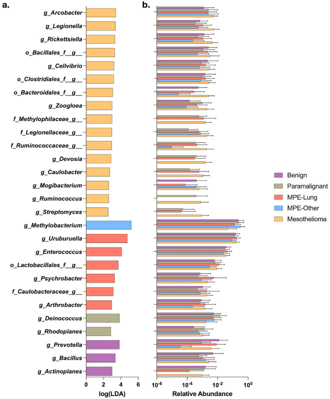
Malignant pleural effusions (MPE) complicate malignancies and portend worse outcomes. MPE is comprised of various components, including immune cells, cancer cells, and cell-free DNA/RNA. There have been investigations into using these components to diagnose and prognosticate MPE. We hypothesize that the microbiome of MPE is unique and may be associated with diagnosis and prognosis. We compared the microbiota of MPE against microbiota of pleural effusions from non-malignant and paramalignant states. We collected a total of 165 pleural fluid samples from 165 subjects; Benign (n = 16), Paramalignant (n = 21), MPE-Lung (n = 57), MPE-Other (n = 22), and Mesothelioma (n = 49). We performed high throughput 16S rRNA gene sequencing on pleural fluid samples and controls. We showed that there are compositional differences among pleural effusions related to non-malignant, paramalignant, and malignant disease. Furthermore, we showed differential enrichment of bacterial taxa within MPE depending on the site of primary malignancy. Pleural fluid of MPE-Lung and Mesothelioma were associated with enrichment with oral and gut bacteria that are commonly thought to be commensals, including Rickettsiella, Ruminococcus, Enterococcus, and Lactobacillales. Mortality in MPE-Lung is associated with enrichment in Methylobacterium, Blattabacterium, and Deinococcus. These observations lay the groundwork for future studies that explore host-microbiome interactions and their influence on carcinogenesis.


Conflict of interest statement

The authors declare no competing interests.

Figures

Figure 1
Figure 1
Microbial compositional differences between groups of pleural fluid. (a) Bacterial load (copies/mL) by ddPCR. p-values by Kruskal–Wallis rank sum test; individual comparisons by Wilcoxon rank sum tests with Benjamini–Hochberg adjustment for multiple comparisons. (b) Alpha diversity (Shannon diversity). p-values by Kruskal–Wallis rank sum test. Individual comparisons by Wilcoxon rank sum tests with Benjamini–Hochberg adjustment for multiple comparisons. (c) Beta diversity (Bray–Curtis dissimilarity index). p-values by PERMANOVA. ns not significant.
Figure 2
Figure 2
Taxonomic differences between groups of pleural fluid. (a) Identification of taxa enriched in group of pleural fluid by linear discriminant analysis effect size (LEfSe). (b) Mean relative abundance for each taxa identified as enriched by LEfSe.
Figure 3
Figure 3
Taxonomic differences based on survival analysis. (a) Kaplan–Meier curves for groups of malignant pleural effusions. (b) Differential enrichment analysis by LEfSe for survival of subjects in the MPE-Lung group at time of median survival (33.6 months), MPE-Other group at time of median survival (36 months), and Mesothelioma group at time of median survival (19.6 months). Taxa colored in purple are potential contaminants.
Figure 4
Figure 4
Random forest classifiers to predict mortality at median time of survival. (a) Area under the curve (AUC) of receiver operator curves (ROC) based on random forest identification of taxonomic classifiers predicting mortality in MPE-Lung median mortality (33.6 months) using the top 1, 5, 10, 20, 50, 75, and 100% of total discriminant taxa based on Gini values (n = 343). (b) ROC for the best-fit random forest classifier in MPE-Lung. (c) Taxa with greatest Gini Index from the random forest classifier with the greatest AUC (10% of taxa). (d) AUC of ROC based on random forest identification of taxonomic classifiers predicting mortality in MPE-Other median mortality (36 months) using the top 1, 5, 10, 20, 50, 75, and 100% of total discriminant taxa based on Gini values (n = 254). (e) ROC for the best-fit random forest classifier in MPE-Other. (f) Taxa with greatest Gini Index from the random forest classifier with the greatest AUC (1% of taxa). The taxa colored in purple is a potential contaminant. (g) AUC of ROC based on random forest identification of taxonomic classifiers predicting median mortality in Mesothelioma (19.6 months) using the top 1, 5, 10, 20, 50, 75, and 100% of total discriminant taxa based on Gini values (n = 503). (h) ROC for the best-fit random forest classifier in Mesothelioma. (i) Taxa with greatest Gini Index from the random forest classifier with the greatest AUC (5% of taxa). Potential contaminants are not shown.

References

    1. Goldstraw P, et al. The IASLC Lung Cancer Staging Project: Proposals for revision of the TNM stage groupings in the forthcoming (eighth) edition of the TNM classification for lung cancer. J. Thorac. Oncol. 2016;11:39–51. doi: 10.1016/j.jtho.2015.09.009. - DOI - PubMed
    1. Grosu HB, Kazzaz F, Vakil E, Molina S, Ost D. Sensitivity of initial thoracentesis for malignant pleural effusion stratified by tumor type in patients with strong evidence of metastatic disease. Respiration. 2018;96:363–369. doi: 10.1159/000490732. - DOI - PubMed
    1. Kim Y, Shin S, Lee KA. Exosome-based detection of EGFR T790M in plasma and pleural fluid of prospectively enrolled non-small cell lung cancer patients after first-line tyrosine kinase inhibitor therapy. Cancer Cell Int. 2021;21:50. doi: 10.1186/s12935-021-01761-x. - DOI - PMC - PubMed
    1. Han YQ, Xu SC, Zheng WQ, Hu ZD. Diagnostic value of microRNAs for malignant pleural mesothelioma: A mini-review. Thorac. Cancer. 2021;12:8–12. doi: 10.1111/1759-7714.13746. - DOI - PMC - PubMed
    1. Bao Q, Xu Y, Ding M, Chen P. Identification of differentially expressed miRNAs in differentiating benign from malignant pleural effusion. Hereditas. 2020;157:6. doi: 10.1186/s41065-020-00119-z. - DOI - PMC - PubMed

Publication types