. 2023 Jan;10(1):013510.
doi: 10.1117/1.NPh.10.1.013510. Epub 2023 Feb 3.

Revealing the spatiotemporal requirements for accurate subject identification with resting-state functional connectivity: a simultaneous fNIRS-fMRI study

Affiliations

Revealing the spatiotemporal requirements for accurate subject identification with resting-state functional connectivity: a simultaneous fNIRS-fMRI study

Sergio L Novi et al. Neurophotonics. 2023 Jan.

Abstract

Significance: Brain fingerprinting refers to identifying participants based on their functional patterns. Despite its success with functional magnetic resonance imaging (fMRI), brain fingerprinting with functional near-infrared spectroscopy (fNIRS) still lacks adequate validation.

Aim: We investigated how fNIRS-specific acquisition features (limited spatial information and nonneural contributions) influence resting-state functional connectivity (rsFC) patterns at the intra-subject level and, therefore, brain fingerprinting.

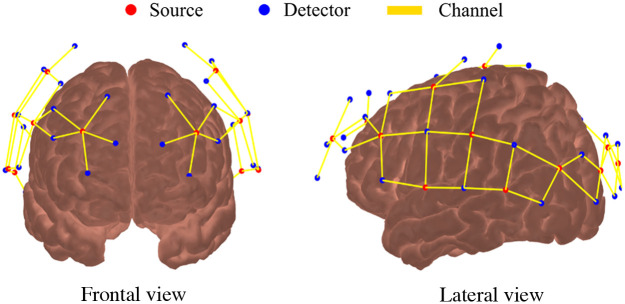
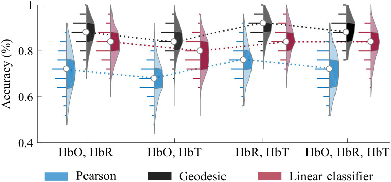
Approach: We performed multiple simultaneous fNIRS and fMRI measurements in 29 healthy participants at rest. Data were preprocessed following the best practices, including the removal of motion artifacts and global physiology. The rsFC maps were extracted with the Pearson correlation coefficient. Brain fingerprinting was tested with pairwise metrics and a simple linear classifier.

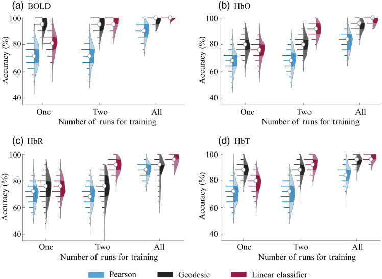
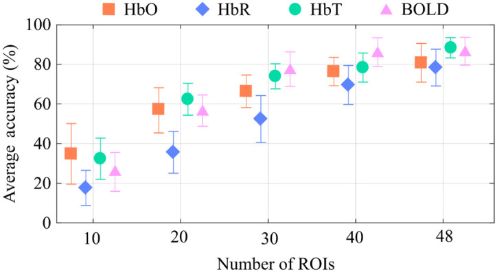
Results: Our results show that average classification accuracy with fNIRS ranges from 75% to 98%, depending on the number of runs and brain regions used for classification. Under the right conditions, brain fingerprinting with fNIRS is close to the 99.9% accuracy found with fMRI. Overall, the classification accuracy is more impacted by the number of runs and the spatial coverage than the choice of the classification algorithm.

Conclusions: This work provides evidence that brain fingerprinting with fNIRS is robust and reliable for extracting unique individual features at the intra-subject level once relevant spatiotemporal constraints are correctly employed.

Keywords: brain fingerprinting; functional magnetic resonance imaging; functional near-infrared spectroscopy; resting-state functional connectivity; subject identification.


Figures

Fig. 1
Fig. 1
Optical probe used for fNIRS. The probe contains 16 sources (red dots) and 32 detectors (blue dots) distributed symmetrically between the 2 hemispheres, covering most of the head and including frontal, temporal, parietal, and occipital lobes. The solid yellow lines represent the 64 source-detector combinations (i.e., channels).
Fig. 2
Fig. 2
Accuracy of subject identification for Pearson, geodesic, and linear classifier approaches as a function of the number of available runs available for training when (a) BOLD, (b) HbO, (c) HbR, and (d) HbT signals are used separately. The condition “All” refers to the case in which one run was used for testing and the remaining runs were used for training. Due to the pruning criteria, the number of runs per participant varied from three to five for BOLD and four to six for fNIRS. Each distribution depicts all observed results in detail (300 values in total). In each violin plot, the white circle represents the median, and shaded regions indicate the first and third quartiles (as a typical boxplot). The horizontal lines indicate the accuracy values obtained across all trials, and their lengths are proportional to the frequency of each obtained accuracy within the distribution.
Fig. 3
Fig. 3
Average geodesic classification accuracy as a function of the number of ROIs when only one run was used for training and another for testing. The error bars represent the standard deviation. The performance on subject identification increases with the number of ROIs used in the classification for all contrasts (BOLD, HbO, HbR, and HbT); in particular, HbT accuracy performed similarly to BOLD when the number of ROIs across the two techniques was equal.
Fig. 4
Fig. 4
Classification accuracy during subject identification when combining fNIRS contrasts. The accuracy was calculated using the Pearson, geodesic, and linear classifier approaches. We used only one run for training and another for testing in this case. Combining HbR and HbT presented the highest performance across all possible combinations. Each distribution depicts all observed results in detail (300 values in total). In each violin plot, the white circle represents the median, and shaded regions indicate the first and third quartiles (as a typical boxplot). The horizontal lines show the observed accuracy values, and their lengths are proportional to the frequency of each finding inside the distribution. The dotted lines are provided to facilitate comparison across the different combinations only.

Similar articles

Cited by

References

    1. Huppert T. J., et al. , “Quantitative spatial comparison of diffuse optical imaging with blood oxygen level-dependent and arterial spin labeling-based functional magnetic resonance imaging,” J. Biomed. Opt. 11(6), 064018 (2006).JBOPFO10.1117/1.2400910 - DOI - PMC - PubMed
    1. Huppert T. J., et al. , “A temporal comparison of BOLD, ASL, and NIRS hemodynamic responses to motor stimuli in adult humans,” Neuroimage 29(2), 368–382 (2006).NEIMEF10.1016/j.neuroimage.2005.08.065 - DOI - PMC - PubMed
    1. Steinbrink J., et al. , “Illuminating the BOLD signal: combined fMRI–fNIRS studies,” Magn. Reson. Imaging 24(4), 495–505 (2006).MRIMDQ10.1016/j.mri.2005.12.034 - DOI - PubMed
    1. Schaeffer S., Iadecola C., “Revisiting the neurovascular unit,” Nat. Neurosci. 24(9), 1198–1209 (2021).NANEFN10.1038/s41593-021-00904-7 - DOI - PMC - PubMed
    1. Mesquita R. C., Huppert T. J., Boas D. A., “Exploring neuro-vascular and neuro-metabolic coupling in rat somatosensory cortex,” Phys. Med. Biol. 54(2), 175–185 (2009).PHMBA710.1088/0031-9155/54/2/001 - DOI - PMC - PubMed