Thoracic Compressive Myelopathy in a Patient With Takayasu Arteritis
- PMID: 36756014
- PMCID: PMC9902057
- DOI: 10.7759/cureus.33496
Thoracic Compressive Myelopathy in a Patient With Takayasu Arteritis
Abstract
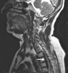
Takayasu arteritis (TA), also known as occlusive thromboaortopathy, is a type of chronic inflammatory arteritis that primarily affects large vessels. Compressive thoracic myelopathy is a rare and distinct manifestation of TA. We present the case of a 60-year-old woman who developed gradually progressive spastic paraplegia over one year. Magnetic resonance imaging revealed a well-defined extra-dual, intensely enhancing ventrodorsal lesion with severe spinal cord impingement. The aortogram revealed dilatation of the aortic arch (with narrowing of arch vessels) and descending aorta, as well as a right paravertebral soft tissue mass at the D4 level. Given the likelihood of TA, the patient underwent decompressive laminectomy and spinal fusion due to severe spinal cord compression. The biopsy of the dural-based lesion revealed an inflammatory granuloma, and the patient was treated postoperatively with oral prednisolone and mycophenolate mofetil. After six months of immunotherapy, there was excellent neurological recovery and near-total resolution of the lesion.
Keywords: compressive myelopathy; decompressive laminectomy; immunotherapy; spastic paraplegia; takayasu arteritis.
Copyright © 2023, Chandrasekar et al.
Conflict of interest statement
The authors have declared that no competing interests exist.
Figures


References
-
- Neurological manifestations of Takayasu arteritis. Li-xin Z, Jun N, Shan G, Bin P, Li-ying C. Chin Med Sci J. 2011;26:227–230. - PubMed
-
- 2022 American College of Rheumatology/EULAR classification criteria for Takayasu arteritis. Grayson PC, Ponte C, Suppiah R, et al. Ann Rheum Dis. 2022;81:1654–1660. - PubMed
-
- Management of Takayasu arteritis: a systematic review. Keser G, Direskeneli H, Aksu K. Rheumatology (Oxford) 2014;53:793–801. - PubMed
-
- An unusual clinical manifestation of Takayasu's arteritis: spinal cord compression. Kim HA, Kim JH, Won JH, Suh CH. Joint Bone Spine. 2009;76:209–212. - PubMed
Publication types
LinkOut - more resources
Full Text Sources
