

Anti-Oxidative, Anti-Apoptotic, and M2 Polarized DSPC Liposome Nanoparticles for Selective Treatment of Atherosclerosis
- PMID: 36756051
- PMCID: PMC9901454
- DOI: 10.2147/IJN.S384675
Anti-Oxidative, Anti-Apoptotic, and M2 Polarized DSPC Liposome Nanoparticles for Selective Treatment of Atherosclerosis
Erratum in
-
Erratum: Anti-Oxidative, Anti-Apoptotic, and M2 Polarized DSPC Liposome Nanoparticles for Selective Treatment of Atherosclerosis [Corrigendum].Int J Nanomedicine. 2023 Sep 4;18:4985-4986. doi: 10.2147/IJN.S431763. eCollection 2023. Int J Nanomedicine. 2023. PMID: 37693884 Free PMC article.
Abstract
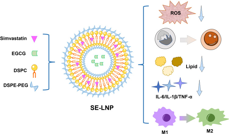
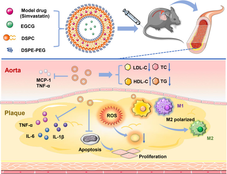
Purpose: Oxidative stress is one of the main pathogenic factors of atherosclerosis. However, no antioxidants have brought positive effects on the treatment of atherosclerosis. To selectively treat atherosclerosis, various means such as antioxidation, anti-apoptosis, and M2 polarization are used. The ultimate goal is that multiple regulatory pathways can help to treat atherosclerosis.
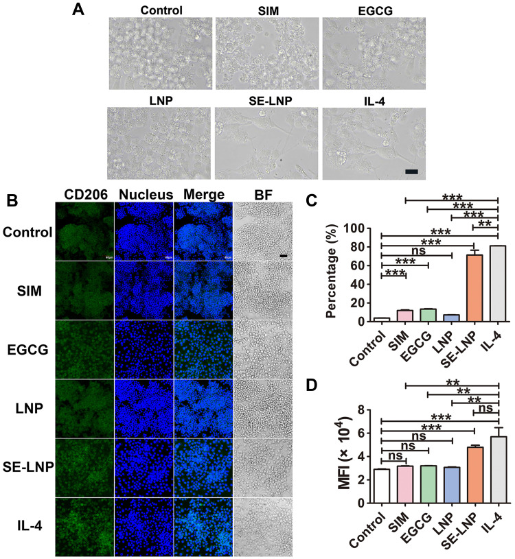
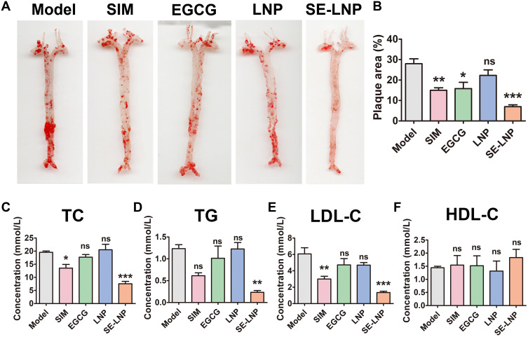
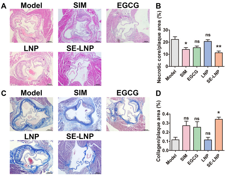
Patients and methods: In this study, Simvastatin (SIM) as a model drug, EGCG as an antioxidant agent, and distearyl phosphatidylcholine (DSPC) as major carriers were used to make liposome nanoparticles (SE-LNPs). The cytotoxicity, phagocytosis, antioxidant, and anti-apoptotic properties of nanoparticles were tested in vitro. ApoE-/- atherosclerotic mice were treated with nanoparticles. The changes of aortic Oil red staining, blood lipid, HE, and Masson sections of the aortic root were observed.
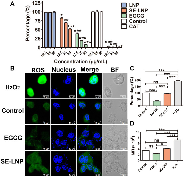
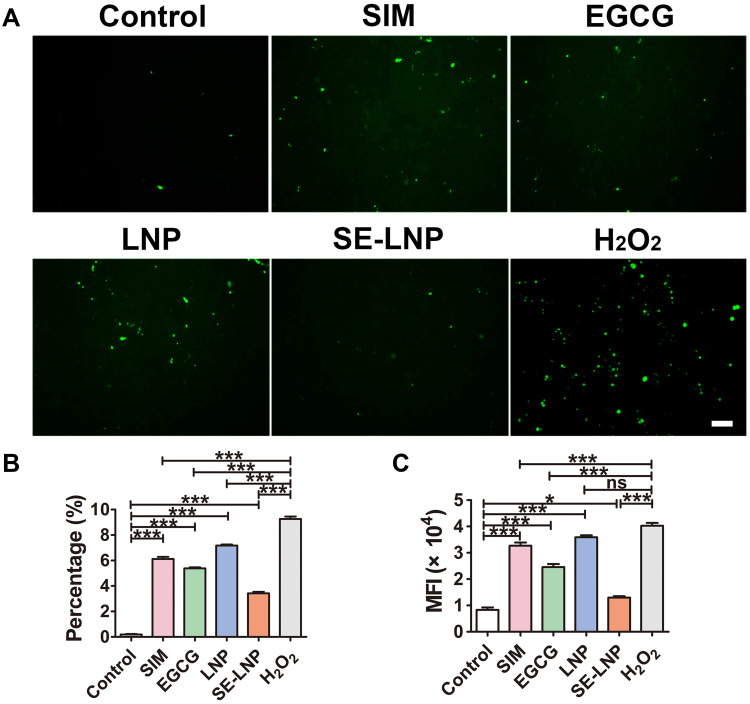
Results: SE-LNPs exhibited a sustained release profile, potentially enabling the accumulation of the majority amount of drugs at the atherosclerotic plaque. The phagocytosis effect was stronger in RAW. The anti-oxidative and anti-apoptotic effects of the formulation were verified in vitro. SE-LNPs promoted the polarization of M2 macrophages. The therapeutic effect of SE-LNPs was assessed in the ApoE-/- mice model of atherosclerosis. SE-LNPs reduced reactive oxygen species and lipids in vivo. The results of Oil red staining, blood lipid, HE, and Masson sections of the aortic root showed the recovery of the focus.
Conclusion: Studies have shown that SE-LNPs could resist oxidation, and apoptosis, promote M2 polarization, and reduce blood lipids and lesions, which is a reliable and selective treatment for atherosclerosis.
Keywords: inhibit inflammation; macrophage polarization; reduce apoptosis; scavenging ROS.
© 2023 Wan et al.
Conflict of interest statement
The authors report no conflicts of interest in this work.
Figures

Similar articles
-
Advances in treatment strategies based on scavenging reactive oxygen species of nanoparticles for atherosclerosis.J Nanobiotechnology. 2023 Aug 17;21(1):271. doi: 10.1186/s12951-023-02058-z. J Nanobiotechnology. 2023. PMID: 37592345 Free PMC article. Review.
-
Synergetic EGCG and coenzyme Q10 DSPC liposome nanoparticles protect against myocardial infarction.Biomater Sci. 2023 Oct 10;11(20):6862-6870. doi: 10.1039/d3bm00857f. Biomater Sci. 2023. PMID: 37646313
-
Hydrogen sulfide regulates vascular endoplasmic reticulum stress in apolipoprotein E knockout mice.Chin Med J (Engl). 2011 Nov;124(21):3460-7. Chin Med J (Engl). 2011. PMID: 22340159
-
Combination of tanshinone IIA and astragaloside IV attenuate atherosclerotic plaque vulnerability in ApoE(-/-) mice by activating PI3K/AKT signaling and suppressing TRL4/NF-κB signaling.Biomed Pharmacother. 2020 Mar;123:109729. doi: 10.1016/j.biopha.2019.109729. Epub 2019 Dec 27. Biomed Pharmacother. 2020. PMID: 31887543
-
Convallatoxin Promotes M2 Macrophage Polarization to Attenuate Atherosclerosis Through PPARγ-Integrin αvβ5 Signaling Pathway.Drug Des Devel Ther. 2021 Feb 23;15:803-812. doi: 10.2147/DDDT.S288728. eCollection 2021. Drug Des Devel Ther. 2021. PMID: 33654384 Free PMC article.
Cited by
-
Reactive oxygen species-scavenging nanomaterials for the prevention and treatment of age-related diseases.J Nanobiotechnology. 2024 May 15;22(1):252. doi: 10.1186/s12951-024-02501-9. J Nanobiotechnology. 2024. PMID: 38750509 Free PMC article. Review.
-
Organic Nanoparticles in Progressing Cardiovascular Disease Treatment and Diagnosis.Polymers (Basel). 2024 May 16;16(10):1421. doi: 10.3390/polym16101421. Polymers (Basel). 2024. PMID: 38794614 Free PMC article. Review.
-
Nanoparticles as a Novel Platform for Cardiovascular Disease Diagnosis and Therapy.Int J Nanomedicine. 2024 Aug 27;19:8831-8846. doi: 10.2147/IJN.S474888. eCollection 2024. Int J Nanomedicine. 2024. PMID: 39220195 Free PMC article. Review.
-
Nanomedicine for Diagnosis and Treatment of Atherosclerosis.Adv Sci (Weinh). 2023 Dec;10(36):e2304294. doi: 10.1002/advs.202304294. Epub 2023 Oct 28. Adv Sci (Weinh). 2023. PMID: 37897322 Free PMC article. Review.
-
Advances in treatment strategies based on scavenging reactive oxygen species of nanoparticles for atherosclerosis.J Nanobiotechnology. 2023 Aug 17;21(1):271. doi: 10.1186/s12951-023-02058-z. J Nanobiotechnology. 2023. PMID: 37592345 Free PMC article. Review.
References
MeSH terms
Substances
LinkOut - more resources
Full Text Sources
Other Literature Sources
Medical
Miscellaneous
