

Sagittal Plane Assessment in Deformity Correction Planning: The Sagittal Joint Line Angle
- PMID: 36756296
- PMCID: PMC9886026
- DOI: 10.5005/jp-journals-10080-1569
Sagittal Plane Assessment in Deformity Correction Planning: The Sagittal Joint Line Angle
Abstract
Aim: Evaluate the validity of a recent approach to calculate the knee flexion or extension contracture contributing to the overall sagittal deformity using the sagittal mechanical axis angle (SMAA) for the overall alignment assessment and sagittal joint line angle (SJLA) for soft tissue contribution. The methods of evaluating these angles and their clinical applications are discussed.
Materials and methods: In total, 107 normal limbs met the criteria and were divided into two groups: skeletally mature and immature. Sagittal alignment was evaluated using the Bone Ninja iPad application, and the posterior distal femoral angle (PDFA), posterior proximal tibial angle (PPTA), SMAA and SJLA were recorded.
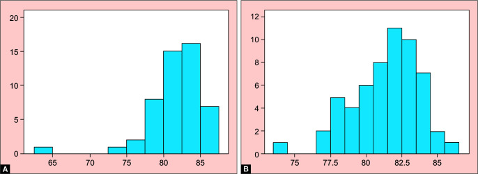
Results: In skeletally immature patients, mean SJLA was 13.46° [standard deviation (SD), 4.55°], and in mature patients, it was 16.91° (SD, 2.948°). The PDFA and PPTA were consistent with previously published measurements.
Conclusion: The SJLA method is a practical way to quantify the soft tissue contribution and degree of contracture. It can also be used for monitoring deterioration or improvement of knee range of motion during lengthening or physical therapy.
Clinical significance: All patients in this study presented to our clinic with symptoms on the contralateral side. This, in addition to the retrospective nature, was a limitation in our study.We recommend a validity study to compare our SJLA method to the classic anterior cortical line angle (ACL) method in addition to an inter-observer and intra-observer reliability study for the SJLA. We also recommend a study on completely normal asymptomatic subjects to better standardise the angle measurements in skeletally immature patients at different ages.
How to cite this article: Abalkhail TB, McClure PK. Sagittal Plane Assessment in Deformity Correction Planning: The Sagittal Joint Line Angle. Strategies Trauma Limb Reconstr 2022;17(3):159-164.
Keywords: Deformity correction; Deformity planning; Knee; Range of motion; Sagittal mechanical axis; Soft tissue contractures.
Copyright © 2022; The Author(s).
Conflict of interest statement
Source of support: Nil Conflict of interest: PKM is a consultant for DePuy Synthes Companies, Novadip, NuVasive Specialized Orthopedics, Orthofix, and Smith & Nephew. The following organizations supported the institution of PKM: DePuy Synthes, NuVasive Specialized Orthopedics, Orthofix, OrthoPediatrics, Paragon 28, Pega Medical, Smith & Nephew, Stryker, Turner Imaging Systems, and WishBone Medical.
Figures


References
-
- Okike K. Preoperative planning in fracture surgery: The end of colored pens and tracing paper?: Commentary on an article by Yanxi Chen, MD, PhD, et al.: “Computer-assisted virtual surgical technology versus three-dimensional printing technology in preoperative planning for displaced three and four-part fractures of the proximal end of the humerus.”. J Bone Joint Surg Am. 2018;100:e146. - PubMed
LinkOut - more resources
Full Text Sources
Miscellaneous
