

Morphological and mechanical alterations in articular cartilage and subchondral bone during spontaneous hip osteoarthritis in guinea pigs
- PMID: 36756384
- PMCID: PMC9900117
- DOI: 10.3389/fbioe.2023.1080241
Morphological and mechanical alterations in articular cartilage and subchondral bone during spontaneous hip osteoarthritis in guinea pigs
Abstract
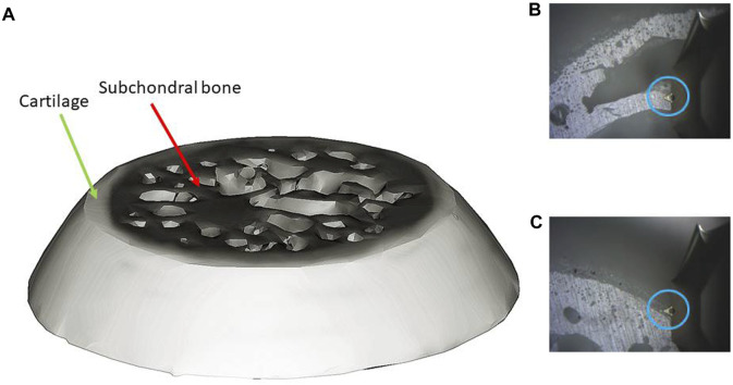
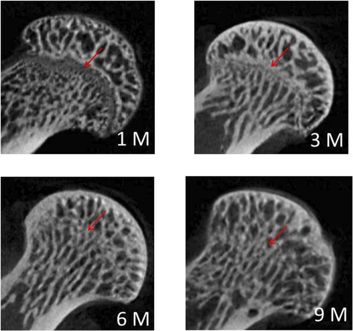
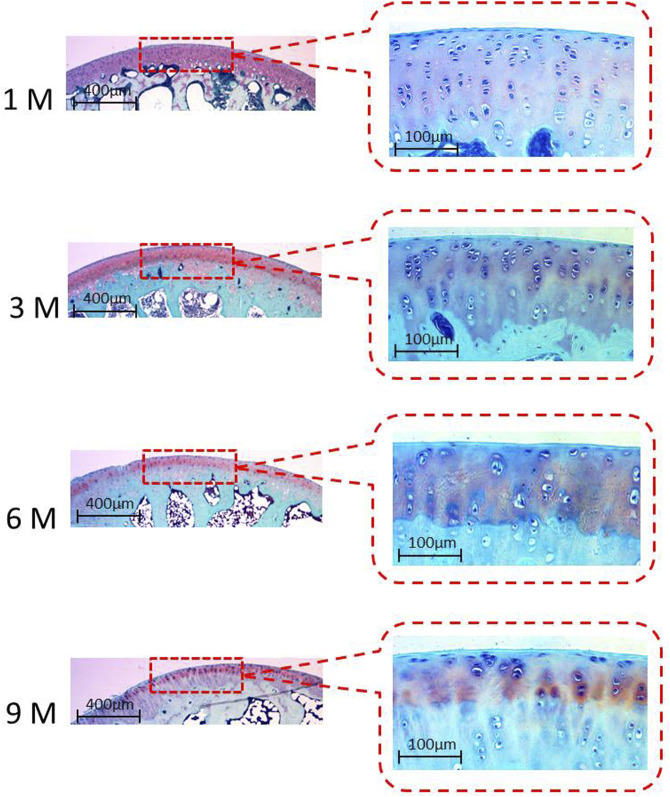
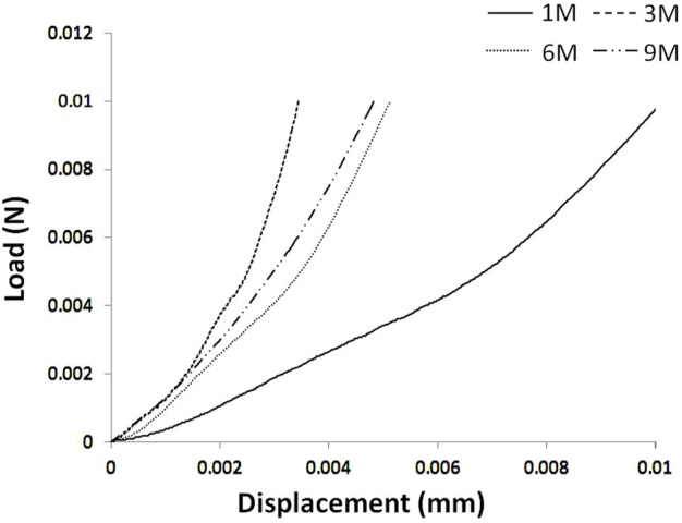
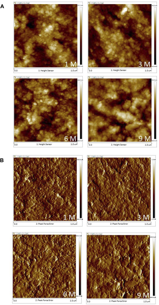
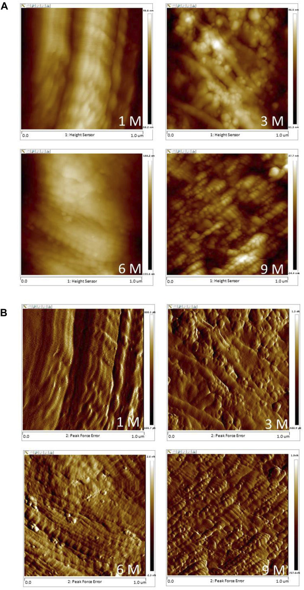
Objectives: This study aimed to investigate the morphological and mechanical changes in articular cartilage and subchondral bone during spontaneous hip osteoarthritis in guinea pigs. Materials and methods: Hip joints of guinea pigs were investigated at 1, 3, 6, and 9 months of age (hereafter denoted as 1 M, 3 M, 6 M, and 9 M, respectively; n = 7 in each group). Morphological and mechanical alterations during spontaneous hip osteoarthritis in guinea pigs were investigated. The alterations included the micromechanical properties of articular cartilage (stiffness and creep deformation), microstructure of the subchondral bone (bone mineral density, bone volume fraction, trabecular thickness, trabecular number, and trabecular separation), micromorphology of the articular cartilage, and surface nanostructure (grain size and roughness) of the articular cartilage and subchondral bone. Results: Micromechanical properties of articular cartilage in 1 M showed the lowest stiffness and highest creep deformation with no significant differences in stiffness or creep deformation amongst 3 M, 6 M, and 9 M. Articular cartilage thickness decreased with age. The earliest degeneration of articular cartilage occurred at 6 months of age, characterised by surface unevenness and evident chondrocytes reduction in micromorphology, as well as increased grain size and decreased roughness in nanostructure. No degeneration at micro- or nanostructure of subchondral bone was observed before 9 months. Conclusion: Morphological degeneration of cartilage occurred before degeneration of mechanical properties. Meanwhile, degeneration of cartilage occurred before degeneration of subchondral bone during hip osteoarthritis. The current study provided novel insights into the structural and micromechanical interaction of hip osteoarthritis, which can serve as a theoretical basis for understanding the formation and progression of osteoarthritis.
Keywords: cartilage; hip osteoarthritis; mechanical properties; morphology; subchondral bone.
Copyright © 2023 Gao, Ren and Gong.
Conflict of interest statement
The authors declare that the research was conducted in the absence of any commercial or financial relationships that could be construed as a potential conflict of interest.
Figures
Similar articles
-
Spatial and temporal changes of subchondral bone proceed to microscopic articular cartilage degeneration in guinea pigs with spontaneous osteoarthritis.Osteoarthritis Cartilage. 2013 Apr;21(4):574-81. doi: 10.1016/j.joca.2013.01.002. Epub 2013 Jan 9. Osteoarthritis Cartilage. 2013. PMID: 23313833
-
Biochemical and Morphological Abnormalities of Subchondral Bone and Their Association with Cartilage Degeneration in Spontaneous Osteoarthritis.Calcif Tissue Int. 2021 Aug;109(2):179-189. doi: 10.1007/s00223-021-00834-3. Epub 2021 Mar 13. Calcif Tissue Int. 2021. PMID: 33715052
-
Role of subchondral bone in osteoarthritis development: a comparative study of two strains of guinea pigs with and without spontaneously occurring osteoarthritis.Arthritis Rheum. 2007 Oct;56(10):3366-74. doi: 10.1002/art.22921. Arthritis Rheum. 2007. PMID: 17907190
-
Bone density and local growth factors in generalized osteoarthritis.Microsc Res Tech. 1997 May 15;37(4):358-71. doi: 10.1002/(SICI)1097-0029(19970515)37:4<358::AID-JEMT10>3.0.CO;2-L. Microsc Res Tech. 1997. PMID: 9185157 Review.
-
Articular cartilage: degeneration and osteoarthritis, repair, regeneration, and transplantation.Instr Course Lect. 1998;47:487-504. Instr Course Lect. 1998. PMID: 9571450 Review.
Cited by
-
Inhibition of complement C3 prevents osteoarthritis progression in guinea pigs by blocking STAT1 activation.Commun Biol. 2024 Mar 27;7(1):370. doi: 10.1038/s42003-024-06051-6. Commun Biol. 2024. PMID: 38538870 Free PMC article.
References
-
- Barr A. J., Campbell T. M., Hopkinson D., Kingsbury S. R., Bowes M. A., Conaghan P. G. (2015). A systematic review of the relationship between subchondral bone features, pain and structural pathology in peripheral joint osteoarthritis. Arthritis Res. Ther. 17 (1), 228. 10.1186/s13075-015-0735-x - DOI - PMC - PubMed
-
- Bartholdy C., Juhl C., Christensen R., Lund H., Zhang W., Henriksen M. (2017). The role of muscle strengthening in exercise therapy for knee osteoarthritis: A systematic review and meta-regression analysis of randomized trials. Semin. Arthritis Rheum. 47 (1), 9–21. 10.1016/j.semarthrit.2017.03.007 - DOI - PubMed
LinkOut - more resources
Full Text Sources