. 2023 Mar;22(3):e13768.
doi: 10.1111/acel.13768. Epub 2023 Feb 8.

DNA repair in cardiomyocytes is critical for maintaining cardiac function in mice

Affiliations

DNA repair in cardiomyocytes is critical for maintaining cardiac function in mice

Martine de Boer et al. Aging Cell. 2023 Mar.

Abstract

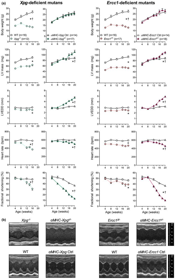
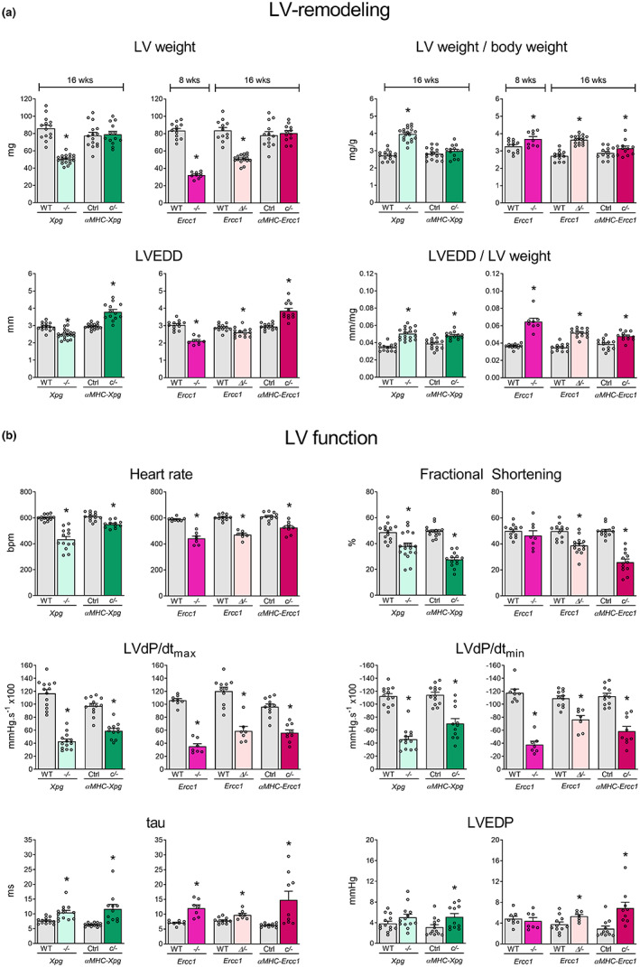
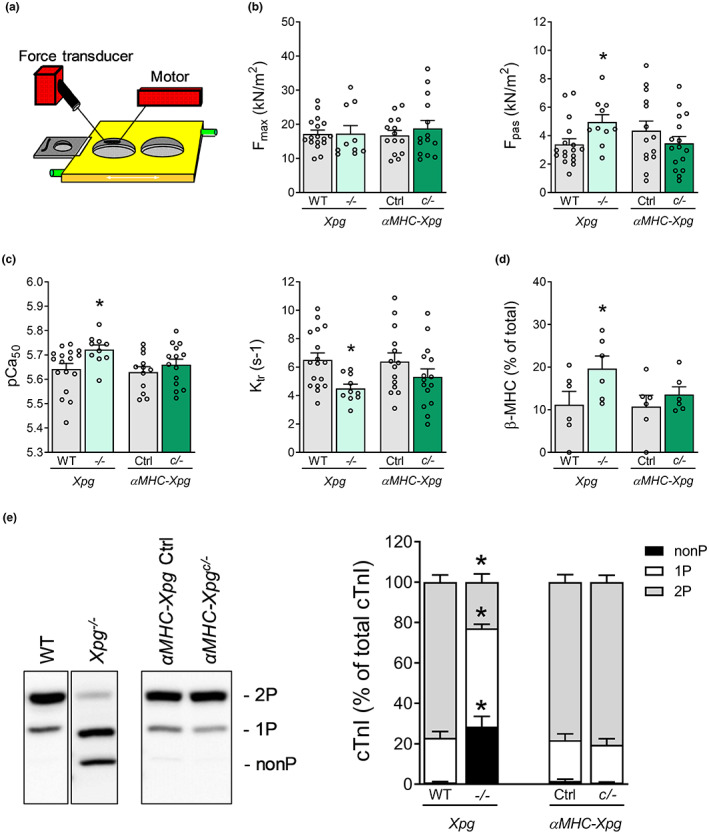
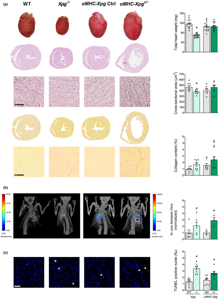
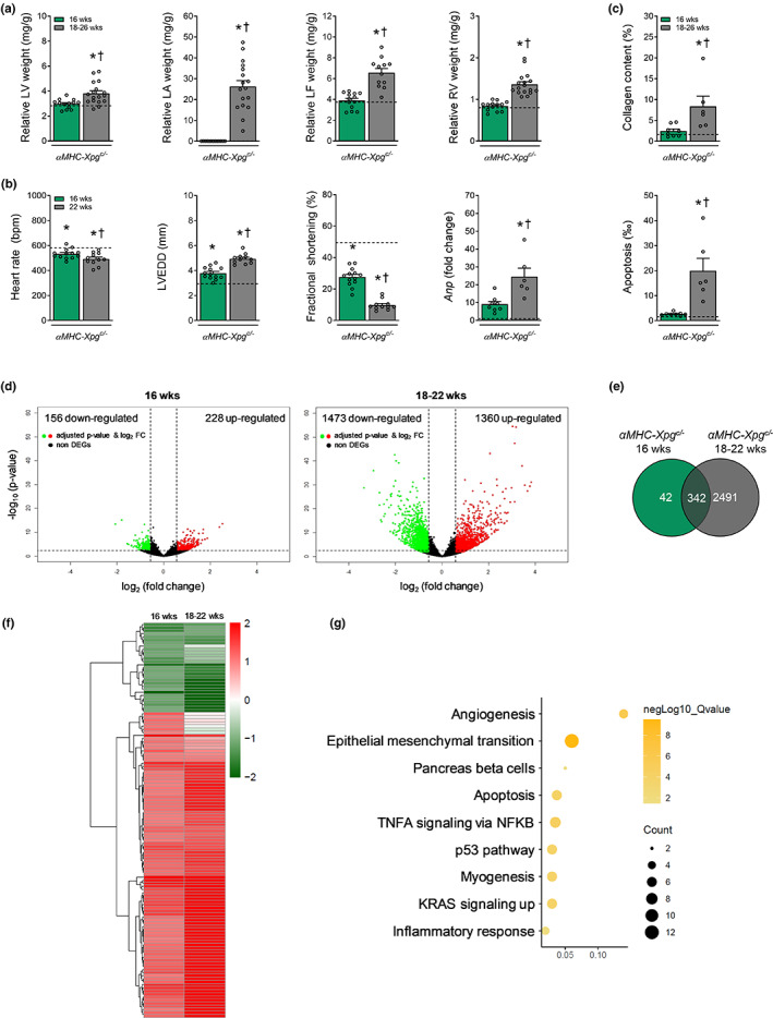
Heart failure has reached epidemic proportions in a progressively ageing population. The molecular mechanisms underlying heart failure remain elusive, but evidence indicates that DNA damage is enhanced in failing hearts. Here, we tested the hypothesis that endogenous DNA repair in cardiomyocytes is critical for maintaining normal cardiac function, so that perturbed repair of spontaneous DNA damage drives early onset of heart failure. To increase the burden of spontaneous DNA damage, we knocked out the DNA repair endonucleases xeroderma pigmentosum complementation group G (XPG) and excision repair cross-complementation group 1 (ERCC1), either systemically or cardiomyocyte-restricted, and studied the effects on cardiac function and structure. Loss of DNA repair permitted normal heart development but subsequently caused progressive deterioration of cardiac function, resulting in overt congestive heart failure and premature death within 6 months. Cardiac biopsies revealed increased oxidative stress associated with increased fibrosis and apoptosis. Moreover, gene set enrichment analysis showed enrichment of pathways associated with impaired DNA repair and apoptosis, and identified TP53 as one of the top active upstream transcription regulators. In support of the observed cardiac phenotype in mutant mice, several genetic variants in the ERCC1 and XPG gene in human GWAS data were found to be associated with cardiac remodelling and dysfunction. In conclusion, unrepaired spontaneous DNA damage in differentiated cardiomyocytes drives early onset of cardiac failure. These observations implicate DNA damage as a potential novel therapeutic target and highlight systemic and cardiomyocyte-restricted DNA repair-deficient mouse mutants as bona fide models of heart failure.

Keywords: DNA damage; DNA repair; apoptosis; cardiac function; congestive heart failure.


Conflict of interest statement

The authors declare no conflict of interest.

Figures

FIGURE 1
FIGURE 1
Impaired DNA repair resulted in time‐dependent deterioration of global LV function in all mutants (Study I, lifespan studies). (a) Effect of Xpg and Ercc1 deficiency on body weight, LV mass and geometry and hemodynamic parameters during age. LV mass, left ventricular mass, calculated using the VisualSonics Cardiac Measurements Package; LVEDD, LV end‐diastolic lumen diameter. The number of animals is indicated in the body weight graph. All mutants have their own corresponding control littermates. Data are presented as mean ± SEM. (b) Representative LV short axis M‐mode images of the different DNA repair‐deficient mutants and corresponding control at age 16 weeks. *p < 0.05 vs. corresponding control; †p < 0.05 genotype × age using mixed linear model–repeated measures.
FIGURE 2
FIGURE 2
LV‐remodeling and LV dysfunction in DNA repair‐deficient mutants at a specific time point (study II). (a) Effect of Xpg and Ercc1 deficiency on LV mass and geometry and (b), hemodynamic parameters in 16‐week‐old Xpg −/− and αMHC‐Xpg c/− , 8‐week‐old Ercc1 −/− , 16‐week‐old Ercc1 Δ/− and αMHC‐Ercc1 c/− mice and corresponding control (n = 7–16 animals/group). LV weight, left ventricular weight; BW, body weight; LVEDD, LV end‐diastolic lumen diameter; LVdP/dtmax, maximum rate of rise of LV pressure; LVdP/dtmin, maximum rate of fall of LV pressure; tau, relaxation time constant; LVEDP, LV end‐diastolic pressure. Data are presented as mean ± SEM. *p < 0.05 vs. corresponding control by two‐way ANOVA followed by SNK post hoc testing.
FIGURE 3
FIGURE 3
Altered cardiomyocyte contractile properties in 16‐week‐old Xpg −/− mice, but not in 16‐week‐old αMHC‐Xpg c/− mice (Study II). (a) Schematic experimental myocyte set‐up. (b) Isometric force measurements in single permeabilized cardiomyocytes (n = 6 animals/group; 1–4 cardiomyocytes/animal). F max, maximal force; F pas, passive force. (c) Determination of Ca2+ sensitivity of force (pCa50) and maximal rate of force redevelopment (Ktr) (n = 6 animals/group; 1–4 cardiomyocytes/animal). (d) Myosin heavy chain isoform composition was determined by protein levels of myosin heavy chain isoforms (n = 6 animals/group). Beta‐myosin heavy chain (β‐MHC) content is expressed as % of total MHC. (e) Phos‐tag analysis of cardiac troponin I (cTnI) species, expressed as % of total phosphorylated cTnI (n = 6–7 animals/group). Phosphorylated cTnI species, of similar molecular weight, were separated into three forms: Non‐phosphorylated (nonP), mono‐phosphorylated (1P) and bis‐phosphorylated (2P). These forms were visualized with a specific antibody against troponin I, which recognizes non‐phosphorylated and phosphorylated forms. Data are presented as mean ± SEM. *p < 0.05 vs. corresponding control by two‐way ANOVA followed by SNK post hoc testing.
FIGURE 4
FIGURE 4
Characterization of LV phenotype in 16‐week‐old Xpg mutants (Study II). (a) Representative images of whole hearts and haematoxylin and eosin‐stained two‐chamber view sections and examination of total heart weight (n = 13–19 animals/group). Representative gomori‐ and picro‐sirius red‐stained LV sections and quantification of, respectively, cardiomyocyte cross‐sectional area and myocardial collagen content (n = 8 animals/group). (b) Representative images and quantification of the in vivo visualization of the membrane‐bound phospholipid phosphatidylserine to detect early apoptosis using FMT combined with μCT (n = 3–6 animals/group). (c) Representative TUNEL‐stained LV sections and quantification of TUNEL‐positive nuclei (indicated by arrows) to detect late apoptotic cells (n = 8 animals/group). Data are presented as mean ± SEM. *p < 0.05, §p = 0.062, #p = 0.087 vs. corresponding control by two‐way ANOVA followed by SNK post hoc testing.
FIGURE 5
FIGURE 5
Progressive congestive heart failure and activation of TP53 in advanced age αMHC‐Xpg c/− mice. (a) Cardiomyocyte‐restricted loss of Xpg resulted in increased LV, LA, LF and RV weight during aging (n = 12–17 animals/group). LV weight, left ventricular weight; LA weight, left atrial weight; LF weight, lung fluid weight; RV weight, right ventricular weight. Weights are normalized to body weight. (b) Echocardiographic analysis at age 22 weeks (Study I, lifespan study) revealed progressive enlarged LV lumen diameter and aggravated loss of factional shortening compared with 16‐week‐old αMHC‐Xpg c/− mice and corresponding control (Study II) (n = 11–15 animals/group). In addition, quantitative real‐time PCR analysis revealed an elevated expression level of Anp (n = 6–8 animals/group). Expression is corrected for Hprt expression and normalized to control. LVEDD, LV end‐diastolic lumen diameter, Anp, atrial natriuretic peptide. (c) Quantification of respectively myocardial fibrosis and TUNEL‐positive nuclei to detect apoptotic cells (n = 6–8 animals/group). (d) Volcano plots present the overall gene expression of 16‐week and 18‐ to 22‐week‐old αMHC‐Xpg c/− compared with 16‐week‐old control (n = 3 animals/group). The dashed lines denote the cut‐off values for a differentially expressed gene (DEG; FDR adjusted P‐value <0.05 and absolute fold change >1.5). Significantly up‐regulated genes are highlighted in red and significantly down‐regulated genes in green. The non‐DEGs are represented in black. (e) Venn diagram display the total amount of overlapping DEGs in both αMHC‐Xpg c/− groups compared with 16‐week‐old control. (f) Heat map of hierarchically clustered overlapping DEGs in 16‐week and 18‐ to 22‐week‐old αMHC‐Xpg c/− mice compared with 16‐week‐old control. (g) Hallmark pathways enriched in αMHC‐Xpg c/− mice. Dot size represents the number of genes in each involved pathway. Dot colour show −log10 (Q‐value) in each term enrichment. X‐axis shows the ratio of the genes to all DEGs. (a–c) For the sake of comparison we have repeated 16‐week‐old αMHC‐Xpg c/− mice (green bars) and corresponding control (black dotted line) in this figure, both from Study II. (d–g) 16‐week‐old αMHC‐Xpg c/− mice: Study II; 18‐ to 22‐week‐old αMHC‐Xpg c/− mice: Study I, lifespan study. Data are presented as mean ± SEM. *p < 0.05 vs. corresponding control; †p < 0.05 vs. αMHC‐Xpg c/− 16 weeks by one‐way ANOVA followed by SNK post hoc testing.

Similar articles

Cited by

References

    1. Agah, R. , Frenkel, P. A. , French, B. A. , Michael, L. H. , Overbeek, P. A. , & Schneider, M. D. (1997). Gene recombination in postmitotic cells. Targeted expression of Cre recombinase provokes cardiac‐restricted, site‐specific rearrangement in adult ventricular muscle in vivo. The Journal of Clinical Investigation, 100(1), 169–179. 10.1172/JCI119509 - DOI - PMC - PubMed
    1. Ataei Ataabadi, E. , Golshiri, K. , van der Linden, J. , de Boer, M. , Duncker, D. J. , Jüttner, A. , de Vries, R. , van Veghel, R. , van der Pluijm, I. , Dutheil, S. , Chalgeri, S. , Zhang, L. , Lin, A. , Davis, R. E. , Snyder, G. L. , Danser, A. H. J. , & Roks, A. J. M. (2021). Vascular ageing features caused by selective DNA damage in smooth muscle cell. Oxidative Medicine and Cellular Longevity, 2021, 2308317. 10.1155/2021/2308317 - DOI - PMC - PubMed
    1. Barnhoorn, S. , Uittenboogaard, L. M. , Jaarsma, D. , Vermeij, W. P. , Tresini, M. , Weymaere, M. , Menoni, H. , Brandt, R. M. , de Waard, M. C. , Botter, S. M. , Sarker, A. H. , Jaspers, N. G. , van der Horst, G. , Cooper, P. K. , Hoeijmakers, J. H. , & van der Pluijm, I. (2014). Cell‐autonomous progeroid changes in conditional mouse models for repair endonuclease XPG deficiency. PLoS Genetics, 10(10), e1004686. 10.1371/journal.pgen.1004686 - DOI - PMC - PubMed
    1. Bartunek, J. , Vanderheyden, M. , Knaapen, M. W. , Tack, W. , Kockx, M. M. , & Goethals, M. (2002). Deoxyribonucleic acid damage/repair proteins are elevated in the failing human myocardium due to idiopathic dilated cardiomyopathy. Journal of the American College of Cardiology, 40(6), 1097–1103; discussion 1104‐1095. 10.1016/s0735-1097(02)02122-8 - DOI - PubMed
    1. Bautista‐Nino, P. K. , Portilla‐Fernandez, E. , Rubio‐Beltran, E. , van der Linden, J. J. , de Vries, R. , van Veghel, R. , de Boer, M. , Durik, M. , Ridwan, Y. , Brandt, R. , Essers, J. , Menzies, R. I. , Thomas, R. , de Bruin, A. , Duncker, D. J. , van HMM, B. , Ghanbari, M. , JHJ, H. , Sedlacek, R. , … Roks, A. J. M. (2020). Local endothelial DNA repair deficiency causes aging‐resembling endothelial‐specific dysfunction. Clinical Science, 134(7), 727–746. 10.1042/CS20190124 - DOI - PubMed

Publication types

Supplementary concepts