. 2023 Mar;27(5):609-621.
doi: 10.1111/jcmm.17638. Epub 2023 Feb 8.

PHF6 promotes the progression of endometrial carcinoma by increasing cancer cells growth and decreasing T-cell infiltration

Affiliations

PHF6 promotes the progression of endometrial carcinoma by increasing cancer cells growth and decreasing T-cell infiltration

Xiaomin Wang et al. J Cell Mol Med. 2023 Mar.

Abstract

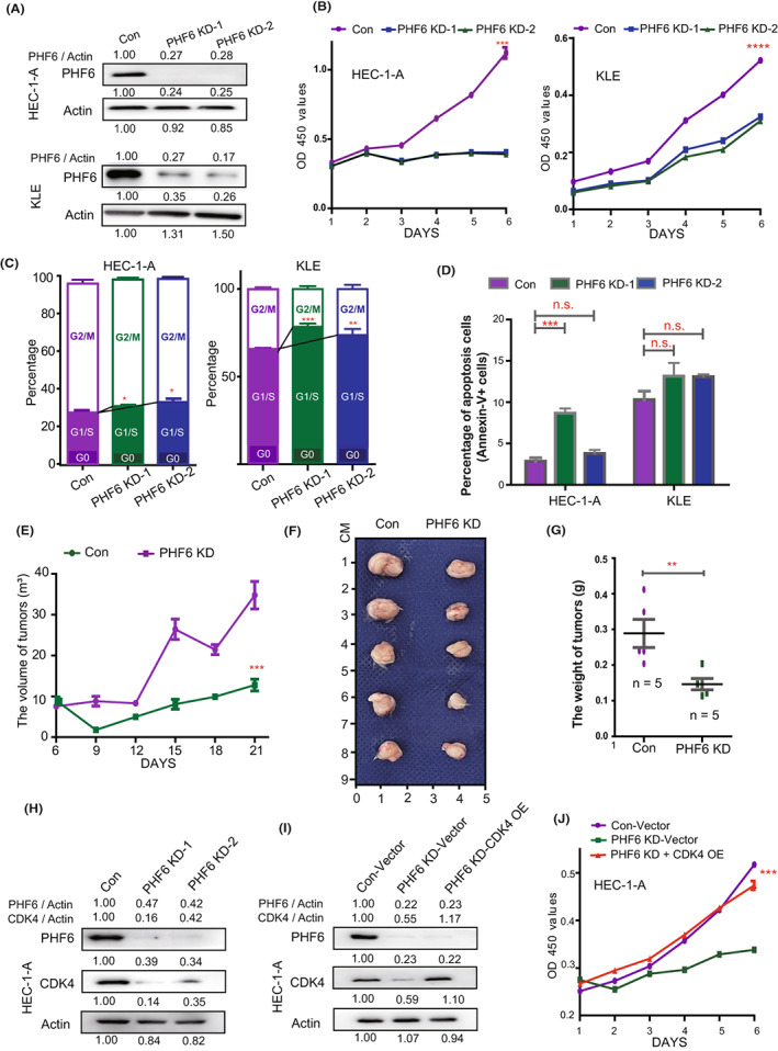
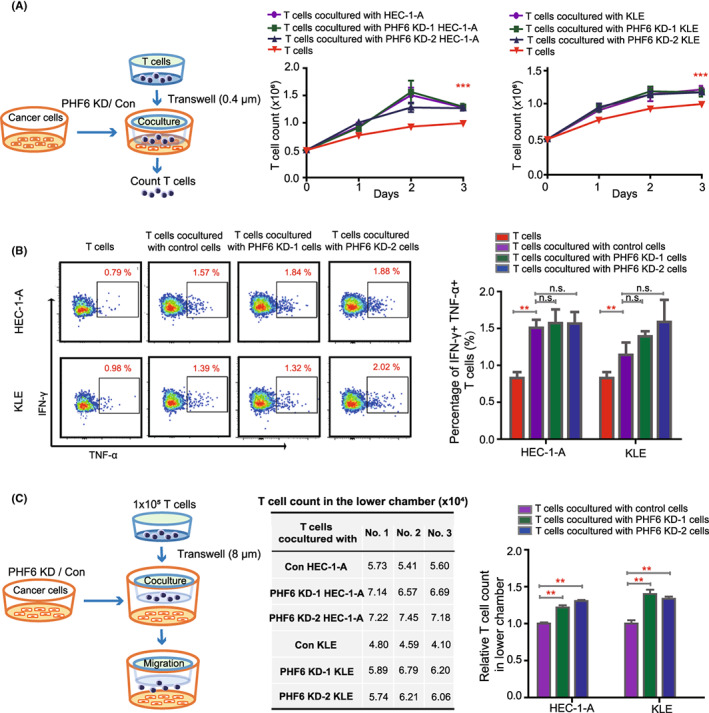
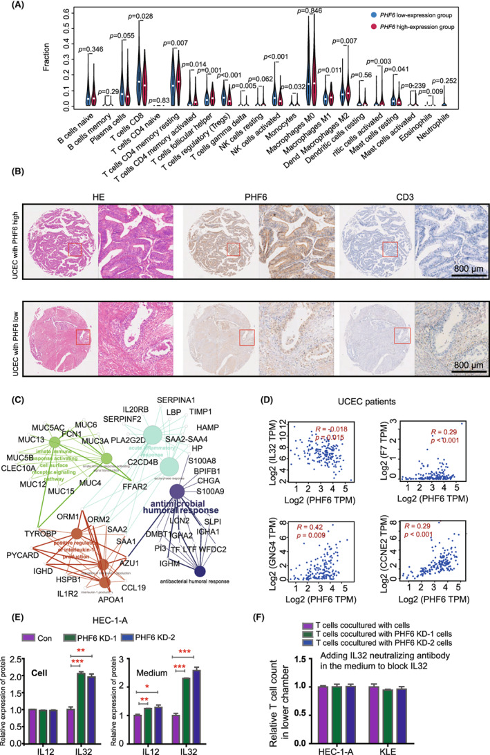
Uterine corpus endometrial carcinoma (UCEC) is the most common cancer of the female reproductive tract. The overall survival of advanced and recurrent UCEC patients is still unfavourable nowadays. It is urgent to find a predictive biomarker and block tumorgenesis at an early stage. Plant homeodomain finger protein 6 (PHF6) is a key player in epigenetic regulation, and its alterations lead to various diseases, including tumours. Here, we found that PHF6 expression was upregulated in UCEC tissues compared with normal tissues. The UCEC patients with high PHF6 expression had poor survival than UCEC patients with low PHF6 expression. PHF6 mutation occurred in 12% of UCEC patients, and PHF6 mutation predicted favourable clinical outcome in UCEC patients. Depletion of PHF6 effectively inhibited HEC-1-A and KLE cell proliferation in vitro and decreased HEC-1-A cell growth in vivo. Furthermore, high PHF6 level indicated a subtype of UCECs characterized by low immune infiltration, such as CD3+ T-cell infiltration. While knockdown of PHF6 in endometrial carcinoma cells increased T-cell migration by promoting IL32 production and secretion. Taken together, our findings suggested that PHF6 might play an oncogenic role in UCEC patients. Thus, PHF6 could be a potential biomarker in predicting the prognosis of UCEC patients. Depletion of PHF6 may be a novel therapeutic strategy for UCEC patients.

Keywords: PHF6; cell cycle; clinical outcome; epigenetic regulated gene; gene regulation; immune infiltration; uterine corpus endometrial carcinoma.


Conflict of interest statement

The authors declare no conflict of interest.

Figures

FIGURE 1
FIGURE 1
High expression of PHF6 predicted the unfavourable survival of UCEC patients. (A) The mRNA expression of PHF6 in UCEC tissues and normal tissues from the TCGA and the GTEx database. (B) The protein expression of PHF6 in tumour tissues and normal tissues in UCEC patients from the TCGA database. (C) Kaplan–Meier survival curves showing the overall survival (OS) of UCEC patients with high PHF6 expression or UCEC patients with low PHF6 expression. (D‐E) The correlation between PHF6 mutation status and OS or progression‐free interval (PFI) of UCEC patients from TCGA dataset. (F) The overall survival curve of UCEC patients with low‐PHF6 + PHF6 mutation, high‐PHF6 + PHF6 mutation, low‐PHF6 + PHF6 non‐mutation, or high‐PHF6 + PHF6 non‐mutation. (G) The ROC curve for predicting the efficacy of PHF6 for UCEC patients. AUC at 5 years of 0.61, 8 years of 0.65, 10 years of 0.64, and 12 years of 0.76. (* represents a significant difference, * p < 0.05)
FIGURE 2
FIGURE 2
Knockdown of PHF6 inhibited the growth of endometrial carcinoma cells by blocking CDK4 expression in vitro and in vivo. (A) Up panel, the expression of PHF6 in PHF6 KD HEC‐1‐A and control cells. Down panel, the expression of PHF6 in PHF6 KD KLE and control cells. (B) The growth curve of PHF6 KD endometrial carcinoma cells and control cells. (C) The cell cycle stage of PHF6 KD endometrial carcinoma cells and control cells. (D) The apoptosis level of PHF6 KD endometrial carcinoma cells and control cells. (E) The growth of PHF6 KD HEC‐1‐A or control cells subcutaneously xenografted tumours in female NSG mice (n = 5). Tumour volume was measured every 3 days. (F) The tumour mass of PHF6 KD HEC‐1‐A or control cells subcutaneously xenografts after harvest. (G) The weight of tumours harvested from NSG mice. (H) The protein expression of CDK4 and PHF6 in PHF6 KD HEC‐1‐A or control cells. (I) The protein expression of PHF6 and CDK4 in Con‐Vector, PHF6 KD‐Vector and PHF6 KD‐CDK4 OE HEC‐1‐A cells. (J) The growth curve of Con‐Vector, PHF6 KD‐Vector and PHF6 KD‐CDK4 OE HEC‐1‐A cells.
FIGURE 3
FIGURE 3
Knockdown of PHF6 in endometrial carcinoma cells promoted T‐cell migration. (A) Left panel, schematic representation of different endometrial carcinoma cells cocultured with T cells. Right panel, the growth curve of T cells cocultured with PHF6 KD endometrial carcinoma cells or control cells. (B) The percentage of IFN‐γ+ and TNF‐α+ cells in T cells when cocultured with PHF6 KD endometrial carcinoma cells or control cells. (C) Left panel, schematic representation of T cells migrated to the lower chamber. Right panel, the absolute number and relative number of T cells in the lower chamber.
FIGURE 4
FIGURE 4
PHF6 inhibited T‐cell infiltration by decreasing the expression of IL32 in UCEC patients. (A) Wilcoxon rank‐sum test revealed the difference in the infiltration levels of 22 immune cells in the low‐PHF6 and the high‐PHF6 UCEC patients. (B) Immunohistochemistry (IHC) verified the correlation between the expression of PHF6 and the infiltration of CD3+ T cells in UCEC tissues. (C) The network of differentially expressed genes between the low‐PHF6 and the high‐PHF6 UCEC patients. (D) The relationship between the expression of PHF6 and immune‐related factors, such as IL32, F7, GNG4 and CCNE4. (E) Left panel, the protein level of IL12 and IL32 in PHF6 KD HEC‐1‐A and control cells by ELISA assay. Right panel, the protein level of IL12 and IL32 in the medium of PHF6 KD HEC‐1‐A and control cells by ELISA assay. (F) IL‐32 neutralizing antibody was added in the medium of PHF6 KD HEC‐1‐A cells, PHF6 KD KLE cells and control cells. The absolute number of T cells in the lower chamber was counted. The relative number of T cells in the lower chamber was evaluated.

References

    1. Najafi M, Majidpoor J, Toolee H, Mortezaee K. The current knowledge concerning solid cancer and therapy. J Biochem Mol Toxicol. 2021;35:e22900. - PubMed
    1. Burstein HJ, Krilov L, Aragon‐Ching JB, et al. Clinical cancer advances 2017: annual report on Progress against cancer from the American Society of Clinical Oncology. J Clin Oncol. 2017;35:1341‐1367. - PubMed
    1. Bennett RL, Licht JD. Targeting epigenetics in cancer. Annu Rev Pharmacol Toxicol. 2018;58:187‐207. - PMC - PubMed
    1. Park JW, Han JW. Targeting epigenetics for cancer therapy. Arch Pharm Res. 2019;42:159‐170. - PMC - PubMed
    1. Yuan S, Wang X, Hou S, et al. PHF6 and JAK3 mutations cooperate to drive T‐cell acute lymphoblastic leukemia progression. Leukemia. 2022;36:370‐382. - PMC - PubMed

Publication types