. 2023 Feb;95(2):e28566.
doi: 10.1002/jmv.28566.

SARS-CoV-2 infection of kidney tissues from severe COVID-19 patients

Affiliations

SARS-CoV-2 infection of kidney tissues from severe COVID-19 patients

Shawn Radovic et al. J Med Virol. 2023 Feb.

Abstract

Background: Coronavirus disease 2019 (COVID-19) caused by infection of severe acute respiratory syndrome coronavirus 2 (SARS-CoV-2) manifests diverse clinical pathologies involving multiple organs. While the respiratory tract is the primary SARS-CoV-2 target, acute kidney injury is common in COVID-19 patients, displaying as acute tubular necrosis (ATN) resulting from focal epithelial necrosis and eosinophilia, glomerulosclerosis, and autolysis of renal tubular cells. However, whether any renal cells are infected by SARS-CoV-2 and the mechanism involved in the COVID-19 kidney pathology remain unclear.

Methods: Kidney tissues obtained at autopsy from four severe COVID-19 patients and one healthy subject were examined by hematoxylin and eosin staining. Indirect immunofluorescent antibody assay was performed to detect SARS-CoV-2 spike protein S1 and nonstructural protein 8 (NSP8) together with markers of different kidney cell types and immune cells to identify the infected cells.

Results: Renal parenchyma showed tissue injury comprised of ATN and glomerulosclerosis. Positive staining of S1 protein was observed in renal parenchymal and tubular epithelial cells. Evidence of viral infection was also observed in innate monocytes/macrophages and NK cells. Positive staining of NSP8, which is essential for viral RNA synthesis and replication, was confirmed in renal parenchymal cells, indicating the presence of active viral replication in the kidney.

Conclusions: In fatal COVID-19 kidneys, there are SARS-CoV-2 infection, minimally infiltrated innate immune cells, and evidence of viral replication, which could contribute to tissue damage in the form of ATN and glomerulosclerosis.

Keywords: COVID-19; SARS-CoV-2; acute tubular necrosis, ATN; glomerulosclerosis; renal cell infection.


Conflict of interest statement

CONFLICT OF INTEREST STATEMENT

The authors declare no conflict of interest.

Figures

FIGURE 1
FIGURE 1
Representative hematoxylin-eosin staining images of postmortem kidney tissues from four COVID-19 patients and one health control shown in both left and right columns. Eosinophilic cast (black asterisk in case 2, right panel), autolysis (white arrow in case 3, right panel), sclerosed glomeruli (red arrow in case 3, left panel), thyroidization (white asterisk in case 5, left panel), nonspecific mononuclear inflammation (black arrow in case 5, left panel), and arterionephrosclerosis (red asterisk in case 5, right panel).
FIGURE 2
FIGURE 2
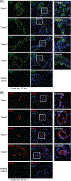
Representative multicolor immunofluorescence staining of severe acute respiratory syndrome coronavirus 2 (SARS-CoV-2) structural and nonstructural proteins in postmortem kidney tissues from coronavirus disease 2019 (COVID-19) patients and a healthy subject. (A) SARS-CoV-2 S1 protein (RBD, pseudo color green) in all four COVID-19 cases but not the healthy control. (B) SARS-CoV-2 NSP8 protein (pseudo color red) in all four COVID-19 cases but not the healthy control. Nuclei were stained with DAPI (pseudo color blue).
FIGURE 3
FIGURE 3
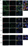
Representative multicolor immunofluorescence staining of severe acute respiratory syndrome coronavirus 2 (SARS-CoV-2) spike protein, and markers of renal and innate immune cells in postmortem kidney tissues from coronavirus disease 2019 patients and a healthy subject. (A) Images of dual immunofluorescence detection of tubular epithelial cells (β-catenin, pseudo color green) and SARS-CoV-2 S1 protein (RBD, pseudo color red) in case 1 and 5 but not the healthy control. (B) Images of dual immunofluorescence detection of monocytes and macrophages (CD68, pseudo color red) and SARS-CoV-2 S1 protein (RBD, pseudo color green) in case 1 and 5 but not the healthy control. (C) Images of dual immunofluorescence detection of NK cells (CD56, pseudo color red) and SARS-CoV-2 S1 protein (RBD, pseudo color green) in case 3 but not the healthy control. Nuclei were stained with DAPI (pseudo color blue).

References

    1. Harrison AG, Lin T, Wang P. Mechanisms of SARS-CoV-2 transmission and pathogenesis. Trends Immunol. 2020;41(12):1100–1115. doi:10.1016/j.it.2020.10.004 - DOI - PMC - PubMed
    1. Lu R, Zhao X, Li J, et al. Genomic characterisation and epidemiology of 2019 novel coronavirus: implications for virus origins and receptor binding. Lancet. 2020;395(10224):565–574. doi:10.1016/S0140-6736(20)30251-8 - DOI - PMC - PubMed
    1. Puelles VG, Lütgehetmann M, Lindenmeyer MT, et al. Multiorgan and renal tropism of SARS-CoV-2. N Engl J Med. 2020;383(6): 590–592. doi:10.1056/NEJMc2011400 - DOI - PMC - PubMed
    1. Ramos da Silva S, Ju E, Meng W, et al. Broad severe acute respiratory syndrome coronavirus 2 cell tropism and immunopathology in lung tissues from fatal coronavirus disease 2019. J Infect Dis. 2021;223(11):1842–1854. doi:10.1093/infdis/jiab195 - DOI - PMC - PubMed
    1. Wu F, Zhao S, Yu B, et al. A new coronavirus associated with human respiratory disease in China. Nature. 2020;579(7798):265–269. doi:10.1038/s41586-020-2008-3 - DOI - PMC - PubMed

Publication types

Substances