. 2023 Feb;3(2):e652.
doi: 10.1002/cpz1.652.

Multiplexed Transgenic Selection and Counterselection Strategies to Expedite Genetic Manipulation Workflows Using Drosophila melanogaster

Affiliations

Multiplexed Transgenic Selection and Counterselection Strategies to Expedite Genetic Manipulation Workflows Using Drosophila melanogaster

Koen J T Venken et al. Curr Protoc. 2023 Feb.

Abstract

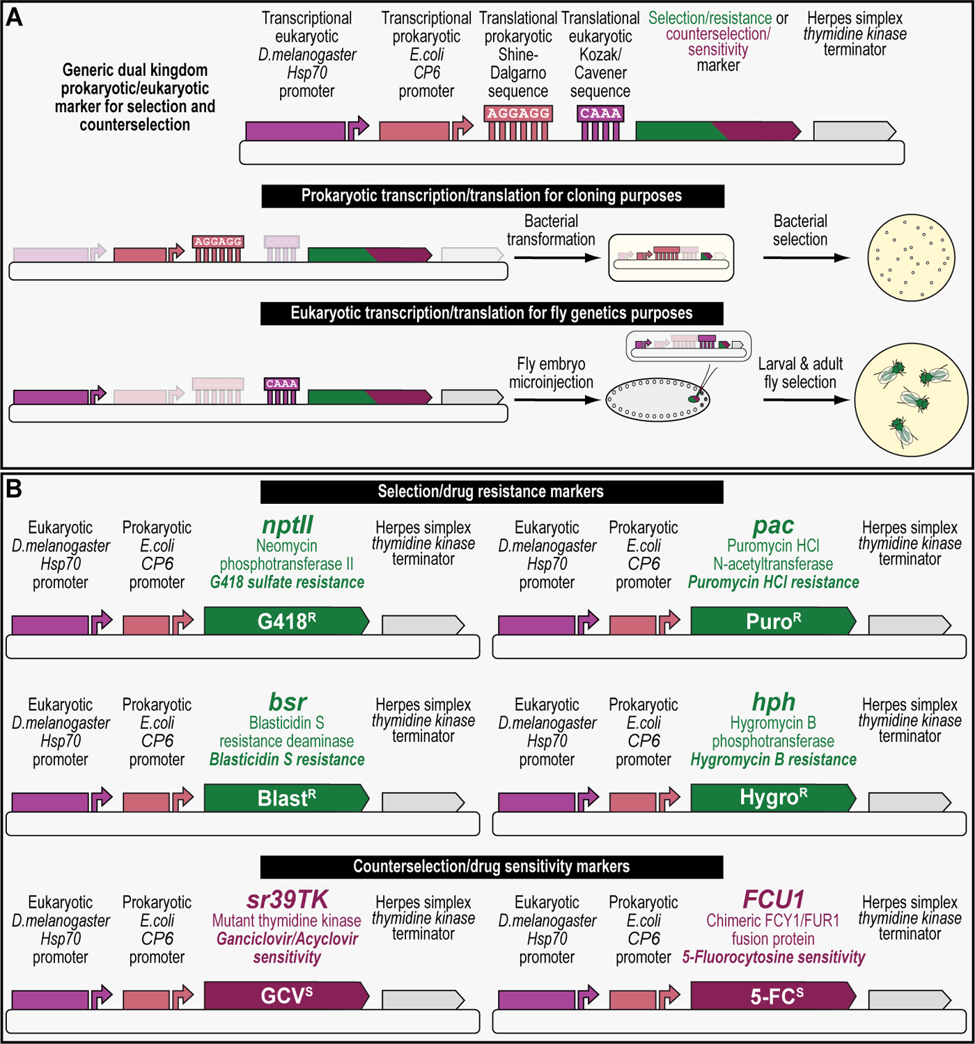
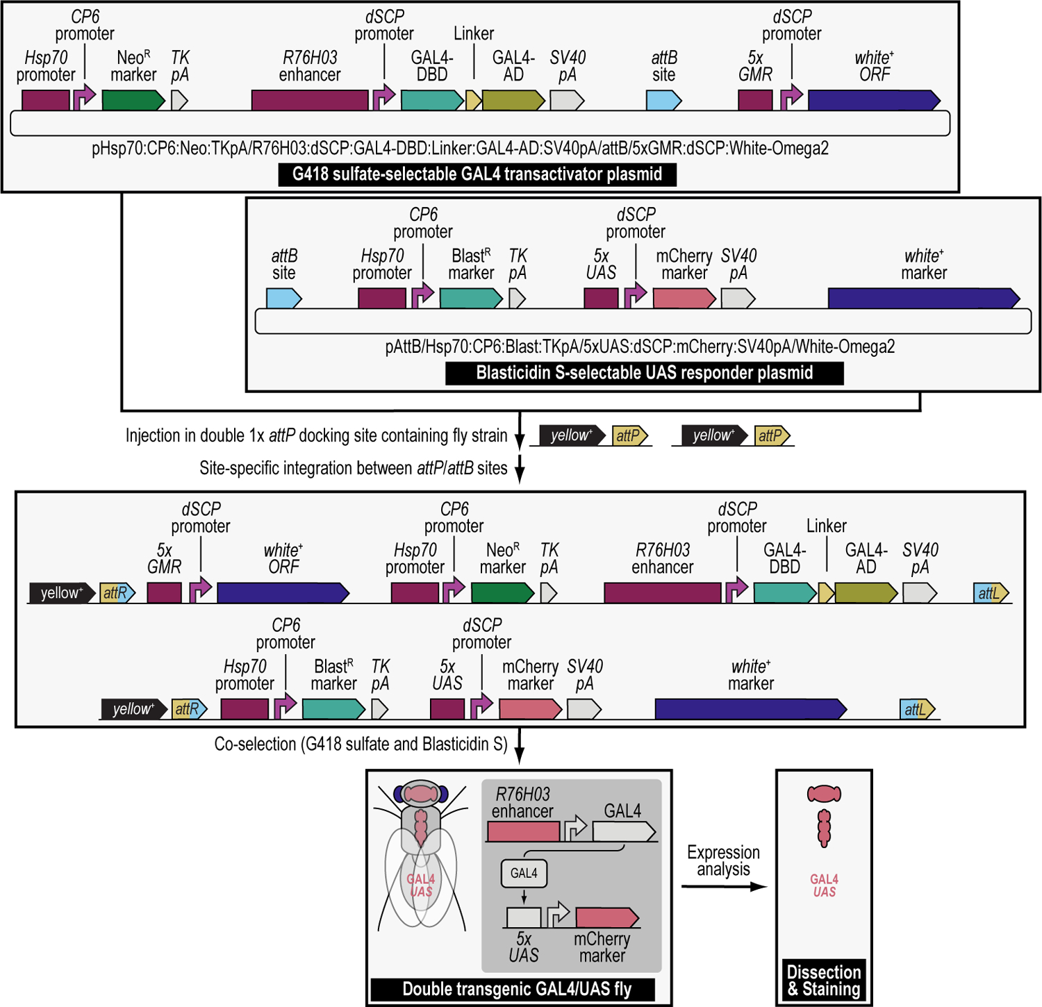
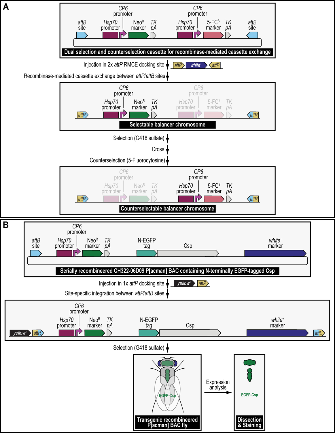
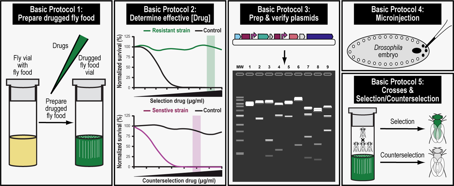
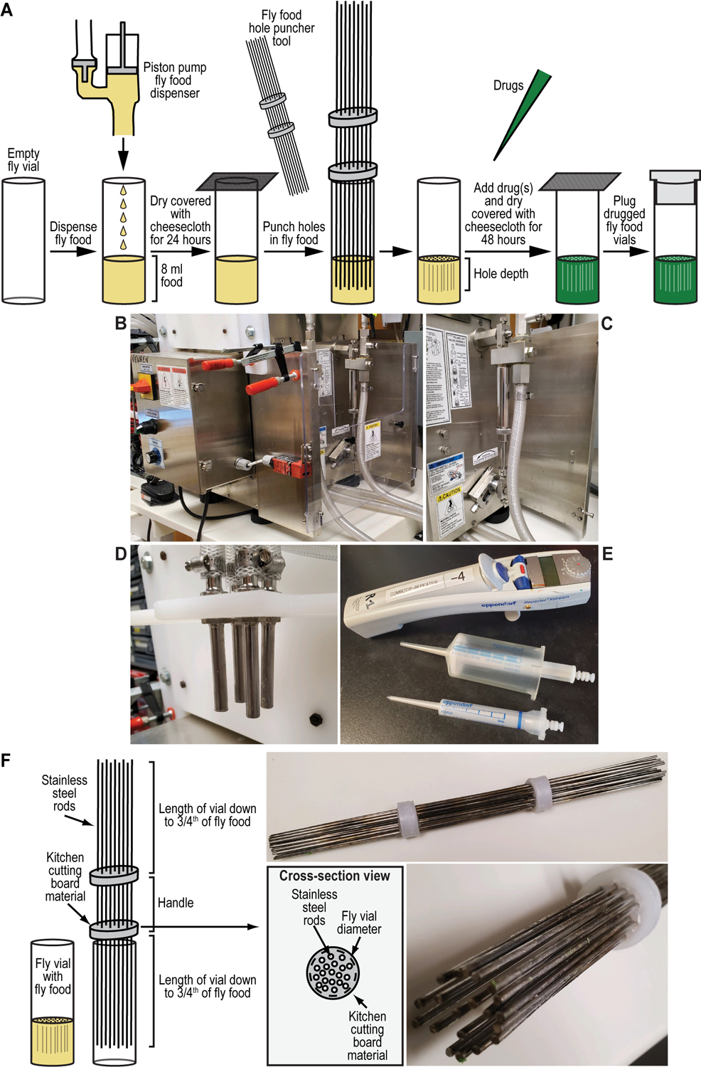
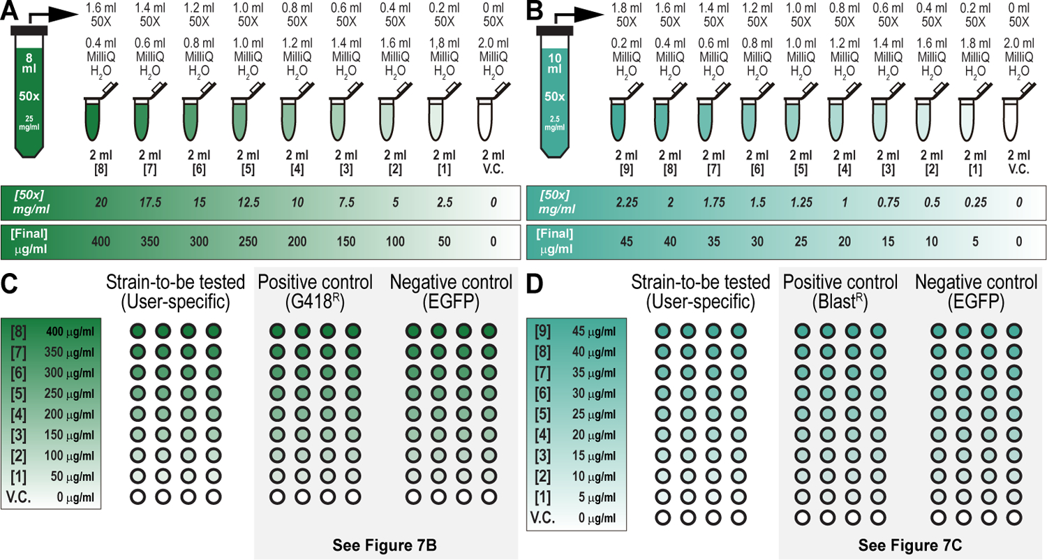
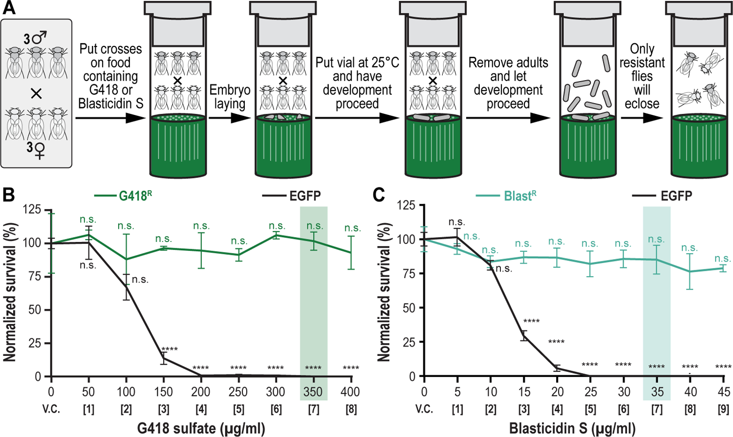
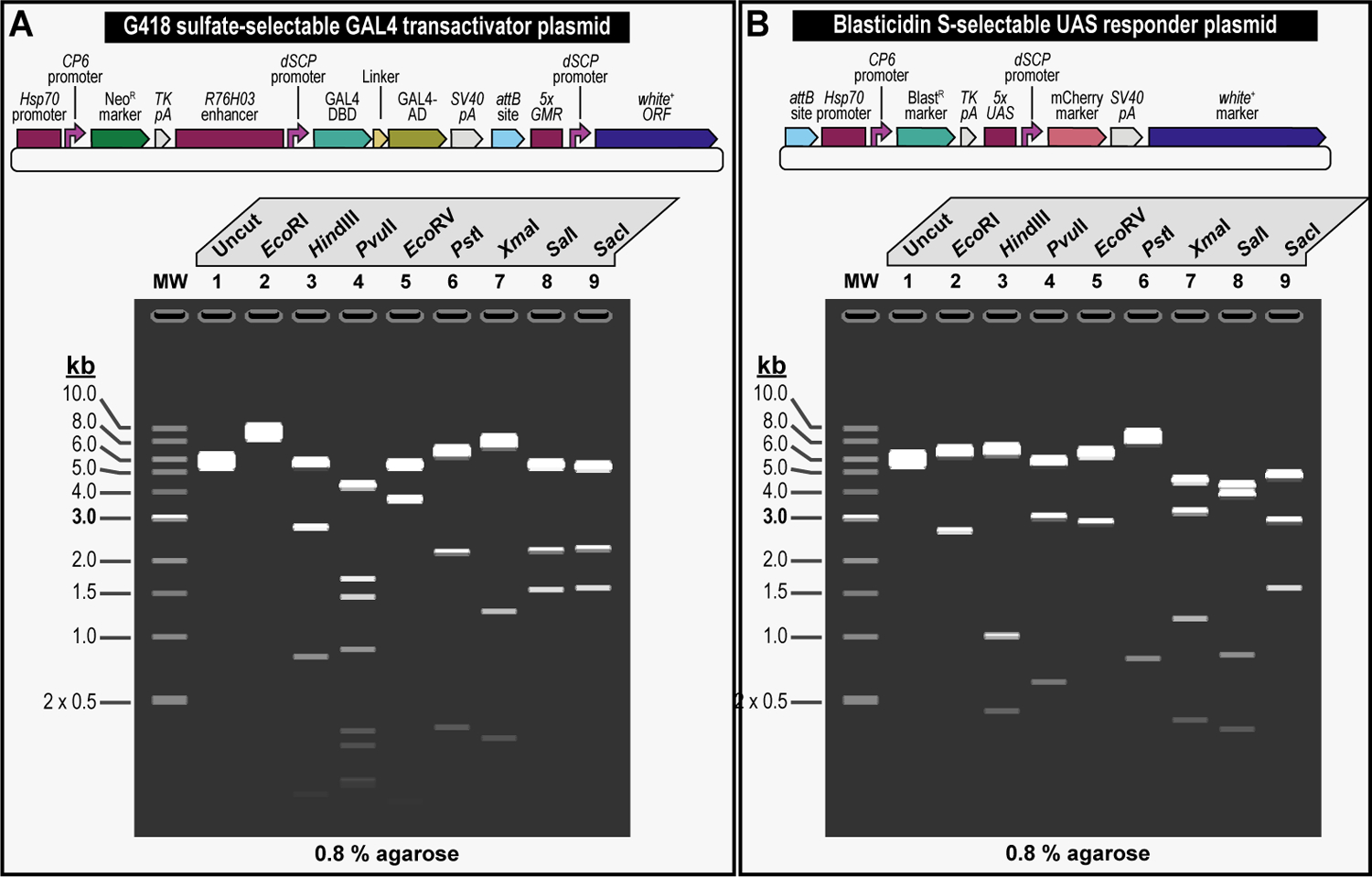
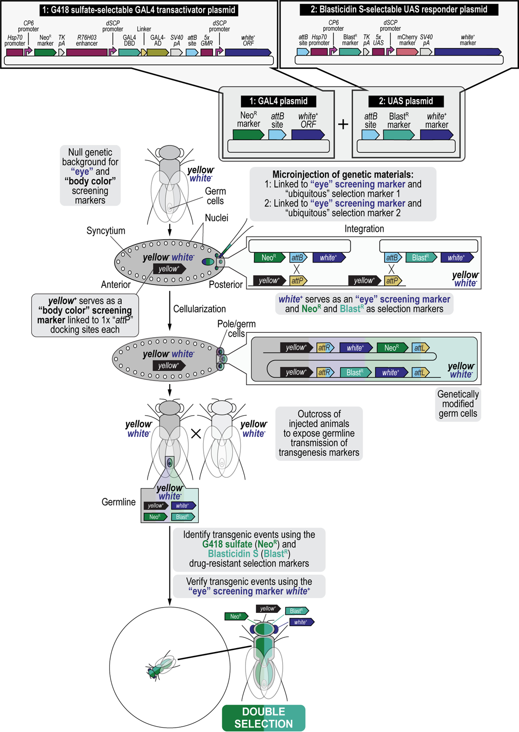
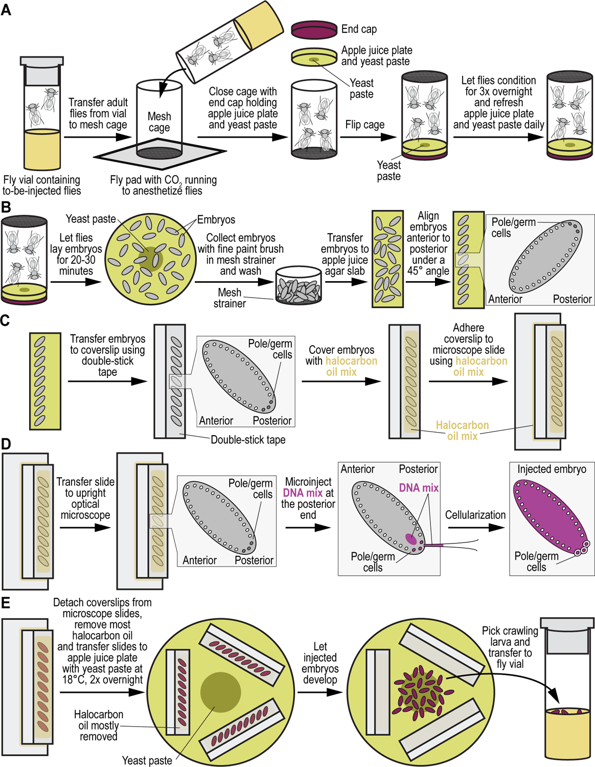
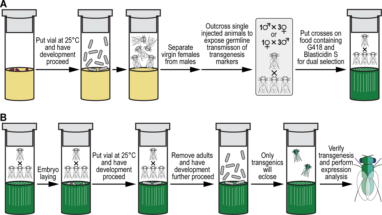
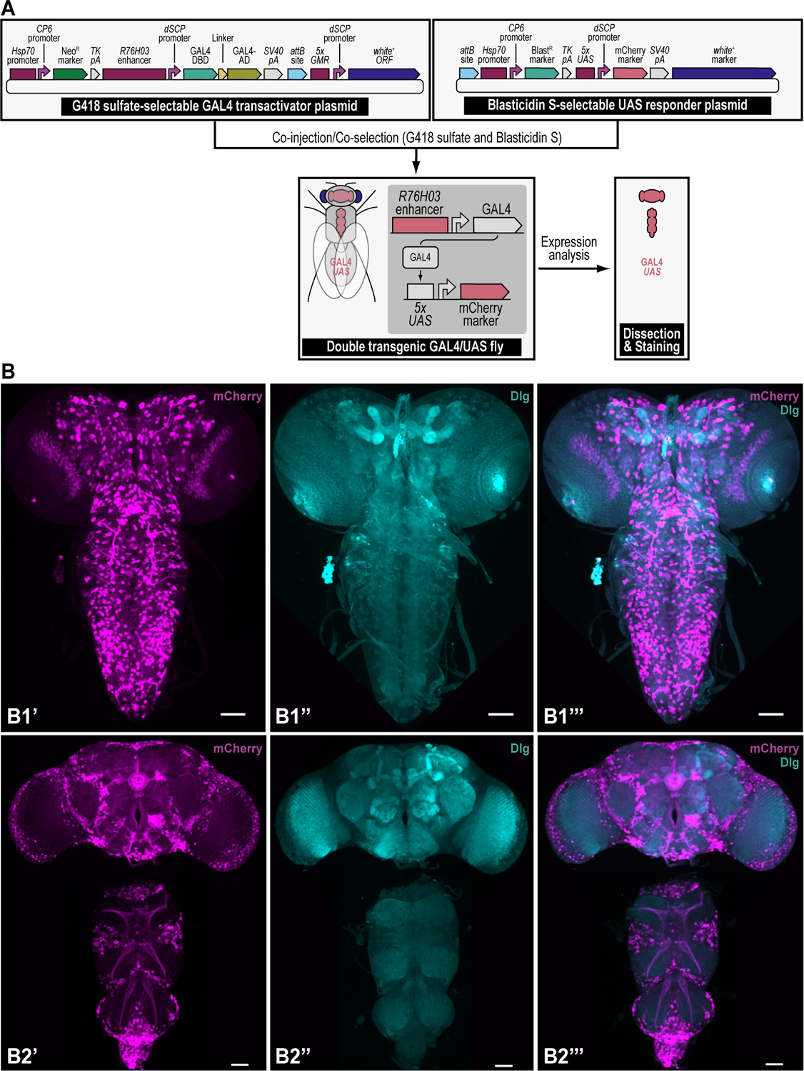
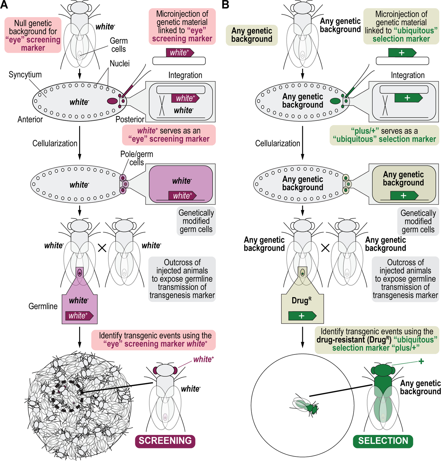
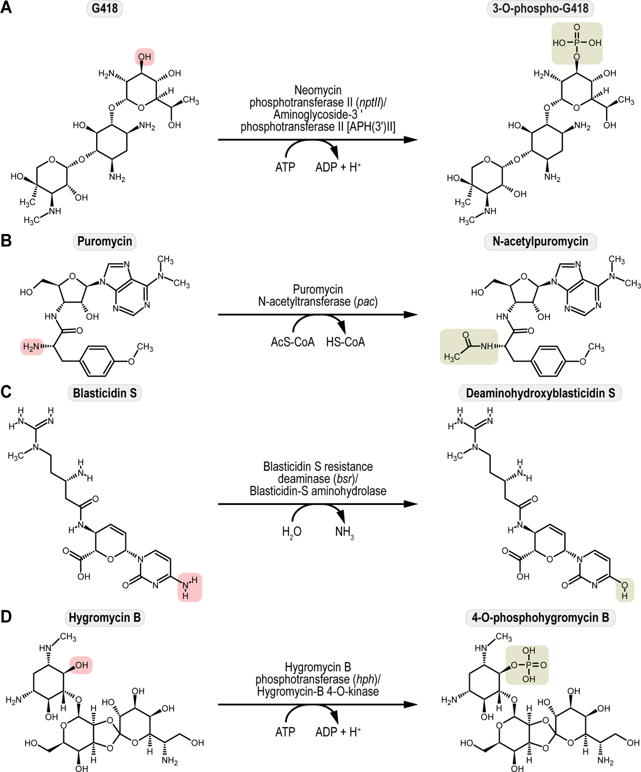
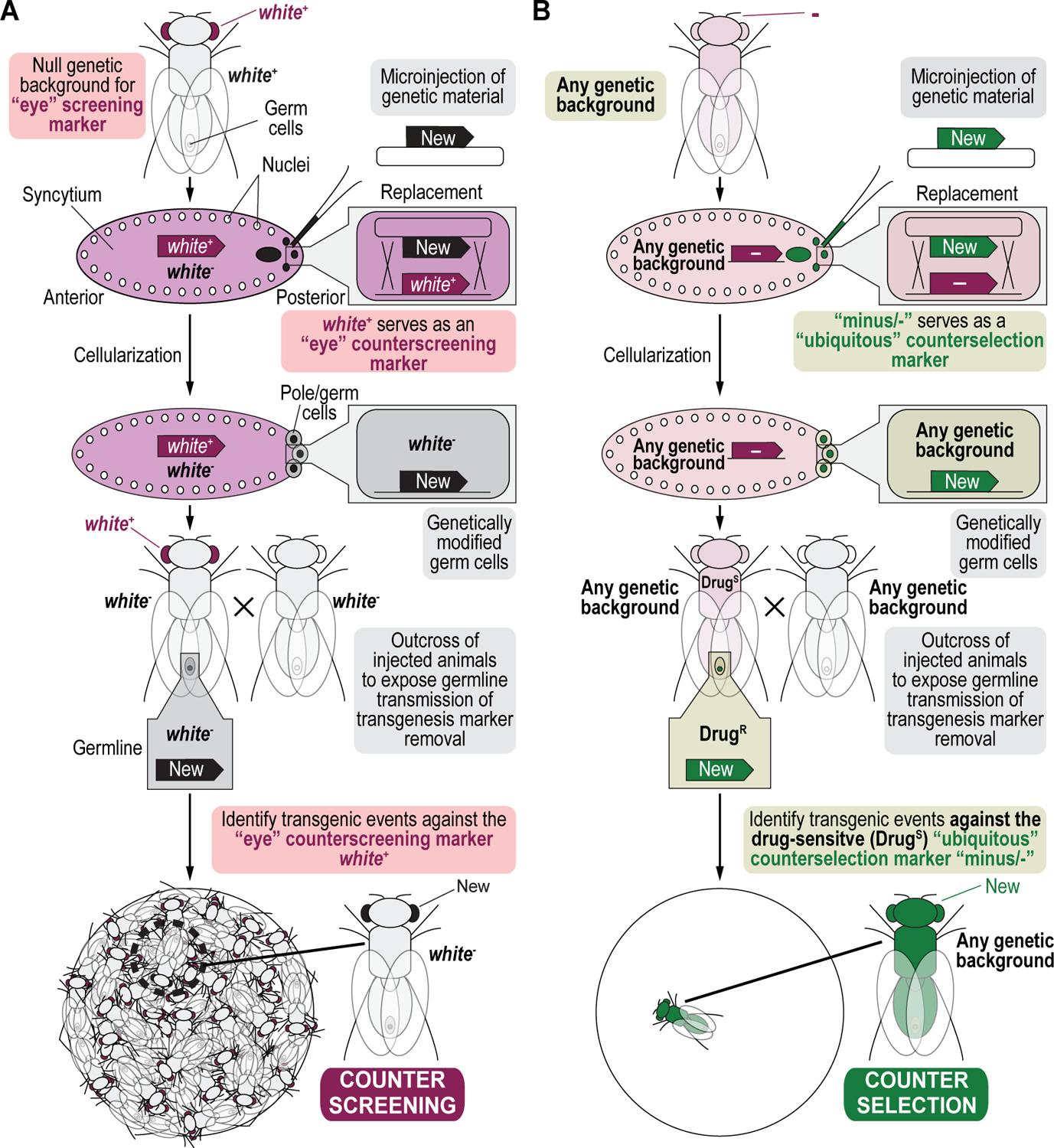
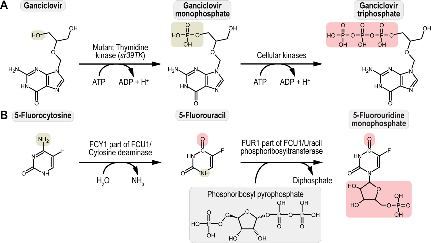
We recently described a set of four selectable and two counterselectable markers that provide resistance and sensitivity, respectively, against their corresponding drugs using the model organism Drosophila melanogaster. The four selectable markers provide animals with resistance against G418 sulfate, puromycin HCl, blasticidin S, or hygromycin B, whereas the two counterselection markers make animals sensitive to ganciclovir/acyclovir or 5-fluorocytosine. Unlike classical phenotypic markers, whether visual or fluorescent, which require extensive screening of progeny of a genetic cross for desired genotypes, resistance and sensitivity markers eliminate this laborious procedure by directly selecting for, or counterselecting against, the desired genotypes. We demonstrated the usefulness of these markers with three applications: 1) generating dual transgenic animals for binary overexpression (e.g., GAL4/UAS) analysis in a single step through the process of co-injection, followed by co-selection resulting in co-transgenesis; 2) obtaining balancer chromosomes that are both selectable and counterselectable to manipulate crossing schemes for, or against, the presence of the modified balancer chromosome; and 3) making both selectable and fluorescently tagged P[acman] BAC transgenic animals for gene expression and proteomic analysis. Here, we describe detailed procedures for how to use these drug-based selection and counterselection markers in the fruit fly D. melanogaster when making dual transgenic animals for binary overexpression as an example. Dual transgenesis integrates site-specifically into two sites in the genome in a single step, namely both components of the binary GAL4/UAS overexpression system, via a G418 sulfate-selectable GAL4 transactivator plasmid and a blasticidin S-selectable UAS responder plasmid. The process involves co-injecting the two plasmids, followed by co-selection using G418 sulfate and blasticidin S, resulting in co-transgenesis of the two plasmids in the fly genome. We demonstrate the functionality of the procedure based on the expression pattern obtained after dual transgenesis of the two plasmids. We provide protocols on how to prepare drugged fly food vials, determine the effective drug concentration for markers used during transgenic selection and counterselection strategies, and prepare and confirm plasmid DNA for microinjection, followed by the microinjection procedure itself and setting up crossing schemes to isolate desired progeny through selection and/or counterselection. These protocols can be easily adapted to any combination of the six selectable and counterselectable markers we described or any new marker that is resistant or sensitive to a novel drug. Protocols on how to build plasmids by synthetic-assembly DNA cloning or modify plasmids by serial recombineering to perform a plethora of selection, counterselection, or any other genetic strategies are presented in two accompanying Current Protocols articles. © 2023 Wiley Periodicals LLC. Basic Protocol 1: Preparing drugged fly food vials for transgenic selection and counterselection strategies using D. melanogaster Basic Protocol 2: Determining the effective drug concentration for resistance and sensitivity markers used during transgenic selection and counterselection strategies using D. melanogaster Basic Protocol 3: Preparing and confirming plasmid DNA for microinjection to perform transgenic selection and counterselection strategies using D. melanogaster Basic Protocol 4: Microinjecting plasmid DNA into fly embryos to perform transgenic selection and counterselection strategies using D. melanogaster Basic Protocol 5: Crossing schemes to isolate desired progeny through transgenic selection and counterselection strategies using D. melanogaster.

Keywords: Drosophila melanogaster; counterselection; genetic manipulation; genetics; multiplexed; selection; transgenesis.


Conflict of interest statement

CONFLICT OF INTEREST STATEMENT

The authors declare no conflict of interest

Figures

Figure 1.
Figure 1.. Schematics of markers used during selection and counterselection genetic strategies using Drosophila melanogaster.
(A) Detailed schematic of a generic dual kingdom eukaryotic/prokaryotic marker to facilitate selection and counterselection genetic strategies using Drosophila melanogaster. The dual kingdom eukaryotic/prokaryotic selection and counterselection markers contain a fusion promoter with both eukaryotic and prokaryotic properties, a gene encoding the selection/resistance or counterselection/sensitivity marker, and the minimal terminator of the thymidine kinase gene of the herpes simplex virus. The fusion promoter consists of the eukaryotic promoter of the Drosophila melanogaster Hsp70 gene for optimal transcription in flies, followed by the prokaryotic synthetic Escherichia coli CP6 promoter for optimal transcription in bacteria. At the 3’ end of the CP6 promoter are a prokaryotic Shine-Dalgarno sequence for optimal translational in bacteria, followed by a eukaryotic Kozak/Cavener sequence for optimal translational in flies, i.e., since the Shine-Dalgarno sequence is located around 8 bases upstream of the start codon, the Kozak/Cavener sequence could be fitted in between Shine-Dalgarno sequence and the translational start codon. For bacterial cloning purposes, CP6-stimulated transcription and Shine-Dalgarno-stimulated translation of the marker provides bacterial selection, while for fly genetics purposes, Hsp70-stimulated transcription and Kozak/Cavener-stimulated translation of the marker provides larval and fly selection. (B) Schematics of all dual kingdom eukaryotic/prokaryotic markers to facilitate selection and counterselection genetic strategies using Drosophila melanogaster. The four selection/drug resistance markers (Table 1) include G418 sulfate resistance (G418R), encoding the protein Neomycin phosphotransferase II (nptII) that can be selected for using G418 sulfate, Puromycin HCl resistance (PuroR), encoding the protein Puromycin HCl N-acetyltransferase (pac) that can be selected for using Puromycin HCl, Blasticidin S resistance (BlastR), encoding the protein Blasticidin S deaminase (bsr) that can be selected for using Blasticidin S, and Hygromycin B resistance (HygroR), encoding the protein Hygromycin B phosphotransferase (hph) that can be selected for using Hygromycin B. The two counterselection/drug sensitivity markers (Table 1) include ganciclovir sensitivity (GCVS), encoding the protein thymidine kinase (sr39TK) that can be counterselected against using ganciclovir or acyclovir, and 5-fluorocytosine sensitivity (5-FCS), encoding the protein FCU1 (FCU1) that can be counterselected against using 5-fluorocytosine.
Figure 2.
Figure 2.. Multiplexed dual selection transgenesis as an example of a selection and counterselection genetic strategy using Drosophila melanogaster.
Using iterative synthetic assembly DNA cloning (see Current Protocols #2), two plasmids needed for a binary GAL4/UAS overexpression expression experiment are generated. The first synthetically assembled “transactivator” transgene provides G418 sulfate selection (NeoR) and encodes the binary GAL4 transactivator driven by the R76H03 enhancer (i.e., the G418 sulfate-selectable GAL4 transactivator plasmid). This plasmid was assembled from DNA parts encoding the Hsp70 promoter from Drosophila melanogaster (Hsp70 promoter), the synthetic Escherichia coli CP6 promoter (CP6 promoter), the Neomycin phosphotransferase II of transposon Tn5 (NeoR marker), the minimal polyadenylation signal of the thymidine kinase gene from the herpes simplex virus (TK pA), the R76H03 enhancer from Drosophila melanogaster (R76H03 enhancer), the Drosophila melanogaster synthetic core promoter (dSCP promoter), the DNA binding domain of the GAL4 activator from Saccharomyces cerevisiae (GAL4-DBD), a (GlyGlyGlySer)4 peptide linker (Linker), the transcription factor activation domain of the GAL4 activator from Saccharomyces cerevisiae (GAL4-AD), the late polyadenylation signal from simian vacuolating virus 40 (SV40 pA), a ФC31 bacteriophage attB attachment site for site-specific transgenesis (attB site), and the open reading frame of the dominant “eye” screening marker called “white+” from Drosophila melanogaster (white+ ORF) driven by enhancer elements from the GMR gene (5xGMR) and the Drosophila melanogaster synthetic core promoter (dSCP promoter). The second synthetically assembled “responder” transgene provides Blasticidin S selection (BlastR) and encodes the binary UAS responder reporter, providing red fluorescent protein (mCherry) reporter expression (i.e., the Blasticidin S-selectable UAS responder plasmid). This plasmid was assembled from DNA parts encoding a ФC31 bacteriophage attB attachment site for site-specific transgenesis (attB site), the Hsp70 promoter from Drosophila melanogaster (Hsp70 promoter), the synthetic Escherichia coli CP6 promoter (CP6 promoter), the Blasticidin S resistance deaminase gene (BlastR marker), the minimal polyadenylation signal of the thymidine kinase gene from the herpes simplex virus (TK pA), 5 copies of the binding site for the GAL4 DNA binding domain (5xUAS), the Drosophila melanogaster synthetic core promoter (dSCP promoter), the red fluorescent protein reporter mCherry (mCherry marker), the late polyadenylation signal from simian vacuolating virus 40 (SV40 pA), and the dominant “eye” screening marker called “mini-white” from Drosophila melanogaster (white+ marker). A double transgenic fly (GAL4/UAS) is obtained in a single step by coinjecting both transgenes in a fly strain containing two 1x attP docking sites (each linked to the dominant body pigmentation marker yellow+), followed by site-specific integration between an attP attachment site located in each docking site and the attB attachment site located in each plasmid, and co-selection using both G418 sulfate and Blasticidin S. The resulting double transgenic fly is used for gene expression analysis (see Figure 12). Site-specific integration between an attP and attB site results in an attR and attL site.
Figure 3.
Figure 3.. Simplified schematics of other examples of transgenic selection and counterselection genetic strategies in Drosophila melanogaster.
(A) Selectable and counterselectable balancer chromosomes to simplify crossing schemes. Synthetic assembly DNA cloning (see Current Protocols #2) is used to generate a plasmid containing both a drug resistance marker for selection genetics, G418R (Hsp70:CP6:Neo:TK), as well as a drug sensitivity marker for counterselection genetics, 5-FCS (Hsp70:CP6:5-FC:TK). Hsp70:CP6:Neo:TK was generated from DNA parts encoding the Hsp70 promoter from Drosophila melanogaster (Hsp70 promoter), the synthetic Escherichia coli CP6 promoter (CP6 promoter), the Neomycin phosphotransferase II of transposon Tn5 (NeoR marker), and the minimal polyadenylation signal of the thymidine kinase gene from the herpes simplex virus (TK pA), while Hsp70:CP6:5-FC:TK was generated from DNA parts encoding the Hsp70 promoter from Drosophila melanogaster (Hsp70 promoter), the synthetic Escherichia coli CP6 promoter (CP6 promoter), 5-fluorocytosine sensitivity marker encoding the protein FCU1 (5-FCS), and the minimal polyadenylation signal of the thymidine kinase gene from the herpes simplex virus (TK pA). The combination of both markers is flanked by inverted attB attachment sites for recombinase-mediated cassette exchange. After microinjection of this plasmid into a fly strain containing a 2xattP docking site (i.e., two inverted attP attachment sites are flanking the dominant eye color marker white+), this plasmid can integrate site-specifically into this docking site using both inverted attB attachment sites present within the plasmid backbone. Site-specific integration between attP and attB sites results in attR (shown) and attL (they are both in the plasmid that gets lost and therefore not shown) sites. The resulting transgenics are selected for using G418 sulfate, removing the white+ marker during the process of recombinase-mediated cassette exchange. This upgraded chromosome can then be selected for further during subsequent crossing schemes, using the Hsp70:CP6:Neo:TK marker (G418R) and fly food supplemented with G418 sulfate. Alternatively, when unwanted in progeny, this chromosome can be counterselected against during subsequent crossing schemes, using the Hsp70:CP6:5-FC:TK marker (5-FCS) on fly food supplemented with 5-Fluorocytosine. (B) Selectable and tagged genomic P[acman] BAC reporter transgenes for gene expression analysis. Serial recombineering (see Current Protocols #3) is used to upgrade the CH322–06D09 P[acman] BAC clone encompassing the gene encoding the synaptic vesicle protein Cysteine string protein (Csp) with a resistance marker for selection genetics, G418R (Hsp70:CP6:Neo:TK), consisting of DNA parts encoding the Hsp70 promoter from Drosophila melanogaster (Hsp70 promoter), the synthetic Escherichia coli CP6 promoter (CP6 promoter), the Neomycin phosphotransferase II of transposon Tn5 (NeoR marker), and the minimal polyadenylation signal of the thymidine kinase gene from the herpes simplex virus (TK pA), as well as a marker for fluorescent tagging (EGFP) at the N-terminus of Csp (N-EGFP tag). After microinjection of this plasmid into a fly strain containing a 1xattP docking site (linked to the dominant body pigmentation marker yellow+), the dually modified P[acman] transgene can integrate site-specifically into this docking site using the attB attachment site present within the plasmid backbone. The resulting transgenics are selected for using G418 sulfate and verified using the dominant “eye” screening marker called “mini-white” from Drosophila melanogaster (white+ marker). Site-specific integration between an attP and attB site results in an attR and attL site. After establishing a stable fly stock, the resulting transgenic flies can be analyzed for gene expression patterns.
Figure 4.
Figure 4.. Experimental steps during a typical selection and counterselection genetic strategy experiment using Drosophila melanogaster.
The first step in this protocol consists of preparing drugged fly food for selection and counterselection genetics (Basic Protocol 1). The second step consists of determining the effective drug concentration needed for successful selection and counterselection genetics (Basic Protocol 2). The third step consists of preparing and verifying the plasmids that will be injected to perform selection and counterselection genetics (Basic Protocol 3). The fourth step describes the microinjection procedure of the prepped and verified plasmids needed to perform selection and counterselection genetics (Basic Protocol 4). The fifth and final step describes the crossing scheme to perform selection and counterselection genetics, resulting in the identification of progeny that survive the selection and/or counterselection procedure (Basic Protocol 5).
Figure 5.
Figure 5.. Preparing drugged fly food vials for selection and counterselection genetic strategies using Drosophila melanogaster.
(A-E) Fly food preparation to perform selection and counterselection genetic strategies using Drosophila melanogaster. (A) Drugged fly food is prepared by precisely dispensing 8 ml of fly food into an empty fly vial using a piston pump food dispenser (see B-D). Alternatively, food can be hand dispensed using an electronic multipipette repeater and accompanying 50-ml combitip (see E). Fly vials filled with fly food are air dried while covered with cheesecloth for at least 12 hours (typically 24 hours). Holes are punched in the dried fly food vials using the fly food hole puncher tool (see F), and drugs added using an electronic multipipette repeater and accompanying 5-ml combitip (see E). Filled and drugged fly vials are air dried while covered with cheesecloth for an additional 48 hours. Drugged fly food vials are plugged to prevent contamination with other insects or other small animals. (B-D) Pictures of a quadruple piston pump (B), zoom in on a single piston pump unit (C), and the quadruple food dispenser allowing food dispensing in four fly vials at once (D). I Electronic multipipette repeater with 50-ml and 5-ml combitips used for precise food or drug dispensing, respectively. (F) Fly food hole puncher tool to facilitate selection and counterselection genetic strategies using Drosophila melanogaster. The fly food hole puncher tool consists of twenty 12” stainless-steel rods hot glued into two plastic discs made of kitchen cutting board material. Plastic discs are obtained from a typical kitchen board using a battery-operated electric drill (Black & Decker MATRIX Quick Connect System) equipped with a hole saw of the desired diameter. Excess plastic is removed from the discs by using a torch lighter. Holes in the discs for the rods are made at twenty matching locations in both discs using the same battery-operated electric drill (see above) but equipped with a drill bit having the same diameter as the 12” stainless-steel rods. All rods are positioned in place before hot gluing to the plastic discs by heating up the rods using a torch lighter. The discs are measured and sawed to be slightly bigger than the diameter of a standard fly vial to block full entry in the fly vial and positioned in such a way that the rods reach only three-fourths of the way down into the food as a compromise between drug percolation and structural integrity of the food plug. The exact dimensions of the discs (and the hole saw used to cut them out of the kitchen board) will depend on the type of vials used (i.e., wide versus narrow).
Figure 6.
Figure 6.. Drug and fly food preparations for drug titration analysis for multiplexed dual selection transgenesis as an example of a selection and counterselection genetic strategy using Drosophila melanogaster.
(A) Drug preparation workflow to perform drug titration analysis for G418 sulfate. Eight ml of 50x master stock of G418 sulfate (25 mg/ml) is made and diluted with MilliQ water over nine 2-ml tubes, as indicated, resulting in the final concentrations (0, 2.5, 5, 7.5, 10, 12.5, 15, 17.5, and 20 mg/ml) and working dilutions (0, 50, 100, 150, 200, 250, 300, 350, and 400 μg/ml), as indicated. (B) Drug preparation workflow to perform drug titration analysis for Blasticidin S. Ten ml of 50x master stock of Blasticidin S (2.5 mg/ml) is made and diluted with MilliQ water over nine 2-ml tubes, as indicated, resulting in the final concentrations (0, 0.25, 0.5, 0.75, 1, 1.25, 1.5, 1.75, 2, and 2.25 mg/ml) and working dilutions (0, 5, 10, 15, 20, 25, 30, 35, 40, and 45 μg/ml), as indicated. (C) Food vial configuration to perform drug titration analysis for G418 sulfate. A user-specific strain (strain-to-be tested) (Left) is examined against a positive control strain (G418R) (Middle), and a negative control strain (EGFP) (Right) (see Table 2). A 9-point (including vehicle control) titration curve with a single experimental strain (user-specific strain), a positive control strain (G418R), and a negative control strain (EGFP control strain) requires a minimum of 108 fly food vials (36 per strain, 4 per drug concentration). Drug titration analysis for G418 sulfate is shown for the G418R and EGFP strains below (see Figure 7B). (D) Food vial configuration to perform drug titration analysis for Blasticidin S. A user-specific strain (strain-to-be tested) (Left) is examined against a positive control strain (BlastR) (Middle), and a negative control strain (EGFP) (Right) (see Table 2). A 10-point (including vehicle control) titration curve with a single experimental strain (user-specific strain), a negative control strain (EGFP control strain), and the positive control strain (BlastR) requires a minimum of 120 fly food vials (40 per strain, 4 per drug concentration). Drug titration analysis for Blasticidin S is shown for the BlastR and EGFP strains below (see Figure 7C).
Figure 7.
Figure 7.. Drug titration survival analysis for G418 sulfate and Blasticidin S to perform multiplexed dual selection transgenesis as an example of a selection and counterselection genetic strategy using Drosophila melanogaster.
(A) Setting up crosses and selection procedure. Three males and females of each strain being tested are put together in drugged fly food vials (G418 sulfate or Blasticidin S). Crosses are allowed to lay embryos while vial is put in an incubator maintained at 25°C to let development proceed through larval stages L1, L2 and L3. Adults are removed after 7 days, while development proceeds further through pupal stage. Only resistant animals will eclose, counted and used to determine drug titration survival analysis. (B) Drug titration survival analysis for G418 sulfate. A G418 sulfate-resistant strain (G418R) was compared against the EGFP control strain (EGFP). The concentration highlighted in green (350 μg/ml) represents the effective selection concentration at which only the G418 sulfate-resistant animals survive while all control animals (EGFP) are eliminated without significantly affecting the survival of the G418 sulfate-resistant strain versus vehicle control. (C) Drug titration survival analysis for Blasticidin S. A Blasticidin S-resistant strain (BlastR) was compared to the EGFP control strain (EGFP). The concentration highlighted in cyan (35 μg/ml) represents the effective selection concentration at which only the Blasticidin S-resistant animals survive while all control animals (EGFP) are eliminated without significantly affecting the survival of the Blasticidin S-resistant strain versus vehicle control.
Figure 8.
Figure 8.. Preparation and verification of plasmids to-be-injected for multiplexed dual selection transgenesis as an example of a selection and counterselection genetic strategy using Drosophila melanogaster.
(A) Simplified schematic of the synthetically assembled transgene providing G418 sulfate selection (NeoR) and encoding the binary GAL4 transactivator driven by the R76H03 enhancer (G418 sulfate-selectable GAL4 transactivator plasmid) (Top). Simulated gel showing restriction enzyme digestions for the G418 sulfate-selectable GAL4 transactivator plasmid. To confirm appropriate assembly, several restriction enzymes are being tested for DNA fingerprinting that once digested, are visualized on a 0.8% agarose gel: EcoRI (Lane 2), HindIII (Lane 3), PvuII (Lane 4), EcoRV (Lane 5), PstI (Lane 6), XmaI (Lane 7), SalI (Lane 8), and SacI (Lane 9). In addition to the digested plasmid, a lane showing uncut clone is shown as well (Lane 1) (Bottom). (B) Simplified schematic of the synthetically assembled transgene providing Blasticidin S selection (BlastR) and encoding the binary UAS responder reporter, reporting red fluorescent protein (mCherry) reporter expression (Blasticidin S-selectable UAS responder plasmid) (Top). Simulated gel showing restriction enzyme digestions for the Blasticidin S-selectable UAS responder plasmid. To confirm appropriate assembly, several restriction enzymes are being tested for DNA fingerprinting that once digested, were visualized on a 0.8% agarose gel: EcoRI (Lane 2), HindIII (Lane 3), PvuII (Lane 4), EcoRV (Lane 5), PstI (Lane 6), XmaI (Lane 7), SalI (Lane 8), and SacI (Lane 9). In addition to the digested plasmid, a lane showing uncut clone is shown as well (Lane 1) (Bottom). Simulated agarose gels were produced using the SnapGene cloning software (v6.1, www.snapgene.com).
Figure 9.
Figure 9.. Schematic overview of the microinjection workflow for multiplexed dual selection transgenesis as an example of a selection and counterselection genetic strategy using Drosophila melanogaster.
Both plasmids, the G418-sulfate selectable GAL4 transactivator plasmid and the Blasticidin S-selectable UAS responder plasmid, each obtained by iterative synthetic assembly DNA cloning (see Current Protocols #2), are coinjected in embryos of a fly strain containing two 1xattP docking sites, each linked to the dominant body pigmentation marker yellow+ and maintained in a double recessive null allele background for yellow and white, potentially followed by site-specific integration between an attP attachment site located in each docking site and the attB attachment site located in each plasmid, resulting in an attR and attL site for each successful transgenesis event. To identify transgenic events, resultant adult animals that may have a modified germline are outcrossed to a marker deficient, recessive null allele background for both yellow and white, to keep tracking the 1xattP docking sites linked to the dominant body pigmentation marker yellow+ and expose germline transmission of the physical eye color marker white+, i.e., both plasmids have, besides their respective selection markers (NeoR and BlastR), the white+ screening marker present for additional confirmation of transgenesis (see Figure 2). Progeny from these crosses is co-selected on food containing both G418 sulfate and Blasticidin S. Only double resistant animals survive treatment. Resulting double transgenic flies are resistant to both G418 sulfate and Blasticidin S, as well as positive for the dominant eye color marker white+. The resulting double transgenic flies are used to generate a stable fly stock which is used for downstream analysis, e.g., gene expression analysis (see Figure 12).
Figure 10.
Figure 10.. Schematic overview of the microinjection procedure for multiplexed dual selection transgenesis as an example of a selection and counterselection genetic strategy using Drosophila melanogaster.
(A) Injection cage preparation. The first step in the generation of transgenic animals is setting up a small embryo collection cage. Adult flies from the to-be-injected fly strain (in our example the double attP docking site containing strain VK00033,VK00020, see Table 2), are anesthetized using CO2 and transferred into a clean small embryo collection cage resting atop a fly pad. To ensure flies remain anesthetized during the transfer, CO2 is run through the pad. Once all flies are inside the cage, an apple juice agar plate with a dab of yeast paste in the center is placed on top of the cage opening and fixed in place with a small end cap. Once flies have recovered from the CO2 exposure, the cage is flipped over and left overnight in a quiet place at room temperature. In the morning, the apple juice agar plate is replaced with a new one containing a fresh dab of yeast paste. This is repeated for three days. (B) Embryo collection and alignment. On the morning of the fourth day, flies are allowed to clear all embryos on a fresh apple juice agar plate for 2–3 hours. From this point forward apple juice agar plates are collected every 20–30 minutes. Embryos are collected from the apple juice agar plates using a fine paint brush. Collected embryos are prepared for injection by first washing them with water in a homemade mesh strainer before being transferred to a small, rectangular slab of apple juice agar. Using the fine paint brush, embryos are aligned in a 45° angle at the edge of the apple juice agar slab with the more acute, anterior end with the respiratory appendages of the embryos facing the front edge of the tape. (C) Preparing aligned embryos for microinjection. Once aligned, embryos are transferred to a coverslip with a piece of double-sided tape on it. Embryos are transferred in such a way as to have the blunt, posterior end of the embryos facing the edge. Care should be taken to keep the edge of the embryos inside the tape edge so prevent optical interference of the tape during injection. The coverslip is prepared for injection by covering the aligned embryos with a mix of halocarbon oils. Oil mix is applied by letting a large drop drip onto the coverslip and allowing gravity to spread the oil along the edge with the embryos. (D) Performing microinjection. The coverslip is then placed onto a glass microscope slide using two small dabs of halocarbon oil mix as adherent. The slide and coverslip are then placed onto the stage of an upright optical microscope and injected with solution containing transgenic DNA material, i.e., G418-sulfate selectable GAL4 transactivator and Blasticidin S-selectable UAS responder plasmids (see Basic Protocol 3), using a glass microcapillary. (E) Post-injection handling. Coverslips with injected embryos are placed onto a fresh apple juice agar plate around a central dab of yeast paste and kept at room temperature. Excess halocarbon oil can be removed from the coverslip using the tip of a Kim Wipe. Surviving L1 stage larvae will hatch and find their way to the yeast in the middle of the apple juice agar plate. After 2 to 3 days larvae are picked with a blunt picking tool or paint brush and transferred to a fly vial containing regular fly food with some fresh yeast paste. Cuts can be made into the fly food with a spatula to make it easier for the transferred larvae to penetrate and consume the food.
Figure 11.
Figure 11.. Schematic overview of the crossing scheme to isolate transgenic events from multiplexed dual selection transgenesis as an example of a selection and counterselection genetic strategy using Drosophila melanogaster.
(A) Setting up crosses. Transferred larvae are allowed to develop in an incubator maintained at 25°C until they eclose. Virgin females and males are separated from each other, and single injected adult animals outcrossed to three flies of the proper sex and with the appropriate genotype (marker deficient, recessive null allele background for both yellow and white) to expose germline transmission of transgenesis markers (NeoR, BlastR, and white+) by putting them on food containing both selection drugs, G418 sulfate and Blasticidin S. (B) Selection procedure. Crosses are allowed to lay embryos while vials are put in an incubator maintained at 25°C to let development proceed through larval stages L1–L3. Adults used to set up the crosses are removed after 7 days, while development proceeds further through pupal stage. Only animals transgenic for both plasmids (i.e., the G418-sulfate selectable GAL4 transactivator and Blasticidin S-selectable UAS responder plasmids) are dual resistant (NeoR and BlastR) and will eclose. Transgenesis is verified (through presence of the white+ marker), followed by generating a stable fly stock which is used for downstream analysis, e.g., gene expression analysis (see Figure 12).
Figure 12.
Figure 12.. Gene expression patterns obtained after multiplexed dual selection transgenesis as an example of a selection and counterselection genetic strategy using Drosophila melanogaster.
(A) Simplified schematic of the experimental workflow to obtain a gene expression pattern after multiplexed dual selection transgenesis as an example of a selection and counterselection genetic strategy using Drosophila melanogaster. Two plasmids, 1) a synthetically assembled transgene providing G418 sulfate selection (NeoR) and encoding the binary GAL4 transactivator driven by the R76H03 enhancer, and 2) a synthetically assembled transgene providing Blasticidin S selection (BlastR) and encoding the binary UAS responder reporter, providing red fluorescent protein (mCherry) reporter expression, are used for multiplexed dual selection transgenesis. A double transgenic fly can be obtained directly by injecting both transgenes followed by co-selection using both G418 sulfate and Blasticidin S (shown), or indirectly by injecting each transgene separately (not shown), followed by selection using G418 sulfate for one transgene (GAL4 transactivator), and Blasticidin S for the other transgene (UAS responder), subsequently followed by crossing both transgenes together and co-selection using both G418 sulfate and Blasticidin S. The resulting double transgenic fly is used to generate a stable fly stock which is used for downstream analysis, e.g., gene expression analysis (see B). (B) Gene expression pattern obtained after multiplexed dual selection transgenesis as an example of a selection and counterselection genetic strategy using Drosophila melanogaster. Expression patterns obtained from the double transgenic fly containing both the G418 sulfate-selectable GAL4 transactivator transgene and the Blasticidin S-selectable UAS responder transgene, shows red fluorescent protein (mCherry) reporter expression in the larval (B1’-B1’”) and in the adult (B2’-B2”’) central nervous systems (CNS). Panels on the left represent mCherry staining (B1’ and B2’), panels in the middle represent staining of the neuropil marker Discs Large (Dlg) (B1”and B2”), and panels on the right represent the merge of both staining patterns (B1”’ and B2”’). Larval CNS staining shows scattered cell bodies and projections throughout the brain lobes and the ventral nerve cord. Detailed patterns of the adult CNS are described elsewhere (Matinyan et al., 2021b). Scale bars represent 50 μm. Primary antibodies used are rat monoclonal anti-mCherry (Invitrogen M-11217, 1/500), and mouse monoclonal anti-Dlg (Developmental Studies Hybridoma Bank, 4F3, 1/100). Secondary antibodies are goat anti-rat AlexaFluor 568 (Invitrogen A-11077, 1/500), and chicken anti-mouse AlexaFluor488 (Invitrogen A-21200, 1/500). The images represent maximum intensity projection stacks obtained on a Zeiss Axio Imager M2 with an ApoTome2 and processed using Zen software Blue Version 2.3 pro HWL. Detailed staining protocols are described elsewhere (Matinyan et al., 2021b; Gnerer et al., 2015).
Figure 13.
Figure 13.. Comparison between phenotypic screening and drug-based selection.
(A) Phenotypic screening. The identification of tractable germline genetic manipulations, including transgenesis and gene targeting events, by phenotypic screening relies on dominantly expressed physical markers, such as the eye color-encoding gene white and the body pigmentation-encoding gene yellow. Tractable germline genetic manipulations involve microinjection of transgenic or other genetic material, coupled to a physical marker (white+) into the posterior end of early-stage embryos targeting the nuclei of the developing germline. Resultant adult animals have a modified germline and must be outcrossed to a marker deficient, recessive null allele background (white), to expose germline transmission of the physical marker. Modified progeny are identified by screening for the presence of the physical marker among otherwise null allele animals. Such screening can be very time-consuming depending on the efficiency of transgenesis or any other tractable genome engineering paradigm. (B) Drug-based selection. Instead, selection-based tractable germline genetic manipulations couple dominantly expressed drug resistance markers (indicated as “+”) to the transgene or other genetic material of interest. Tractable germline selection marker-based genetic manipulations also involve microinjection of transgenic or other genetic material followed by a cross to expose germline transmission of the selection marker. However, as selection markers are heterologous to Drosophila melanogaster, a marker deficient, recessive null allele background is not required, and any genetic background can be used in the crossing scheme. Progeny are selected on food with drug and only resistant animals survive treatment, eliminating the need to screen modified animals reducing the workload even if transgenesis or another tractable genome engineering paradigm occurs at (very) low frequency.
Figure 14.
Figure 14.. Drug inactivation/resistance reactions catalyzed by the four selection markers.
(A) G418 sulfate inactivation/resistance catalyzed by Neomycin phosphotransferase II (nptII). Neomycin phosphotransferase II, a aminoglycoside-3’-phosphotransferase II, APH(3’)II, inactivates the toxic G418 compound into the non-toxic 3-O-phospho-G418. (B) Puromycin HCl inactivation/resistance catalyzed by Puromycin N-acetyltransferase (pac). Puromycin N-acetyltransferase inactivates the toxic Puromycin compound into the non-toxic N-acetylpuromycin. (C) Blasticidin S inactivation/resistance catalyzed by Blasticidin S resistance deaminase (bsr). Blasticidin S resistance deaminase, a Blasticidin-S aminohydrolase, inactivates the toxic Blasticidin S compound into the non-toxic deaminohydroxyblasticidin S. (D) Hygromycin B inactivation/resistance catalyzed by Hygromycin B phosphotransferase (hph). Hygromycin B phosphotransferase, a Hygromycin-B 4-O-kinase, inactivates the toxic Hygromycin B compound into the non-toxic 4-O-phosphohygromycin B. Toxic chemical groups (highlighted in red) are converted into non-toxic versions (highlighted in green). Chemical structures of toxic drug substates, G418 sulfate, Puromycin HCl, Blasticidin S, and Hygromycin B, were downloaded as “.mol” files from the free chemical structure database, ChemSpider (https://www.chemspider.com/) (Williams, 2008), and imported in the chemical structure drawing program, ChemSketch (https://www.acdlabs.com/) (Li et al., 2004), to generate the non-toxic reaction products catalyzed by each selection marker, Neomycin phosphotransferase II (nptII), Puromycin N-acetyltransferase (pac), Blasticidin S resistance deaminase (bsr), and Hygromycin B phosphotransferase (hph), respectively. Chemical structures of drug substrates and reaction products were each exported as a pdf file and edited using Adobe Illustrator, Adobe Creative Cloud.
Figure 15.
Figure 15.. Comparison between phenotypic counterscreening and drug-based counterselection.
(A) Phenotypic counterscreening. Phenotypic counterscreening involves screening against an undesired dominantly expressed physical marker associated with an unwanted genotype. Typically, this process involves replacement of one marker, already present within the genome. e.g., white+, with a new marker (New), such as during a recombinase-mediated cassette exchange (where the white+ would be flanked by inverted recombinase/integrase sites) and then screening, if possible, for the presence of the new marker, while counterscreening against the original marker (white+) in a recessive null allele background (white). Such counterscreening can be very time consuming as well, depending on the efficiency of transgenesis or any other tractable genome engineering paradigm. (B) Drug-based counterselection. Instead counterselection couples an undesired dominantly expressed drug sensitivity marker (indicated as “-”) to an unwanted genotype. Since counterselection markers are heterologous to Drosophila melanogaster as well, a marker deficient, recessive null allele background is not required, and any genetic background can be used in the crossing scheme. Replacement of the counterselection marker removes the sensitivity allowing desired modified progeny to survive counterselection whereas animals carrying the original counterselection marker retain drug sensitivity and will not survive.
Figure 16.
Figure 16.. Drug activation/sensitivity reactions catalyzed by the two counterselection markers.
(A) Ganciclovir activation/sensitivity catalyzed by the mutant Thymidine kinase sr39TK (sr39TK). The mutant Thymidine kinase sr39TK converts the non-toxic Ganciclovir to the non-toxic Ganciclovir monophosphate, which is then further phosphorylated into the toxic compound Ganciclovir triphosphate by cellular kinases. (B) 5-Fluorocytosine activation/sensitivity catalyzed by the FCY1/FUR1 fusion protein FCU1 (FCU1). The FCY1 part of FCU1, a cytosine deaminase, converts the non-toxic 5-Fluorocytosine to the non-toxic 5-Fluorouracil, which is then further modified by the FUR1 part of FCU1, a uracil phosphoribosyltransferase, into the toxic compound 5-Fluorouridine monophosphate. Non-toxic chemical groups (highlighted in green) are converted into toxic versions (highlighted in red). Chemical structures of drug substates, Ganciclovir and 5-Fluorocytosine, were downloaded as “.mol” files from the free chemical structure database, ChemSpider (https://www.chemspider.com/) (Williams, 2008), and imported in the chemical structure drawing program, ChemSketch (https://www.acdlabs.com/) (Li et al., 2004), to generate reaction products catalyzed by each selection marker, the mutant Thymidine kinase sr39TK (sr39TK), and the FCY1/FUR1 fusion protein FCU1 (FCU1), respectively. Chemical structures of drug substrates and reaction products were each exported as a pdf file and edited using Adobe Illustrator, Adobe Creative Cloud.

Similar articles

References

LITERATURE CITED

    1. Bateman JR, Lee AM, and Wu C. -ting 2006. Site-Specific Transformation of Drosophila via ϕC31 Integrase-Mediated Cassette Exchange. Genetics 173:769–777. - PMC - PubMed
    1. Bellen HJ, Wangler MF, and Yamamoto S 2019. The fruit fly at the interface of diagnosis and pathogenic mechanisms of rare and common human diseases. Human Molecular Genetics 28:R207–R214. - PMC - PubMed
    1. Benedict MQ, Salazar CE, and Collins FH 1995. A new dominant selectable marker for genetic transformation; Hsp70-opd. Insect Biochemistry and Molecular Biology 25:1061–1065. - PubMed
    1. Beumer KJ, and Carroll D 2014. Targeted genome engineering techniques in Drosophila. Methods 68:29–37. - PMC - PubMed
    1. Bier E 2005. Drosophila, the golden bug, emerges as a tool for human genetics. Nature Reviews Genetics 6:9–23. - PubMed

INTERNET RESOURCES

    1. Public plasmid repository, Addgene. https://www.addgene.org/
    1. DNA manipulation software, SnapGene. https://www.snapgene.com/
    1. Public Drosophila fly strain repository, Bloomington Drosophila Stock Center. https://bdsc.indiana.edu/
    1. Free chemical structure database, ChemSpider. https://www.chemspider.com/
    1. Chemical structure drawing program with a free academic version, ChemSketch. https://www.acdlabs.com/

LinkOut - more resources