

Association of Blood Pressure With Rates of Macular Ganglion Cell Complex Thinning in Patients With Glaucoma
- PMID: 36757702
- PMCID: PMC9912170
- DOI: 10.1001/jamaophthalmol.2022.6092
Association of Blood Pressure With Rates of Macular Ganglion Cell Complex Thinning in Patients With Glaucoma
Abstract
Importance: There are scarce data on the association of blood pressure measures with subsequent macular structural rates of change in patients with glaucoma.
Objective: To investigate the association of baseline blood pressure measures with rates of change of the macular ganglion cell complex in patients with central or moderate to advanced glaucoma damage at baseline.
Design, setting, and participants: This prospective cohort study, conducted from August 2021 to August 2022, used data from patients in the Advanced Glaucoma Progression Study at the University of California, Los Angeles. Participants were between 39 and 80 years of age and had more than 4 macular imaging tests and 2 or more years of follow-up.
Exposures: A diagnosis of glaucoma with either central damage or a visual field mean deviation worse than -6 dB.
Main outcomes and measures: The main outcome was the association of blood pressure measures with ganglion cell complex rates of change. Macular ganglion cell complex thickness rates of change were estimated with a bayesian hierarchical model. This model included relevant demographic and clinical factors. Blood pressure measures, intraocular pressure, and their interactions were added to the model to assess the association of baseline blood pressure measures with global ganglion cell complex rates of change.
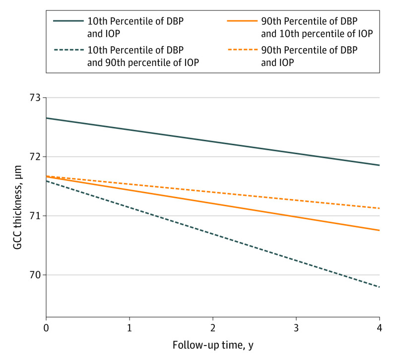
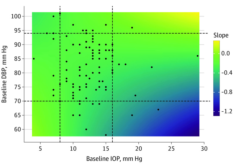
Results: The cohort included 105 eyes from 105 participants. The mean (SD) age, 10-2 visual field mean deviation, and follow-up time were 66.9 (8.5) years, -8.3 (5.3) dB, and 3.6 (0.4) years, respectively, and 67 patients (63.8%) were female. The racial and ethnic makeup of the cohort was 15 African American (14.3%), 23 Asian (21.9%), 12 Hispanic (11.4%), and 55 White (52.4%) individuals based on patient self-report. In multivariable analyses, female sex, history of taking blood pressure medications, higher intraocular pressure, thicker central corneal thickness, shorter axial length, higher contrast sensitivity at 12 cycles per degree, and higher baseline 10-2 visual field mean deviation were associated with faster ganglion cell complex thinning. Lower diastolic blood pressure was associated with faster rates of ganglion cell complex thinning at higher intraocular pressures. For intraocular pressures of 8 and of 16 mm Hg (10% and 90% quantiles, respectively), every 10 mm Hg-lower increment of diastolic blood pressure was associated with 0.011 μm/y slower and -0.130 μm/y faster rates of ganglion cell complex thinning, respectively.
Conclusions and relevance: In this cohort study, a combination of lower diastolic blood pressure and higher intraocular pressure at baseline was associated with faster rates of ganglion cell complex thinning. These findings support consideration of evaluating and addressing diastolic blood pressure as a therapeutic measure in patients with glaucoma if supported by appropriate clinical trials.
Conflict of interest statement
Figures
Comment in
-
Blood Pressure and Glaucoma-A Complex Relationship.JAMA Ophthalmol. 2023 Mar 1;141(3):258-259. doi: 10.1001/jamaophthalmol.2022.6515. JAMA Ophthalmol. 2023. PMID: 36757692 No abstract available.
References
Publication types
MeSH terms
Grants and funding
LinkOut - more resources
Full Text Sources
Medical
Miscellaneous
