. 2023 Aug 1;146(8):3273-3288.
doi: 10.1093/brain/awad039.

The clinical and genetic spectrum of autosomal-recessive TOR1A-related disorders

Afshin Saffari  1   2 Tracy Lau  3 Homa Tajsharghi  4 Ehsan Ghayoor Karimiani  5   6 Ariana Kariminejad  7 Stephanie Efthymiou  3 Giovanni Zifarelli  8 Tipu Sultan  3 Mehran Beiraghi Toosi  9   10 Sahar Sedighzadeh  11   12 Victoria Mok Siu  13 Juan Darío Ortigoza-Escobar  14 Aisha M AlShamsi  15 Shahnaz Ibrahim  16 Nouriya Abbas Al-Sannaa  17 Walla Al-Hertani  18 Whalen Sandra  19 Mark Tarnopolsky  20 Shahryar Alavi  3 Chumei Li  20 Debra-Lynn Day-Salvatore  21 Maria Jesús Martínez-González  22 Kristin M Levandoski  21 Emma Bedoukian  23 Suneeta Madan-Khetarpal  24 Michaela J Idleburg  24 Minal Juliet Menezes  25   26 Aishwarya Siddharth  18 Konrad Platzer  27 Henry Oppermann  27 Martin Smitka  28 Felicity Collins  26   29 Monkol Lek  30 Mohmmad Shahrooei  31   32 Maryam Ghavideldarestani  31 Isabella Herman  33   34   35   36 John Rendu  37 Julien Faure  37 Janice Baker  38 Vikas Bhambhani  38 Laurel Calderwood  39   40 Javad Akhondian  41 Shima Imannezhad  42 Hanieh Sadat Mirzadeh  42 Narges Hashemi  9 Mohammad Doosti  6 Mojtaba Safi  6 Najmeh Ahangari  43 Paria Najarzadeh Torbati  6 Soheila Abedini  3 Vincenzo Salpietro  3 Elif Yilmaz Gulec  44 Safieh Eshaghian  45 Mohammadreza Ghazavi  46 Michael T Pascher  47 Marina Vogel  47   48 Angela Abicht  47   49 Sébastien Moutton  50 Ange-Line Bruel  51   52 Claudine Rieubland  53 Sabina Gallati  53 Tim M Strom  54 Hanns Lochmüller  55   56 Mohammad Hasan Mohammadi  57 Javeria Raza Alvi  58 Elaine H Zackai  59 Beth A Keena  59 Cara M Skraban  59 Seth I Berger  60 Erin H Andrew  60 Elham Rahimian  61 Michelle M Morrow  62 Ingrid M Wentzensen  62 Francisca Millan  62 Lindsay B Henderson  62 Hormos Salimi Dafsari  63   64   65 Heinz Jungbluth  65   66 Natalia Gomez-Ospina  67 Anne McRae  68 Merlene Peter  68 Danai Veltra  69 Nikolaos M Marinakis  69 Christalena Sofocleous  69 Farah Ashrafzadeh  42 Davut Pehlivan  33   34   35 Johannes R Lemke  27   70 Judith Melki  71 Audrey Benezit  72 Peter Bauer  8 Denisa Weis  73 James R Lupski  34   35   74   75 Jan Senderek  47 John Christodoulou  26   76 Wendy K Chung  77 Rose Goodchild  78   79 Amaka C Offiah  80 Andres Moreno-De-Luca  81 Mohnish Suri  82 Darius Ebrahimi-Fakhari  1   83   84   85 Henry Houlden  3 Reza Maroofian  3
Affiliations

The clinical and genetic spectrum of autosomal-recessive TOR1A-related disorders

Afshin Saffari et al. Brain. .

Abstract

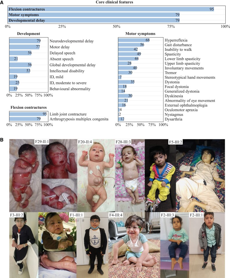
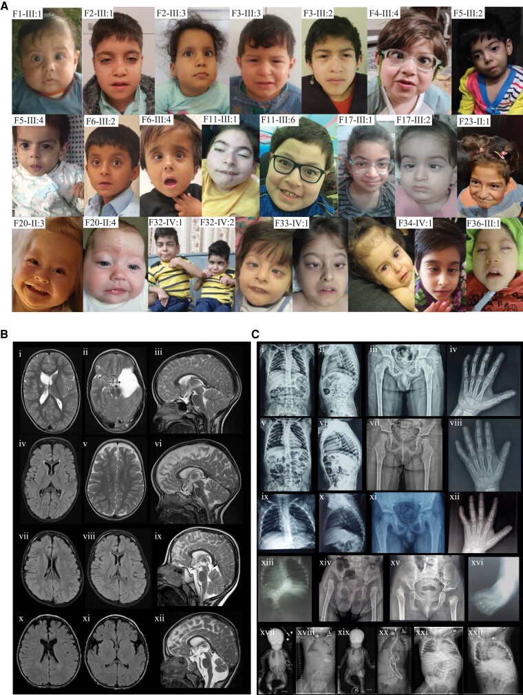
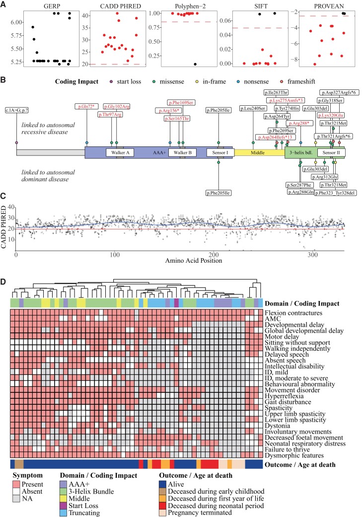
In the field of rare diseases, progress in molecular diagnostics led to the recognition that variants linked to autosomal-dominant neurodegenerative diseases of later onset can, in the context of biallelic inheritance, cause devastating neurodevelopmental disorders and infantile or childhood-onset neurodegeneration. TOR1A-associated arthrogryposis multiplex congenita 5 (AMC5) is a rare neurodevelopmental disorder arising from biallelic variants in TOR1A, a gene that in the heterozygous state is associated with torsion dystonia-1 (DYT1 or DYT-TOR1A), an early-onset dystonia with reduced penetrance. While 15 individuals with AMC5-TOR1A have been reported (less than 10 in detail), a systematic investigation of the full disease-associated spectrum has not been conducted. Here, we assess the clinical, radiological and molecular characteristics of 57 individuals from 40 families with biallelic variants in TOR1A. Median age at last follow-up was 3 years (0-24 years). Most individuals presented with severe congenital flexion contractures (95%) and variable developmental delay (79%). Motor symptoms were reported in 79% and included lower limb spasticity and pyramidal signs, as well as gait disturbances. Facial dysmorphism was an integral part of the phenotype, with key features being a broad/full nasal tip, narrowing of the forehead and full cheeks. Analysis of disease-associated manifestations delineated a phenotypic spectrum ranging from normal cognition and mild gait disturbance to congenital arthrogryposis, global developmental delay, intellectual disability, absent speech and inability to walk. In a subset, the presentation was consistent with foetal akinesia deformation sequence with severe intrauterine abnormalities. Survival was 71%, with higher mortality in males. Death occurred at a median age of 1.2 months (1 week-9 years), due to respiratory failure, cardiac arrest or sepsis. Analysis of brain MRI studies identified non-specific neuroimaging features, including a hypoplastic corpus callosum (72%), foci of signal abnormality in the subcortical and periventricular white matter (55%), diffuse white matter volume loss (45%), mega cisterna magna (36%) and arachnoid cysts (27%). The molecular spectrum included 22 distinct variants, defining a mutational hotspot in the C-terminal domain of the Torsin-1A protein. Genotype-phenotype analysis revealed an association of missense variants in the 3-helix bundle domain to an attenuated phenotype, while missense variants near the Walker A/B motif as well as biallelic truncating variants were linked to early death. In summary, this systematic cross-sectional analysis of a large cohort of individuals with biallelic TOR1A variants across a wide age-range delineates the clinical and genetic spectrum of TOR1A-related autosomal-recessive disease and highlights potential predictors for disease severity and survival.

Keywords: AMC5; NDD; Torsin-1A; arthrogryposis multiplex congenita 5; biallelic variation.


Conflict of interest statement

M.M.M., I.M.W, L.B.H. and F.M. are employees of GeneDx, LLC. J.R.L. has stock ownership in 23andMe, is a paid consultant for Regeneron Genetics Center, and is a co-inventor on multiple US and European patents related to molecular diagnostics for inherited neuropathies, eye diseases, genomic disorders and bacterial genomic fingerprinting. The Department of Molecular and Human Genetics at Baylor College of Medicine receives revenue from clinical genetic and genomic testing conducted at Baylor Genetics (BG); J.R.L. serves on the Scientific Advisory Board (SAB) of BG. W.K.C. is on the scientific advisory board of the Regeneron Genetics Center and the Board of Directors of Prime Medicine. L.C. has served as a paid consultant for Horizon Therapeutics and PTC Therapeutics. M.T. has served as Ontario Ministry of Health Exceptional Access Program member for genetic testing approvals. N.G.-O. is a consultant and has equity interest in Graphite Bio and Codexis. All other authors declare no conflict of interest.

Figures

Figure 1
Figure 1
Pedigrees and segregation results. Individuals with TOR1A-related disorders are indicated by filled black shapes with an arrow pointing to the proband. Open shapes represent unaffected individuals. Diagonal lines across indicate deceased individuals. Squares represent males, circles represent females, diamonds indicate unknown gender, triangles without a diagonal line represent miscarriages, while triangles with a diagonal line represents termination of pregnancy. Grey shaded shapes indicate individuals with hereditary spastic paraplegia. Two interconnected lines for II:3 and II:4 in Family 20 as well as V:1 and V:2 in Family 39 indicate monozygotic twins. The presence of consanguinity between two individuals is represented by double lines. Below each individual, A = Generation number; B = Number of that individual in that generation. Segregation results for all individuals tested are indicated with either red (presence of the TOR1A variant) and/or black (presence of the reference allele). RED/RED text indicates the presence of TOR1A variants in a homozygous or compound heterozygous state, while RED/BLACK text indicates the presence of the TOR1A variant in a heterozygous state. The inheritance of the compound heterozygous TOR1A variants for Families 9, 10, 20, 23, 24 and 36 are indicated by maternal allele (M), paternal allele (F) or unknown inheritance (?). For previously reported families, a reference to the original report has been included in the family identifier. Families 29–31 have been only briefly reported as part of large heterogenous cohorts with suspected genetic aetiologies. Clinical data for those families have been collected for this study. In Family 29, only individual II:2 has been previously reported. For previously reported families, 32–40, follow-up data have been added where available.
Figure 2
Figure 2
Clinical spectrum. (A) A total of 57 individuals were analysed. Frequencies of core clinical features were present in the majority of individuals in our cohort. Below, a detailed breakdown of HPO derived phenotypic features for the core symptom categories ‘Development’, ‘Flexion contractures’ and ‘Motor symptoms’ is shown. Frequencies were printed on the respective bars. (B) Photographs of individuals with biallelic TOR1A variants depicting the spectrum of the disease. (F29-II:1) Foetal akinesia deformation sequence. (F20-II:4 and F28-III:3) Congenital arthrogryposis, umbilical hernia. (F5-III:2) Severe arthrogryposis, minimal motor function. (F3-III:2) Arthrogryposis and scoliosis significantly impacting gait. (F1-III:1) Congenital contractures with improvement in childhood. (F4-III:4) Congenital contractures affecting the lower limbs, right club foot. (F2-III:1) and sibling (F2-III:3) with mild motor impairment, preserved ambulation.
Figure 3
Figure 3
Facial features, neuroimaging and skeletal radiographs. (A) Facial photographs of individuals with biallelic TOR1A variants at different ages, showing the characteristic facial features, which include narrow forehead (bifrontal/bitemporal narrowing), broad/full nasal tip, full cheeks, thick/full vermilion of lower lip and broad, tall or pointed chin. For individuals F33-IV:1 and F34-IV:1, photographs at different ages are provided. (B) Neuroimaging findings in individuals with TOR1A variants. Findings for F2-III:1 (iiii) include an arachnoid cyst in left middle and anterior cranial fossae (ii), areas of cystic encephalomalacia in the right basal ganglia with ex-vacuo dilation of the right lateral ventricular body (i), white matter volume loss (i) and Chiari malformation type 1 (iii). Findings for F2-III:3 (ivvi) include foci of T2 hyperintense signal in the bilateral subcortical and periventricular white matter (iv and v), white matter volume loss (iv) and hypoplastic corpus callosum posterior body and isthmus (vi). Findings for F32-IV:1 (viiix) include small foci of fluid-attenuated inversion recovery hyperintense signal in the periventricular white matter (vii and viii), prominent posterior fossa cisterns and mega cisterna magna (ix). Findings for F29-II:2 (xxii) include foci of T2 hyperintense signal in the bilateral subcortical and periventricular white matter (x), parenchymal volume loss of the frontal lobes with prominence of the sylvian fissures (xi), mildly hypoplastic corpus callosum, vermian hypoplasia, prominent posterior fossa cisterns and mega cisterna magna (xii). (C) Skeletal radiographs of individuals with TOR1A variants. Findings for F2-III:1 (iiv) include mild scoliosis and thoracic kyphosis centred at the level of a hypoplastic T12 vertebra (i and ii), mild flattening of the capital femoral epiphyses (iii) and fifth finger clinodactyly (iv). Findings for F2-III:3 (vviii) include mild scoliosis and thoracic kyphosis centred at the level of a hypoplastic T12 vertebra (v and vi), bilateral coxa valga with loss of height of the capital femoral epiphyses and slender femoral shafts (vii) and fifth finger clinodactyly (viii). Findings for F1-III:1 (ixxii) include mild thoracic kyphosis (ix and x), mild flattening of the capital femoral epiphyses (xi) and fifth finger clinodactyly (xii). Findings for F3-III:2 (xiii and xiv) include 11 ossified ribs on the right and 12 on the left, thoracolumbar scoliosis (xiii) and dysplastic acetabula with dislocated capital femoral epiphyses and early formation of pseudo acetabula, absent ossification of both capital femoral epiphyses and short femoral necks (xiv). F3-III:3 (xv) had dysplastic acetabula, right hip dislocation with early formation of a pseudo acetabulum, absent and delayed ossification of the left capital femoral epiphysis, mild irregularity and sclerosis of the left acetabulum and short femoral necks. F33-IV:1 (xvi) had talipes equinovarus. Findings for F22-III:1 at birth (xvii) and 17 months of age (xviii). (xvii) Anteroposterior radiograph of the baby. The long tubular bones are slender and somewhat overmodelled. Allowing for patient positioning, there is no scoliosis and the hip joints are not dislocated. Umbilical vein catheter, endotracheal tube and nasogastric tube noted. (xviii) Anteroposterior radiograph of chest and abdomen. There are 12 pairs of ribs and normal segmentation. There is now a significant thoracolumbar scoliosis concave to the right. There is absent ossification of bilateral dislocated femoral heads, with hypoplastic acetabula and early pseudoacetabula formation. The long tubular bones are gracile. Tracheostomy and gastrostomy noted. Findings for F22-III:2 at birth (xix) and 2 years of age (xx). (xix) Anteroposterior radiograph of the baby. There are 12 pairs of ribs and normal segmentation. There is a thoracolumbar scoliosis concave to the left. The long tubular bones are slender and somewhat overmodelled. The hip joints do not appear dislocated. Umbilical vein catheter and nasogastric tube noted. (xx) Anteroposterior radiograph of chest and abdomen. The thoracolumbar scoliosis has progressed. There is absent ossification of bilateral dislocated femoral heads, with hypoplastic acetabula and pseudoacetabula formation. The long tubular bones are gracile and appear of reduced radiodensity. Tracheostomy, ventriculoperitoneal shunt and gastrostomy noted. Findings for F23-II:1 (xxi and xxii). Anteroposterior radiograph of chest and abdomen at 1 year 1 month (xxi) and 8 years 5 months (xxii) demonstrate a progressive and significant thoracolumbar scoliosis concave to the right. The earlier radiograph shows dislocation of the right hip joint; the left hip joint does not appear to be dislocated on the limited view available.
Figure 4
Figure 4
Molecular characteristics. (A) Variant effect prediction scores for all 22 distinct TOR1A variants. Genomic Evolutionary Rate Profiling (GERP) scores ranged from −12.3 to 6.17 show the level of conservation, with scores close to 6.17 indicating a high level of conservation. CADD Phred, Polyphen-2, SIFT and PROVEAN scores inferred the level of deleteriousness. Recommended thresholds are indicated with dashed red lines. Variants rated as likely to be deleterious are coloured in red. (B) Schematic of the Torsin-1A primary protein structure with its functional domains. Amino acid positions of variants linked to autosomal-recessive TOR1A-related disorders are plotted above and variants reported in autosomal-dominant DYT-TOR1A torsion dystonia below the Torsin-1A protein. Of note, in autosomal-dominant cases, only the p.Glu303del variant has been verified to cause classic early onset dystonia. The other variants reported for DYT-TOR1A have either been listed as pathogenic or likely pathogenic on ClinVar or have been reported in single cases. Variants printed in red text were likened to FADS or associated with early death. Coding impacts of variants are colour-coded. (C) CADD Phred scores for all possible missense variants were computed and mapped to the linear protein structure. A generalized additive model was used to predict the tolerance for genetic variation across the protein (blue line). The red line indicates the consensus cut off value for deleteriousness. (D) Exploration of genotype-phenotype correlations. Affected individuals were grouped based on the presence of core symptoms using a hierarchical clustering approach. The spectrum of core symptoms for each individual was visualized using a heatmap. Genetic information about the affected Torsin-1A domains (in case of missense or in-frame variants) or coding impacts of the individual TOR1A variants (in case of truncating or the start loss variant) was annotated above the heatmap. Domains included individuals with at least one missense or in-frame variant in the ‘3-Helix Bundle’ (n = 25), ‘AAA+’ (n = 9) and ‘Middle’ (n = 8) domain of Torsin-1A. Individuals with biallelic frameshift or nonsense variants were assigned to the ‘Truncating’ group (n = 14). The individual with the homozygous start loss variant was assessed separately. Due to the small number of cases within the AAA+ domain, further stratification into subdomains (‘Walker A’ or ‘Walker B’) was not done. Clinical outcomes were annotated blow the heatmap.

References

    1. Granata A, Watson R, Collinson LM, Schiavo G, Warner TT. The dystonia-associated protein torsinA modulates synaptic vesicle recycling. J Biol Chem. 2008;283:7568–7579. - PubMed
    1. Hewett JW, Zeng J, Niland BP, Bragg DC, Breakefield XO. Dystonia-causing mutant torsinA inhibits cell adhesion and neurite extension through interference with cytoskeletal dynamics. Neurobiol Dis. 2006;22:98–111. - PubMed
    1. Nery FC, Zeng J, Niland BP, et al. Torsina binds the KASH domain of nesprins and participates in linkage between nuclear envelope and cytoskeleton. J Cell Sci. 2008;121:3476–3486. - PMC - PubMed
    1. Torres GE, Sweeney AL, Beaulieu JM, Shashidharan P, Caron MG. Effect of torsinA on membrane proteins reveals a loss of function and a dominant-negative phenotype of the dystonia-associated DeltaE-torsinA mutant. Proc Natl Acad Sci U S A. 2004;101:15650–15655. - PMC - PubMed
    1. Lange LM, Junker J, Loens S, et al. Genotype-phenotype relations for isolated dystonia genes: MDSGene systematic review. Mov Disord. 2021;36:1086–1103. - PubMed

Publication types