Review
. 2023 Jan;75(1):62-158.
doi: 10.1124/pharmrev.122.000618. Epub 2022 Dec 8.

Dopamine, Immunity, and Disease

Affiliations
Review

Dopamine, Immunity, and Disease

Breana Channer et al. Pharmacol Rev. 2023 Jan.

Abstract

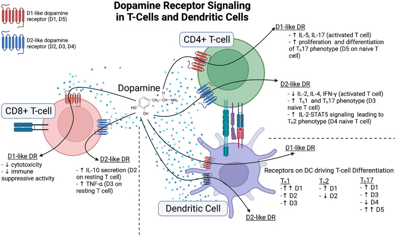
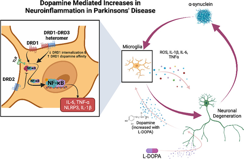
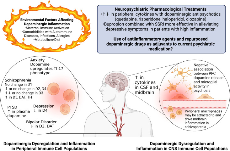
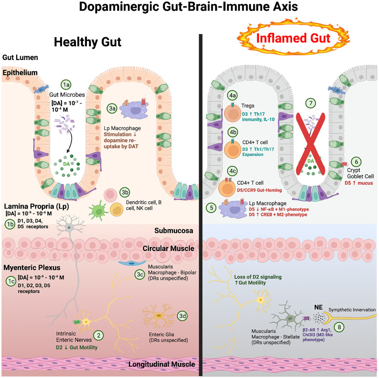
The neurotransmitter dopamine is a key factor in central nervous system (CNS) function, regulating many processes including reward, movement, and cognition. Dopamine also regulates critical functions in peripheral organs, such as blood pressure, renal activity, and intestinal motility. Beyond these functions, a growing body of evidence indicates that dopamine is an important immunoregulatory factor. Most types of immune cells express dopamine receptors and other dopaminergic proteins, and many immune cells take up, produce, store, and/or release dopamine, suggesting that dopaminergic immunomodulation is important for immune function. Targeting these pathways could be a promising avenue for the treatment of inflammation and disease, but despite increasing research in this area, data on the specific effects of dopamine on many immune cells and disease processes remain inconsistent and poorly understood. Therefore, this review integrates the current knowledge of the role of dopamine in immune cell function and inflammatory signaling across systems. We also discuss the current understanding of dopaminergic regulation of immune signaling in the CNS and peripheral tissues, highlighting the role of dopaminergic immunomodulation in diseases such as Parkinson's disease, several neuropsychiatric conditions, neurologic human immunodeficiency virus, inflammatory bowel disease, rheumatoid arthritis, and others. Careful consideration is given to the influence of experimental design on results, and we note a number of areas in need of further research. Overall, this review integrates our knowledge of dopaminergic immunology at the cellular, tissue, and disease level and prompts the development of therapeutics and strategies targeted toward ameliorating disease through dopaminergic regulation of immunity. SIGNIFICANCE STATEMENT: Canonically, dopamine is recognized as a neurotransmitter involved in the regulation of movement, cognition, and reward. However, dopamine also acts as an immune modulator in the central nervous system and periphery. This review comprehensively assesses the current knowledge of dopaminergic immunomodulation and the role of dopamine in disease pathogenesis at the cellular and tissue level. This will provide broad access to this information across fields, identify areas in need of further investigation, and drive the development of dopaminergic therapeutic strategies.


Figures

Fig. 1
Fig. 1
Dopamine signaling through cognate receptors. Dopamine signaling is mediated through its GPCRs. D1-like receptors (D1 and D5, red) classically couple to Gas to mediate activation of adenylate cyclase, leading to cAMP production, PKA activation, and downstream activation of PKA targets. The D2-like receptors (D2, D3, and D4, blue) couple to the Gai pathway to inhibit adenylate cyclase production and oppose D1-like signaling. The D1-like receptors can also lead to activation of PLCβ, thus enhancing calcium flux and PKC activation. The D2-like receptors can also activate this pathway via Gbg. D2-like stimulation can additionally inhibit AKT phosphorylation through the formation of a b-arrestin/PP2A signaling complex. Both D1-like and D2-like stimulation leads to AKT phosphorylation through its activity on the phosphatidylinositol 3-kinase (PI3K)/Akt signaling axis, but the mechanisms behind this are not clear. Downstream, both receptors can activate members of the MAPK family. This occurs through various mechanisms including, but not limited to, cAMP activation, PKC and calcium signaling, and activation of the PI3K/Akt signaling cascade. Created with BioRender.com.
Fig. 2
Fig. 2
Metabolic pathway of dopamine biosynthesis and degradation. Dopamine synthesis is initiated with the hydroxylation of tyrosine by the enzyme TH to generate L-DOPA. L-DOPA is converted to dopamine by AADC. Dopamine beta hydroxylase (DBH) hydroxylates dopamine to form norepinephrine, which is converted to epinephrine by phenylethanolamine-N-methyltransferase (PNMT). Dopamine is primarily metabolized by two enzymatic pathways, COMT and MAO. COMT converts dopamine to 3-methoxytyramine, which is subsequently converted to 3-methoxy-4-hydroxyacetaldehyde by MAO. In contrast, MAO converts dopamine to 3,4-dihydroxyphenylacetaldehyde, which is then converted by aldehyde dehydrogenase (ALDH) to DOPAC. In the final steps of dopamine degradation, ALDH and COMT convert 3-methoxy-4-hydroxyacetaldehyde and DOPAC to HVA, respectively. Created with BioRender.com
Fig. 3
Fig. 3
Dopamine receptor signaling in myeloid cells. Current knowledge of immunomodulatory effects of dopamine signaling in monocytes, macrophages, and microglia are summarized. In monocytes, dopamine signaling through the dopamine receptors leads to an increase in chemokine production, chemokinesis, and transmigration. Studies in macrophages show that dopamine can have bidirectional effects on phagocytosis and NO production, while stress response genes, NF-kB activation, and release of proinflammatory mediators are all increased in response to dopamine. In microglia, dopamine signaling through its receptors increases chemotaxis, phagocytosis, formation of extracellular traps and pro-inflammatory mediator production while decreasing NO production. Additionally, in microglia, in general D1-like receptor stimulation inhibits NF-kB while D2-like receptor stimulation activates NF-kB. Created with BioRender.com.
Fig. 4
Fig. 4
Dopamine receptor signaling in granulocytes. Current knowledge of the immunomodulatory effects of dopamine signaling in neutrophils, eosinophils, and mast cells is summarized. In neutrophils, dopamine acts through its receptors to increase formation of extracellular traps and to decrease NO production and transendothelial migration, while having reported bidirectional effects on neutrophil phagocytosis. Dopamine signaling in eosinophils has been shown to affect eosinophil counts with high doses of dopamine leading to eosinopenia and low doses of dopamine leading to eosinophilia. In mast cells, dopamine acting at D1-like receptors has been shown to increase degranulation while D2-like receptor stimulation decreases cytokine production. Created with BioRender.com.
Fig. 5
Fig. 5
Dopamine receptor signaling in T-cells dendritic cells. Current knowledge of dopaminergic immunomodulation in dendritic cells and T-cells. Dopamine receptor balance on dendritic cells appear to impact T-cell differentiation. Increased expression of D1, D2, and D3 receptors on dendritic cells induces a Th1 phenotype in T-cells. Increased D1 expression with low D2 expression drives Th2 phenotype while increased D1, D3, and D5 along with low D4 expression has been linked to Th17 phenotype. Double arrows before receptor expression indicate a stronger correlation of the corresponding receptor on dendritic cells that drive T-cell differentiation. On activated CD4+ T-cells, dopamine stimulates D1-like receptors to increase IL-5 and IL-17 production and stimulates D2-like receptors leading to reduced IL-2, IL-4, and IFN-y production. On CD8+ T-cells, dopamine stimulation of D1-like receptors leads to decrease cytotoxicity and decreased immune suppressive activity. By acting on D2-like receptors on resting CD8+ T-cells, dopamine increases IL-10 and TNF-α production. Created with BioRender.com.
Fig. 6
Fig. 6
Dopamine-mediated increases in neuroinflammation in PD. The feed-forward cycle in PD starts with the disruption of the dopaminergic system that increases neuroinflammation, which, in turn, exacerbates dopaminergic neuronal dysfunction and death, further disrupting the dopaminergic system. Specifically, α-synuclein aggregates trigger microglial immune activation, leading to the production and secretion of neurotoxic factors (ROS, IL-1β, IL-6 and TNF-α) that result in loss of dopaminergic neurons and additional α-synuclein aggregation. The dysregulation of dopaminergic signaling increases neuroinflammation and feeds further into the cycle. D1/D3 receptor heteromers in PD decrease D1 internalization and increase the affinity of D1 to dopamine. Treatment with L-DOPA also increases the sensitivity of D1 to dopamine. As a result, there is excessive activation of proinflammatory D1 signaling, due to the D1/D3 receptor heteromers and L-DOPA treatment. This is concurrent with the observed downregulation of anti-inflammatory D2 signaling that can promote neuroinflammation and thus enhance neuronal degeneration in PD. Created with BioRender.com.
Fig. 7
Fig. 7
Dopaminergic inflammation in neuropsychiatric diseases. Changes in dopaminergic activity in neuropsychiatric diseases may drive pathogenesis by influencing immune function. As depicted, there are many interactions between pathologic changes in dopaminergic regulation and dopamine-mediated immunomodulatory changes associated with the development of several neuropsychiatric disorders, including schizophrenia, bipolar disorder, depression, and anxiety disorders. In peripheral immune cells such as T-cells and monocytes, alterations in the expression levels of dopamine receptors and associated dopamine proteins are observed and dependent on symptom severity and medication status. In the CNS, increases in inflammation are found in dopaminergic regions such as the midbrain, and dysregulation of CNS dopamine may drive microglial activity and, in turn, regulate peripheral immune responses. In addition, environmental factors associated with predicting neuropsychiatric disease, such as MIA, comorbidities with immune-related disorders, metabolism, and diet appear to be shared among neuropsychiatric disorders. This may contribute to and influence dopaminergic inflammation through common pathways. Finally, research regarding neuropsychiatric pharmacological treatments that are used as first-line treatments for numerous disorders suggests that these drugs, which can target the dopamine system, directly modify inflammation. Thus, they may be preferred in treating certain individuals depending on their baseline inflammatory status. More nuanced usage of novel and repurposed anti-inflammatory and/or dopaminergic drugs as adjuvant therapies could be relevant for many neuropsychiatric disorders and could provide novel targets and substantial improvements in treatments and symptomatology. Created with BioRender.com.
Fig. 8
Fig. 8
Dopaminergic gut–brain–immune axis. In the healthy gut (left), a significant portion of dopamine is produced by intestinal microbes (1a) in the gut lumen, which may be shuttled into the lamina propria (Lp) and submucosa. Dopamine concentrations vary from the nanomolar to the micromolar range in the Lp and submucosa of the intestine (1b), which express all dopamine receptors except D2. Dopaminergic neurons in the myenteric plexus produce similar concentrations of dopamine locally (1c), and this layer has immunoreactivity for all dopamine receptors except D4. Activation of D2 in the myenteric plexus reduces GI motility (2). Stimulation of macrophages in the mucosal layer reduces their capacity for dopamine reuptake by DAT (3a) and may increase extracellular dopamine concentrations for autocrine/paracrine signaling. The impact of dopamine in GI homeostasis is not well understood in several other cell types such as dendritic cells, B-cells, and NK cells (3b); bipolar macrophages (3c); enteric glia (3d); and stellate macrophages (8). Several models of inflammatory bowel disease (right) have shown an inflammatory role of dopamine, primarily through D3 and D5 on Tregs (4a) and CD4+ T-cells (4b and 4c). D5 stimulation is associated with polarization of Lp macrophages to M2-phenotype (5) and increased mucus production by intestinal goblet cells (6). Depletion of the microbiome reduces dopamine levels in the mucosa and exacerbates gut inflammation, which may be attenuated by D1-like agonists (7). Stellate macrophages (8) express the β2-adrenergic receptor, which may be activated by high concentrations of dopamine, and form neuroimmune connections with extrinsic catecholaminergic neurons and intrinsic enteric neurons. Created with BioRender.com.
Fig. 9
Fig. 9
Hypothesized role for dopamine in pathogenesis in the skin. Dopaminergic signaling has been hypothesized to drive pathogenesis in the skin by immune regulation. Keratinocytes, the most abundant cell type in the epidermis, mediate inflammation in the local environment from autocrine and paracrine signaling of dopamine through β adrenergic or D2 receptors. Although an unclear pathway (depicted by dashed arrows), stress hormones such as cortisol can increase Kruppel-like factor 9 transcription factor expression to then induce the upregulation of cytochrome P450 2D6 (CYP2D6). CYP2D6 within keratinocytes can then metabolize tyramine to dopamine which can contribute to the elevated serum/skin dopamine observed in pathology. Melanin synthesis is connected to dopamine synthesis as melanocytes convert L-tyrosine to L-DOPA using the rate limiting enzyme tyrosinase which also converts L-DOPA to DOPAquinone in these cells. DOPAquinone is metabolized to eumelanin and pheomelanin by tyrosinase-related proteins (TYRP1, TYRP2) or cysteine and oxidation reactions, respectively. As depicted by the dashed lines, there is an unclear mechanism of how dopamine is produced in these cells to contribute to the elevated serum dopamine in pathology. Elevated levels of dopamine in the serum and skin in pathology has been shown to act on D1-like receptors on macrophages to increase production of chemokines CXCL9 and CXCL10, which recruits reactive CD8+ T-cells. In the presence of oxidative stress, melanocytes undergo apoptosis and are phagocytized by antigen presenting cells that interact with the recruited CD8+ T-cells. Dashed lines represent pathways that have been hypothesized by some groups and solid arrows represent more established pathways. Created with BioRender.com.
Fig. 10
Fig. 10
Dopaminergic modulation of osteogenesis. Dopamine activity may drive osteogenesis by regulation of osteoclasts and osteoblasts. The macrophage-like osteoclasts respond to dopamine and dopaminergic agents such as bromocriptine and ropinirole at the D2-like receptors. Increased D2-like signaling leads to a reduction in osteoclastogenesis via reduction of cAMP and PKA activity leading to inhibition of CREB phosphorylation and subsequent transcription of osteoclastic markers c-FOS, Nfatc1, tartrate-resistant acid phosphatase, and Ctsk. Dopamine in the bone and through D2-like signaling on osteoclasts inhibits RANK-L and M-CSF induced osteoclastogenesis and therefore bone resorption. Osteoblast activity in response to dopamine appears to be regulated by D1-like receptor signaling. Dopamine or the D1-like receptor agonist SKF38393 act on the D1-like receptors to mediate signaling through ERK1/2, which induces osteogenesis and inhibits bone loss. SCH23390, a D1-receptor antagonist, and high concentrations of dopamine can both inhibit this process. Dashed lines represent pathways that have been hypothesized and solid arrows represent more established pathways. Red arrows depict inhibitory pathways and black arrows are stimulating/activating pathways. Created with BioRender.com.

References

    1. aan het Rot M, Collins KA, Murrough JW, Perez AM, Reich DL, Charney DS, Mathew SJ (2010) Safety and efficacy of repeated-dose intravenous ketamine for treatment-resistant depression. Biol Psychiatry 67:139–145. - PubMed
    1. Abreu CMVeenhuis RTAvalos CRGraham SParrilla DRFerreira EAQueen SEShirk ENBullock BTLi M, et al. (2019) Myeloid and CD4 T cells comprise the latent reservoir in antiretroviral therapy-suppressed SIVmac251-infected macaques. MBio 10:e01659-19. - PMC - PubMed
    1. Abreu P, Llorente E, Hernández MM, González MC (1994) Interleukin-1 beta stimulates tyrosine hydroxylase activity in the median eminence. Neuroreport 5:1356–1358. - PubMed
    1. Acharjee S, Branton WG, Vivithanaporn P, Maingat F, Paul AM, Dickie P, Baker GB, Power C (2014) HIV-1 Nef expression in microglia disrupts dopaminergic and immune functions with associated mania-like behaviors. Brain Behav Immun 40:74–84. - PubMed
    1. Aggeletopoulou I, Assimakopoulos SF, Konstantakis C, Triantos C (2018) Interleukin 12/interleukin 23 pathway: biological basis and therapeutic effect in patients with Crohn’s disease. World J Gastroenterol 24:4093–4103. - PMC - PubMed

MeSH terms

LinkOut - more resources