. 2023 Feb 9;21(2):e3001967.
doi: 10.1371/journal.pbio.3001967. eCollection 2023 Feb.

Fibroblast-expressed LRRC15 is a receptor for SARS-CoV-2 spike and controls antiviral and antifibrotic transcriptional programs

Affiliations

Fibroblast-expressed LRRC15 is a receptor for SARS-CoV-2 spike and controls antiviral and antifibrotic transcriptional programs

Lipin Loo et al. PLoS Biol. .

Abstract

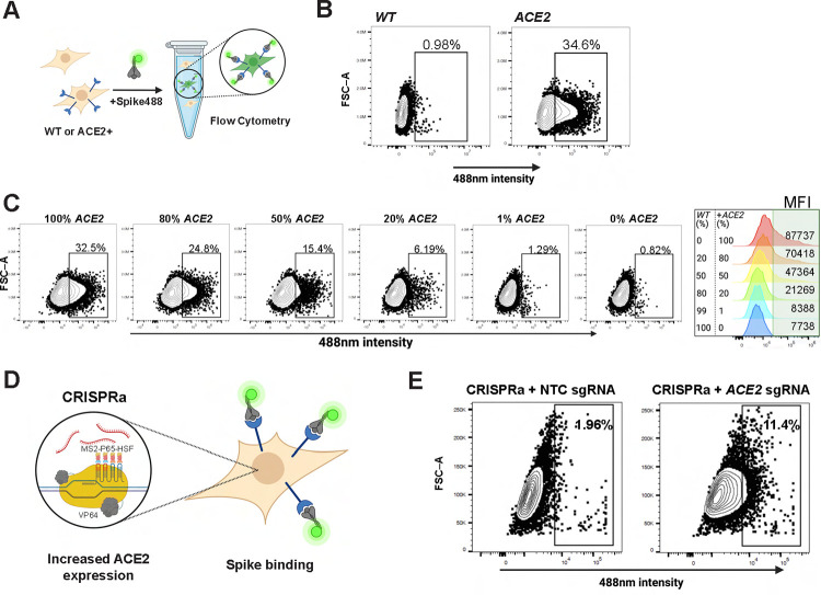
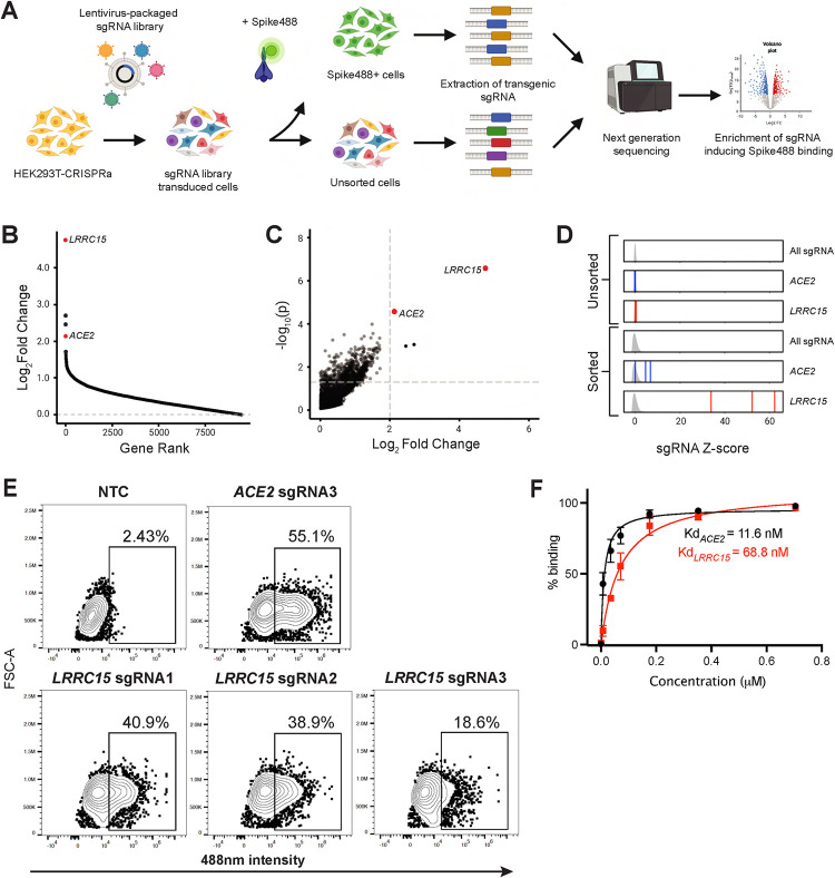
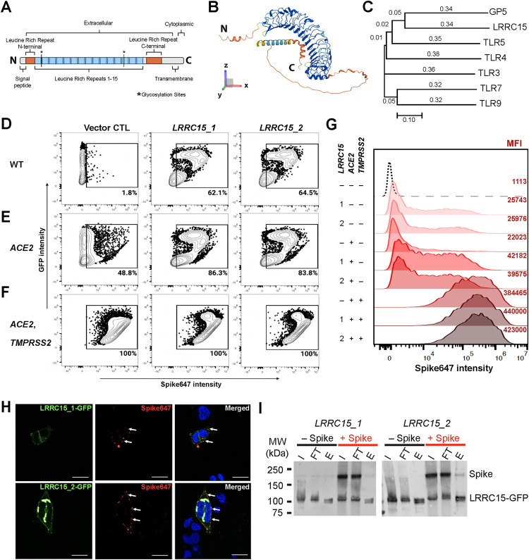
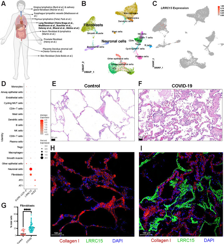
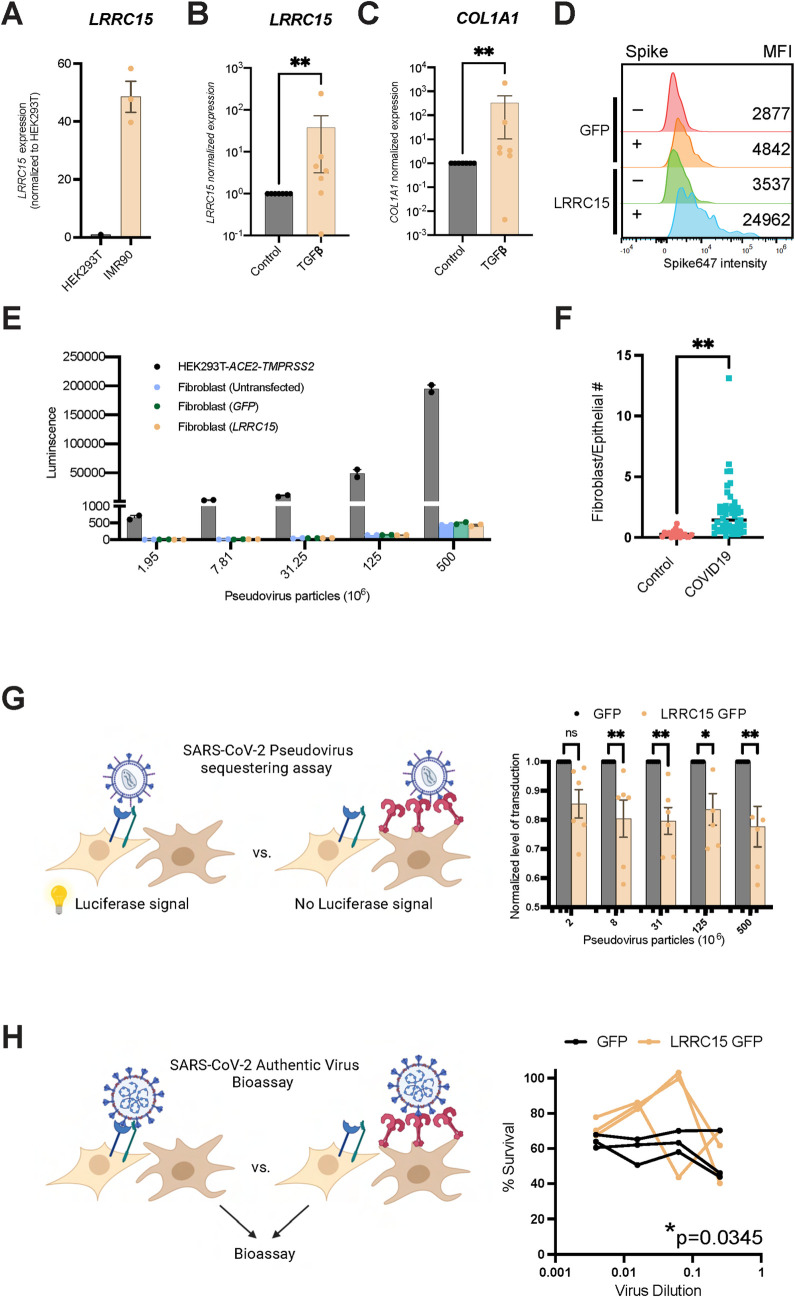
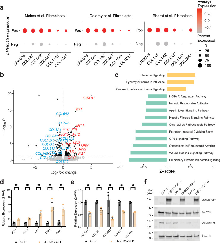
Although ACE2 is the primary receptor for Severe Acute Respiratory Syndrome Coronavirus 2 (SARS-CoV-2) infection, a systematic assessment of host factors that regulate binding to SARS-CoV-2 spike protein has not been described. Here, we use whole-genome CRISPR activation to identify host factors controlling cellular interactions with SARS-CoV-2. Our top hit was a TLR-related cell surface receptor called leucine-rich repeat-containing protein 15 (LRRC15). LRRC15 expression was sufficient to promote SARS-CoV-2 spike binding where they form a cell surface complex. LRRC15 mRNA is expressed in human collagen-producing lung myofibroblasts and LRRC15 protein is induced in severe Coronavirus Disease 2019 (COVID-19) infection where it can be found lining the airways. Mechanistically, LRRC15 does not itself support SARS-CoV-2 infection, but fibroblasts expressing LRRC15 can suppress both pseudotyped and authentic SARS-CoV-2 infection in trans. Moreover, LRRC15 expression in fibroblasts suppresses collagen production and promotes expression of IFIT, OAS, and MX-family antiviral factors. Overall, LRRC15 is a novel SARS-CoV-2 spike-binding receptor that can help control viral load and regulate antiviral and antifibrotic transcriptional programs in the context of COVID-19 infection.


Conflict of interest statement

The authors declare no competing interests.

Figures

Fig 1
Fig 1. A sensitive FACS-based SARS-CoV-2 spike-binding assay amenable to high-throughput screening.
(A) Schematic of SARS-CoV-2 spike-binding assay. HEK293T cells with stable integration of ACE2 cDNA for overexpression (HEK293T-ACE2) are incubated with Alexa Fluor 488-conjugated SARS-CoV-2 spike protein (Spike488). Spike488-binding cells are then detected by flow cytometry. (B) Representative flow cytometry plots for WT HEK293T and HEK293T-ACE2 incubated with Spike488 (N = 3). See also S1B Fig for gating strategy. (C) Titration of HEK293T-ACE2 (ACE2) cells with WT HEK293T cells. Approximately 1% HEK293T-ACE2 cells showed a difference to baseline non-specific binding. Histogram summary showing MFI of flowed cells. (D) Schematic of CRISPRa system used. (E) Representative plot of flow cytometry for a clonal HEK293T-CRISPRa cell line transduced with NTC sgRNA or ACE2 sgRNA (expression confirmation via RT-qPCR in S1A Fig). The data underlying all panels in this figure can be found in DOI: 10.5281/zenodo.7416876. ACE2, angiotensin-converting enzyme 2; CRISPRa, CRISPR activation; MFI, mean fluorescence intensity; NTC, non-targeting control; SARS‑CoV‑2, Severe Acute Respiratory Syndrome Coronavirus 2; sgRNA, single-guide RNA.
Fig 2
Fig 2. Whole-genome CRISPRa screening identified LRRC15 as a novel SARS-CoV-2 spike-binding protein.
(A) Schematic of CRISPRa screen used to identify regulators of SARS-CoV-2 spike binding. (B) Ranking of all genes in screen 1 by log2 fold change (LFC) calculated using MAGeCK and plotted using MAGeCKFlute. See also S1 Table. (C) Gene enrichment analysis of screen 1 performed using MAGeCK. Horizontal dotted line indicates p-value = 0.05. Vertical dotted line indicates LFCs of 2. P-values and LFCs for all genes are reported in S1 Table. (D) sgRNA Z-scores for screen 1 unsorted and sorted samples. Density curve for all sgRNA Z-scores in sample (i.e., sorted or unsorted) is shown in gray. Z-scores for each guide are indicated by vertical lines (blue ACE2, red LRRC15). (E) Flow cytometry analysis of HEK293T-CRISPRa cells transduced with 3 independent LRRC15 sgRNAs. HEK293T-CRISPRa transduced with ACE2 sgRNA3 were used as a positive control and NTC sgRNA-transduced HEK293T-CRISPRa cells were used as a negative control (N = 3). (F) Quantification of Spike647 binding in ACE2 sgRNA3 and LRRC15 sgRNA1 cells via flow cytometry. Dissociation constant (Kd) was calculated by fitting with nonlinear regression (1 site—specific binding). N = 3, error bars represent SD. The data underlying all panels in this figure can be found in DOI: 10.5281/zenodo.7416876. ACE2, angiotensin-converting enzyme 2; CRISPRa, CRISPR activation; LFC, log2 fold change; LRRC15, leucine-rich repeat-containing protein 15; NTC, non-targeting control; SARS‑CoV‑2, Severe Acute Respiratory Syndrome Coronavirus 2; sgRNA, single-guide RNA.
Fig 3
Fig 3. Confirmation that LRRC15 binds to SARS-CoV-2 spike protein.
(A) LRRC15 contains 15 leucine-rich repeats, a short cytoplasmic C-terminus, and 2 glycosylation sites. (B) Predicted protein structure of LRRC15 (from alpha fold). (C) LRRC15 is part of the LRR-Tollkin family. (D) Flow cytometry analysis of Alexa Fluor-647 (Spike647) binding in WT HEK293T cells, (E) HEK293T-ACE2, and (F) HEK293T cells with stable expression of ACE2 cDNA and TMPRSS2 cDNA (HEK293T-ACE2-TMPRSS2). Each cell line was transfected with plasmids encoding cDNA for GFP-tagged LRRC15 (transcript 1 or 2) or with empty GFP vector as negative control plasmid. (G). Histogram summary shows MFI of (D–F). (H) Representative images of interaction between LRRC15-GFP and Alexa Fluor 647-conjugated SARS-CoV-2 HexaPro spike protein in HEK293T cells (N = 2). Images were taken at 40× magnification. Green = LRRC15-GFP, red = Spike647, blue = Hoechst-stained nuclei. Scale bar = 25 μm. (I) Immunoprecipitation of LRRC15 with spike protein. Lysates of HEK293T cells transfected with GFP-tagged LRRC15 (transcript 1 or 2, LRRC15_1 and LRRC15_2, respectively) incubated with SARS-CoV-2 HexaPro spike protein were immunoprecipitated using anti-LRRC15 primary antibody. Immunoblots were performed for LRRC15 and for SARS-CoV-2 HexaPro spike. I = input, FT = flow-through, E = elute. The data underlying all panels in this figure can be found in DOI: 10.5281/zenodo.7416876. ACE2, angiotensin-converting enzyme 2; GP5, glycoprotein V platelet; LRRC15, leucine-rich repeat-containing protein 15; MFI, mean fluorescence intensity; SARS‑CoV‑2, Severe Acute Respiratory Syndrome Coronavirus 2; TLR, toll-like receptor; TMPRRS2, Transmembrane serine protease 2.
Fig 4
Fig 4. LRRC15 is expressed in lung fibroblasts and lines the airways in COVID-19 patients.
(A) Overview of cell types expressing LRRC15 from existing single-cell RNA-sequencing datasets. (B) UMAP plot of lung single-nucleus RNA seq dataset (Melms and colleagues). (C) Feature plot and (D) dotplot shows LRRC15 is expressed in fibroblasts and neuronal cells. Expression of LRRC15 is also observed in fibroblasts of separate studies (See S5 Fig). (E) Proportion of cells that are lung fibroblasts increases with COVID lungs (7.9% in control (N = 19) and 22.9% in COVID (N = 47); unpaired t test, p < 0.0001). (F and G) Representative micrograph of HE-stained lung tissue section obtained from (F) a human donor without COVID-19 and (G) donor diagnosed with COVID-19. Imaging performed at 200× magnification (scale bar = 50 m). All images in S6A Fig (Control, N = 1; COVID-19, N = 4). (H and I) Representative micrograph of immunofluorescence staining in human lung tissue section obtained from (H) donor without COVID-19 and (I) donor diagnosed with COVID-19. Images were taken at 200× magnification (scale bar = 100 m). Red = Collagen I, green = LRRC15, blue = DAPI. All images in S6B Fig (Control, N = 3; COVID-19, N = 4). The data underlying all panels in this figure can be found in DOI: 10.5281/zenodo.7416876. ACE2, angiotensin-converting enzyme 2; COVID-19, Coronavirus Disease 2019; HE, hematoxylin and eosin; LRRC15, leucine-rich repeat-containing protein 15.
Fig 5
Fig 5. LRRC15 is not a SARS-CoV-2 entry receptor but inhibits infection in trans.
(A) IMR90 fibroblasts express LRRC15, quantified via RT-qPCR. N = 3 per cell line. (B and C) TGFβ increased (B) LRRC15 and (C) COL1A1 in fibroblasts, quantified via RT-qPCR. N = 7 for each group. Significance was determined by Mann–Whitney one-tailed test, **p < 0.01. (D) IMR90 fibroblasts expressing LRRC15 bind spike, MFI = mean fluorescence intensity. (E) Fibroblasts do not have innate tropism for SARS-CoV-2 and overexpression of LRRC15 does not mediate infection. Transduction efficiency (luciferase luminescence) was compared to permissive cell line HEK293T-ACE2-TMPRSS2. N = 2 independent replicates for each group. (F) Pooled analysis of 3 independent studies indicate ratio of fibroblasts to epithelial cells in COVID-19 lungs is approx. 2:1 (0.3 in control (n = 19) and 2.06 in COVID-19 (n = 47); unpaired two-tailed t test, p < 0.0001). (G) LRRC15 expressing fibroblasts can suppress SARS-CoV-2 spike pseudovirus infection of HEK293T-ACE2-TMPRSS2 cells. Significance was determined by two-way ANOVA, Sidak’s multiple comparison test, **p < 0.01,*p < 0.05. N = 6 per condition. (H) LRRC15 expressing fibroblasts can suppress authentic SARS-CoV-2 infection of HEK293T-ACE2-TMPRSS2 cells. Significance was determined by two-way ANOVA, Sidak’s multiple comparison test, *p < 0.05. N = 3 per condition. The data underlying all panels in this figure can be found in DOI: 10.5281/zenodo.7416876. ACE2, angiotensin-converting enzyme 2; COL1A1, collagen type I alpha 1 chain; LRRC15, leucine-rich repeat-containing protein 15; SARS‑CoV‑2, Severe Acute Respiratory Syndrome Coronavirus 2; TMPRRS2, Transmembrane serine protease 2.
Fig 6
Fig 6. LRRC15 expression is correlated with collagen and antiviral gene signatures.
(A) LRRC15+ fibroblasts have an enhanced collagen gene signature. Dotplots generated from 3 separate studies. Pos = LRRC15+, Neg = LRRC15-. (B) Volcano plot of DEGs from fibroblasts ectopically expressing either LRRC15 or GFP. N = 3 biological replicates for each group. A subset of DEGs is labeled, including collagen genes and genes related to antiviral signaling. Blue labels indicate down-regulation, while red labels indicate up-regulation. (C) DEG-associated canonical pathways as determined by Ingenuity Pathway Analysis. Canonical pathways were filtered to show only those with p-value < 0.05 and with a nonzero Z-score. (D and E) LRRC15 overexpression causes (D) up-regulation of antiviral transcripts and (E) down-regulation of collagen transcripts by RT-qPCR. Results were calculated using the ΔΔCT method, normalized to the average of control GFP-only cells. Significance was assessed using one-tailed Mann–Whitney test, *p < 0.05. N = 3 per condition. (F) Ectopic expression of LRRC15 in fibroblasts decreases Collagen VI protein expression compared to GFP-only control cells. Western blots for LRRC15 and Collagen VI. The data underlying all panels in this figure can be found in DOI: 10.5281/zenodo.7416876. DEG, differentially expressed gene; IFIT, interferon-induced proteins with tetratricopeptide repeats; LRRC15, leucine-rich repeat-containing protein 15 MX, Myxovirus resistance family; OAS, 2-prime, 5-prime oligoadenylate synthetase family.

References

    1. Li W, Moore MJ, Vasilieva N, Sui J, Wong SK, Berne MA, et al.. Angiotensin-converting enzyme 2 is a functional receptor for the SARS coronavirus. Nature. 2003. Nov 27;426(6965):450–4. doi: 10.1038/nature02145 - DOI - PMC - PubMed
    1. Kuba K, Imai Y, Rao S, Gao H, Guo F, Guan B, et al.. A crucial role of angiotensin converting enzyme 2 (ACE2) in SARS coronavirus-induced lung injury. Nat Med. 2005. Aug;11(8):875–9. doi: 10.1038/nm1267 - DOI - PMC - PubMed
    1. Zhou P, Yang XL, Wang XG, Hu B, Zhang L, Zhang W, et al.. A pneumonia outbreak associated with a new coronavirus of probable bat origin. Nature. 2020. Mar;579(7798):270–3. doi: 10.1038/s41586-020-2012-7 - DOI - PMC - PubMed
    1. Lu R, Zhao X, Li J, Niu P, Yang B, Wu H, et al.. Genomic characterisation and epidemiology of 2019 novel coronavirus: implications for virus origins and receptor binding. Lancet. 2020. Feb 22;395(10224):565–74. doi: 10.1016/S0140-6736(20)30251-8 - DOI - PMC - PubMed
    1. Sanson KR, Hanna RE, Hegde M, Donovan KF, Strand C, Sullender ME, et al.. Optimized libraries for CRISPR-Cas9 genetic screens with multiple modalities. Nat Commun. 2018. Dec 21;9(1):5416. doi: 10.1038/s41467-018-07901-8 - DOI - PMC - PubMed

Publication types