. 2023 Mar;299(3):102992.
doi: 10.1016/j.jbc.2023.102992. Epub 2023 Feb 8.

The small molecule inhibitor NAV-2729 has a complex target profile including multiple ADP-ribosylation factor regulatory proteins

Affiliations

The small molecule inhibitor NAV-2729 has a complex target profile including multiple ADP-ribosylation factor regulatory proteins

Eric M Rosenberg Jr et al. J Biol Chem. 2023 Mar.

Abstract

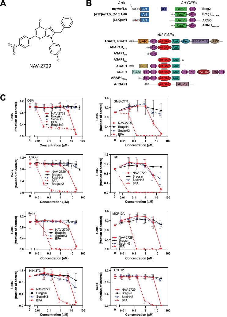
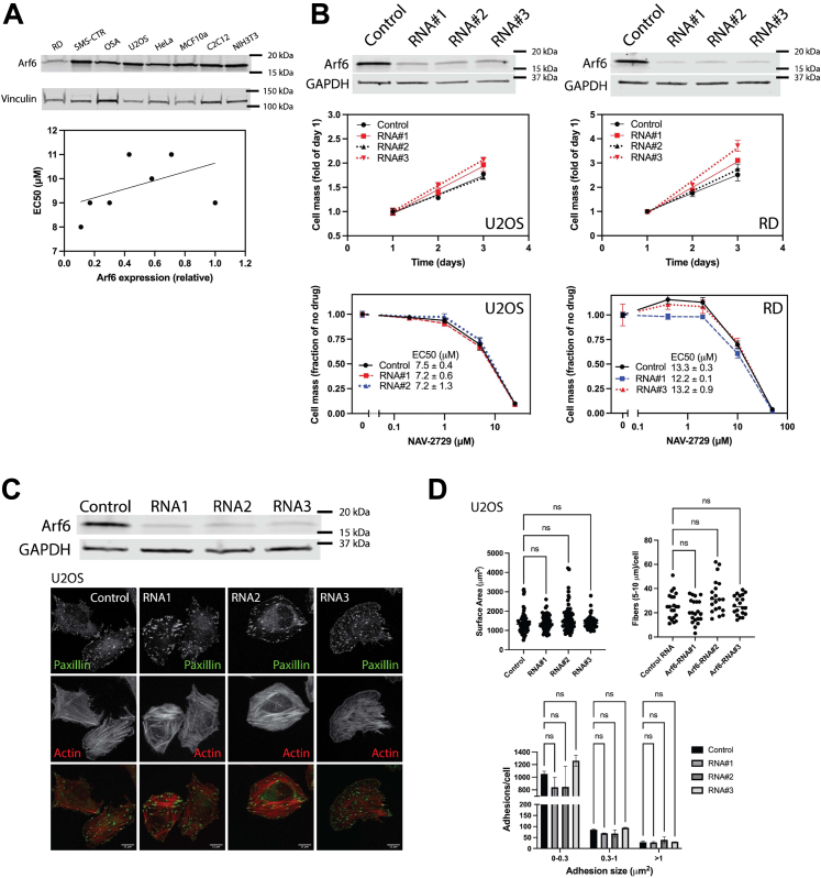
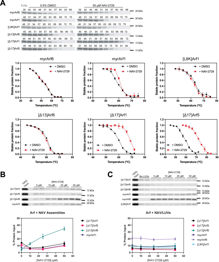
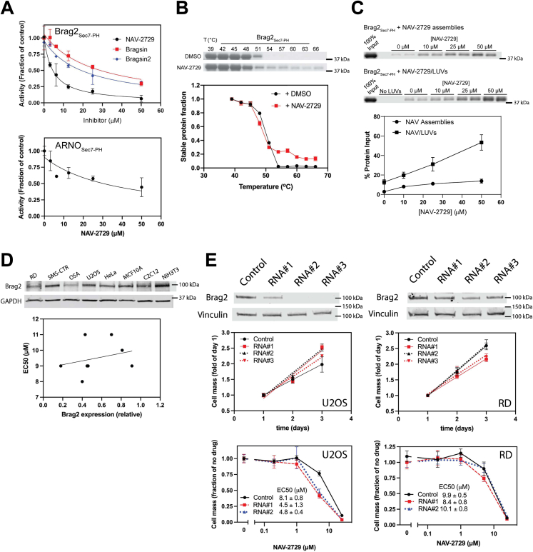
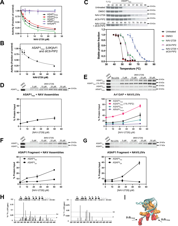
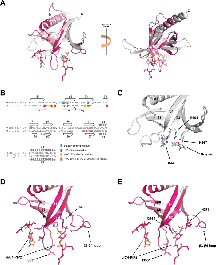
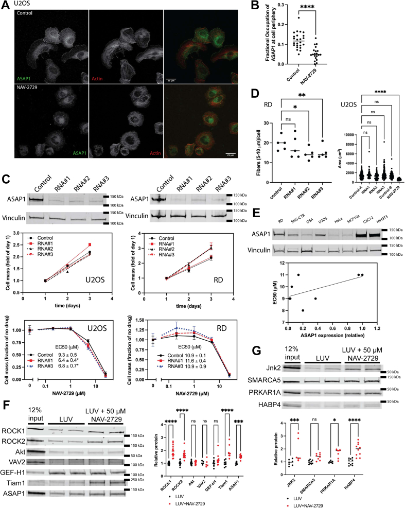
The ADP-ribosylation factor (Arf) GTPases and their regulatory proteins are implicated in cancer progression. NAV-2729 was previously identified as a specific inhibitor of Arf6 that reduced progression of uveal melanoma in an orthotopic xenograft. Here, our goal was to assess the inhibitory effects of NAV-2729 on the proliferation of additional cell types. We found NAV-2729 inhibited proliferation of multiple cell lines, but Arf6 expression did not correlate with NAV-2729 sensitivity, and knockdown of Arf6 affected neither cell viability nor sensitivity to NAV-2729. Furthermore, binding to native Arf6 was not detected; however, we determined that NAV-2729 inhibited both Arf exchange factors and Arf GTPase-activating proteins. ASAP1, a GTPase-activating protein linked to cancer progression, was further investigated. We demonstrated that NAV-2729 bound to the PH domain of ASAP1 and changed ASAP1 cellular distribution. However, ASAP1 knockdown did not fully recapitulate the cytoskeletal effects of NAV-2729 nor affect cell proliferation. Finally, our screens identified 48 other possible targets of NAV-2729. These results illustrate the complexities of defining targets of small molecules and identify NAV-2729 as a model PH domain-binding inhibitor.

Keywords: ADP-ribosylation factor; ASAP1; GTPase; GTPase-activating protein; enzyme inhibitor; guanine nucleotide exchange factor.


Conflict of interest statement

Conflict of interest The authors declare that they have no conflicts of interest with the contents of this article.

Figures

Figure 1
Figure 1
NAV-2729 reduces proliferation of sarcomas, carcinomas, and nontransformed cells with similar potency and efficiency.A, the structure of the small molecule NAV-2729. B, proteins and constructs used in this paper. Domains of each protein are shown (schematics are not to scale). Bolded text indicates proteins or constructs that were recombinantly purified. N-terminal helices are shown for myrArf1, myrArf6, and [L8K]Arf1; the former are myristoylated (shown as lines), whereas [L8K]Arf1 (mutation point shown as red star) is not. When present, His tags used for purification are shown on the proteins’ N- or C-termini; His tags on full-length proteins are shown in parentheses to indicate that they are not normally present in the natural protein sequence. C, effect of Arf GEF inhibitors on proliferation. The effect of NAV-2729, Brefeldin A, Bragsin, SecinH3, or Bragsin2 on relative cell mass was determined using Cell Titer Glo reagent (Promega). Summaries of three (2 for Bragsin2) experiments are shown. Arf, ADP-ribosylation factor; GEF, guanine nucleotide exchange factor.
Figure 2
Figure 2
NAV-2729 does not affect the Golgi apparatus but does affect the actin cytoskeleton.A, effect of NAV-2729 and Brefeldin A on RD and U2OS cells. Cells were treated with DMSO, NAV-2729, or Brefeldin A, then stained using DAPI and immunostained for GM130 and β-COP. Scale bars are 25 μm. B, NAV-2729 does not affect the Golgi in RD cells. Surface area and volume of the Golgi in the cells treated as described in (A) were determined using GM130 as a marker. C, NAV-2729 does not affect Arf1•GTP binding to GGA3. myrArf1•GTP binding to the VHS-GAT tandem from GGA3 fused to GST in the presence or absence of 50 μM NAV-2729 was determined. GST was the negative control. Representative experiment of 3 is shown. D, NAV-2729 and Bragsin effects on the actin cytoskeleton. RD and U2OS cells were plated on fibronectin-coated coverslips and treated with either DMSO, NAV-2729, or Bragsin. Cells were immunostained for paxillin and for filamentous actin. Scale bars are 25 μm for all images except the far right images, which are an approximately 2.7-factor zoom of the images for paxillin within the white boxes. The scale bars are 10 μm in length for the zoomed images. Arrows point to example focal adhesions. E, quantification of actin stress fibers (RD and U2OS), focal adhesions (U2OS), and surface area (U2OS) of the cells shown in (D). ∗p < 0.05; ∗∗p < 0.01; ∗∗∗∗p <0.0001. Arf, ADP-ribosylation factor.
Figure 3
Figure 3
Arf6 might not be the primary target of NAV-2729.A, expression of Arf6 and sensitivity to NAV-2729 do not correlate. Relative expression levels were plotted against EC50 values for inhibition of cell proliferation (Fig. 1C and Table 1). B, knockdown of Arf6 affected neither proliferation nor NAV-2729 sensitivity of RD and U2OS cells. Three different diRNA targeting Arf6 were used to knockdown Arf6 for proliferation assays and two to test NAV-2729 sensitivity. Relative cells mass was determined as in Figure 1C. Figures show summarized results from two experiments. C, knockdown of Arf6 does not affect actin stress fibers or focal adhesions. U2OS cells treated with three different diRNA targeting Arf6 were plated and immunostained as described in Figure 2D. Scale bars are 11 μms in length. D, quantification of the surface area, actin stress fibers, and focal adhesions in U2OS cells shown in (C). Arf, ADP-ribosylation factor.
Figure 4
Figure 4
NAV-2729 binds to Arf1, Arf5, and Arf6 in different contexts.A, thermal shift assays. The effect of NAV-2729 on thermal stability of the indicated proteins was determined and described in Experimental procedures. Representative gels and summaries of three experiments are shown. B, cosedimentation of Arfs with NAV-2729 supramolecular assemblies. NAV-2729 supramolecular assemblies were incubated with protein, then pelleted in an ultracentrifuge. Protein in the pellet was analyzed by SDS-PAGE. Representative gels and summary of two experiments, three experiments for [Δ13]Arf6, are shown. C, cosedimentation of Arfs with LUVs containing NAV-2729. Proteins associating with NAV-2729 containing LUVs were analyzed as described for supramolecular assemblies in (B). Representative gels and summary of two experiments with myrArf1, myrArf6, and [L8K]Arf1 and three with truncated Arfs are shown. Arf, ADP-ribosylation factor; LUV, large unilamellar vesicle.
Figure 5
Figure 5
NAV-2729 binds to and inhibits Brag2.A, inhibition of Brag2 and ARNO. GEF activity was measured as described in Experimental procedures. Bragsin and Bragsin2 were used as positive controls. IC50 values are reported in Table 1. A representative experiment of three is shown for Brag2 and four for ARNO. B, thermal shift to determine binding to Brag2. The steps for the reaction are the same as described in Figure 4A. Representative gels and summary of two experiments are shown. C, cosedimentation with NAV-2729 supramolecular assemblies and with LUVs containing NAV-2729. The steps for the reaction are the same as described in Figure 4, B and C. Representative gels and summary of three experiments for each condition are shown. D, expression of Brag2 does not correlate with sensitivity to NAV-2729. The indicated cell lines were probed for their expression of Brag2 using immunoblotting, and the relative expression levels were plotted against EC50 values for cell killing (Fig. 1C and Table 1). E, effect of reduced expression of Brag2 on proliferation and NAV-2729 sensitivity in RD and U2OS cells. Three different diRNA targeting Brag2 were used to knockdown Brag2 for proliferation assays and two to test NAV-2729 sensitivity. Relative cells mass was determined as in Figure 1C. Results shown are the summary of two experiments. GEF, guanine nucleotide exchange factor; LUV, large unilamellar vesicle.
Figure 6
Figure 6
NAV-2729 binds to and inhibits specific Arf GAPs.A, effect on GAP activity. NAV-2729 was titrated into reactions containing the indicated Arf GAPs, myrArf1•GTP, and LUVs as described in Experimental procedures. A representative experiment of 4 with ASAP1 and ASAP3, and 5 with AGAP1, are shown. B, NAV-2729 inhibits ASAP1 in the absence of a membrane. GAP activity was determined using ASAP1 as the GAP, [L8K]Arf1•GTP as the substrate and diC8-PIP2 as the activating ligand. Results are representative of six experiments. C, ASAP1 binds to NAV-2729 and diC8-PIP2. Thermal shift assays were conducted as described in Figure 4A. Representative gels and the summary of six experiments are shown. D, ASAP1 binds NAV-2729 supramolecular assemblies. ASAP1 was tested for binding as described in Figure 4B. Representative gel and summary of four experiments are shown. E, ASAP1 and ASAP3, but not ARAP1, bind to LUVs containing NAV-2729. These assays were the same as described in Figure 4C. In addition to the 0% PIP2 condition, ASAP1 was tested with LUVs containing 1% PIP2. Summaries of ≥3 experiments are shown. F, the PH domain, but not Arf GAP or ankyrin repeats (ZA construct) of ASAP1, binds to NAV-2729 supramolecular assemblies. Binding was determined as described in Figure 4B. Summary of three experiments is shown. G, the PH domain, but not Arf GAP or ankyrin repeats (ZA construct) of ASAP1, binds to NAV-2729 in LUVs. These assays were the same as described in Figure 4C. Three experiments are summarized. H, interaction between NAV-2729 and ASAP1PH at the membrane interface. The top of each chart shows a summary of data collected. Black bars correspond to residues where data could be collected. Most of the missing residues come from exposed HN exchanging with the solvent at pH 7.4 (unstructured N-terminal stretch between residues 325 and 339 and loops). Charts show chemical shift perturbation differences (△CSP) between 1H-15N CS (Left) or 1H-13C CS (Right) for ASAP1 PH bound to nanodiscs with NAV-2729 and nanodiscs without NAV-2729. Nanodiscs with NAV-2729 contained four NAV-2729 molecules per nanodisc. I, △CSPs are mapped on the crystal structure of the ASAP1 PH domain (PDB 5C79) (43). Residues colored in red have △CSP larger than 2σ and residues colored in orange have △CSP larger than σ, where σ is the SD calculated over all △CSPs. Arf, ADP-ribosylation factor; CSP, chemical shift perturbation; GAP, GTPase-activating protein; LUV, large unilamellar vesicle; PH, pleckstrin homology; PIP2, phosphatidylinositol 4,5-bisphosphate.
Figure 7
Figure 7
Superposition and primary sequence alignment of Brag2PH:Bragsin and ASAP1PH:diC4-PIP2 structures.A, overall superposition of Brag2PH:Bragsin (gray) and ASAPPH:diC4-PIP2 (pink). Bragsin is shown as gray sticks and the two diC4-PIP2 molecules are shown as pink sticks. N- and C-termini are labeled. B, alignment of Brag2PH (6FNE_630-746) and ASAP1PH (5C79_339-437) residues based on structural superposition. Brag2PH residues that bind to Bragsin are colored teal. ASAP1PH residues that bind to PIP2 are shown in red, while those that exhibit chemical shift perturbations upon binding to nanodiscs containing NAV-2729 are shown in orange. The single ASAP1PH residue that binds to both PIP2 and exhibits chemical shift perturbations upon binding NAV-2729 nanodiscs (I353) is shown in a red/orange gradient. Secondary structure elements are labeled. C, Brag2PH residues that bind to Bragsin. R654 is labeled to indicate its positioning relative to R360 in (D). H652 and K667 are labeled to indicate their positioning relative to Q358 and H373 in (E). Secondary structure elements are labeled. D, ASAP1PH residues that bind to diC4-PIP2. R360 is labeled to indicate its positioning relative to R654 in (C). I353 is labeled as it is the sole residue that binds diC4-PIP2 and exhibited chemical shift perturbations upon binding nanodiscs containing NAV-2729. Secondary structure elements are labeled. E, ASAP1PH residues that exhibit chemical shift perturbations upon binding to nanodiscs containing NAV-2729. Q358 and H373 are labeled to indicate their positioning relative to H652 and K667 in (C). All other features are labeled as described in (D). PH, pleckstrin homology; PIP2, phosphatidylinositol 4,5-bisphosphate.
Figure 8
Figure 8
ASAP1 is a target of NAV-2729 but NAV-2729 may bind to additional proteins.A, NAV-2729 reduces ASAP1 in ruffles. U2OS cells were stained with Alexa568-phalloidin, as described in Figure 2D, and immunostained for ASAP1. Scale bars are 25 μms in length. B, fractional occupancy of ASAP1 in the cells shown in (A). The intensity of the ASAP1 signal at the cell edge was compared to total ASAP1. C, reduced expression of ASAP1 did not affect proliferation in RD or U2OS cells. Three diRNA targeting ASAP1 were used to knockdown ASAP1 for proliferation assays, and two were used to test NAV-2729 sensitivity. Relative cells mass was determined as in Figure 1C. Figures summarize two experiments. D, quantification of the number of actin fibers in RD cells and the cell surface area in U2OS cells plated and immunostained as described in Figure 2D. E, ASAP1 expression and NAV-2729 sensitivity are not correlated. Relative expression of ASAP1 was plotted against the EC50 for inhibition of cell proliferation. (Fig, 1C and Table 1). F, candidate approach identifies ROCK1, ROCK2, and Tiam1 as possible binding partners. Binding of proteins from lysates of RD cells to LUVs with and without NAV-2729 was determined as described in Experimental procedures. Each protein was measured in ≥3 experiments performed in duplicate. All data points were plotted. The data were analyzed by two-way ANOVA followed by Sidak’s multiple comparison test implemented in GraphPad Prism. G, validation of proteins identified in cellular thermal shift assay screen. Experiment performed as described in (F). Each protein was measured in ≥3 experiments performed in duplicate. A similar analysis was performed as described in (F). ∗p < 0.05; ∗∗p < 0.01; ∗∗∗p < 0.001; ∗∗∗∗p < 0.0001. LUV, large unilamellar vesicle.

References

    1. Kahn R.A., Cherfils J., Elias M., Lovering R.C., Munro S., Schurmann A. Nomenclature for the human Arf family of GTP-binding proteins: ARF, ARL, and SAR proteins. J. Cell Biol. 2006;172:645–650. - PMC - PubMed
    1. Sztul E., Chen P.W., Casanova J.E., Cherfils J., Dacks J.B., Lambright D.G., et al. ARF GTPases and their GEFs and GAPs: concepts and challenges. Mol. Biol. Cell. 2019;30:1249–1271. - PMC - PubMed
    1. East M.P., Kahn R.A. Models for the functions of arf GAPs. Semin. Cell Dev. Biol. 2011;22:3–9. - PMC - PubMed
    1. D'Souza-Schorey C., Chavrier P. ARF proteins: roles in membrane traffic and beyond. Nat. Rev. Mol. Cell Biol. 2006;7:347–358. - PubMed
    1. Casalou C., Ferreira A., Barral D.C. The role of ARF family proteins and their regulators and effectors in cancer progression: a therapeutic perspective. Front. Cell Dev. Biol. 2020;8:217. - PMC - PubMed

Publication types