. 2023 Mar;299(3):102990.
doi: 10.1016/j.jbc.2023.102990. Epub 2023 Feb 8.

The main protease of SARS-CoV-2 cleaves histone deacetylases and DCP1A, attenuating the immune defense of the interferon-stimulated genes

Affiliations

The main protease of SARS-CoV-2 cleaves histone deacetylases and DCP1A, attenuating the immune defense of the interferon-stimulated genes

Liu Song et al. J Biol Chem. 2023 Mar.

Abstract

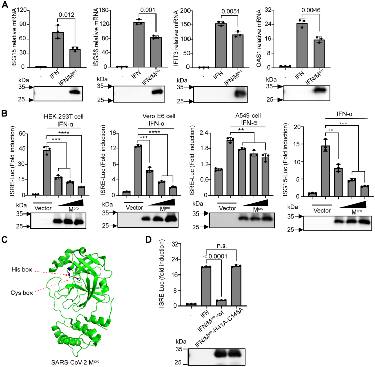
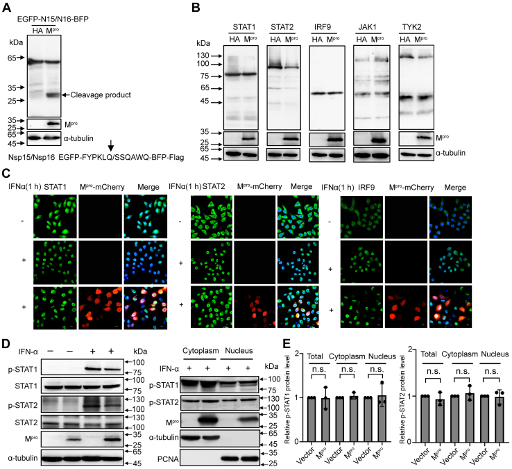
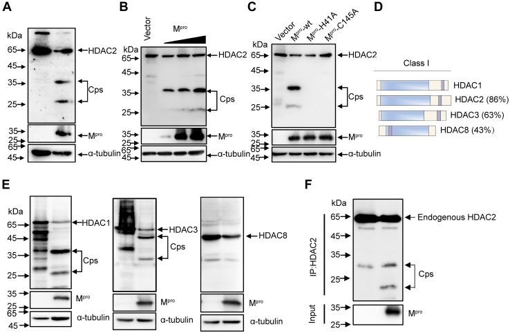
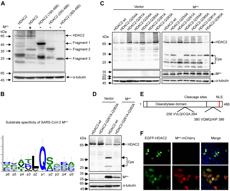
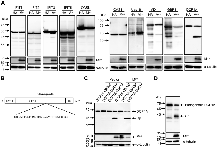
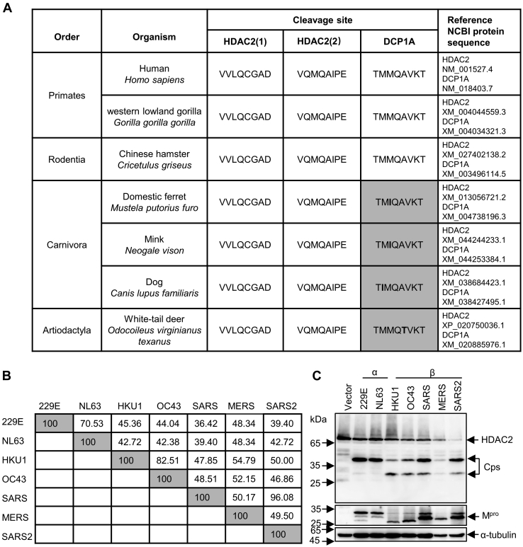
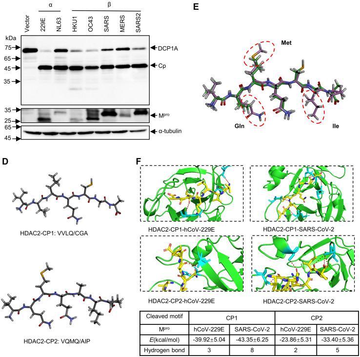
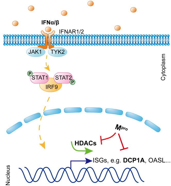
Severe acute respiratory syndrome coronavirus 2 (SARS-CoV-2), which causes coronavirus disease 2019, constitutes an emerging human pathogen of zoonotic origin. A critical role in protecting the host against invading pathogens is carried out by interferon-stimulated genes (ISGs), the primary effectors of the type I interferon (IFN) response. All coronaviruses studied thus far have to first overcome the inhibitory effects of the IFN/ISG system before establishing efficient viral replication. However, whether SARS-CoV-2 evades IFN antiviral immunity by manipulating ISG activation remains to be elucidated. Here, we show that the SARS-CoV-2 main protease (Mpro) significantly suppresses the expression and transcription of downstream ISGs driven by IFN-stimulated response elements in a dose-dependent manner, and similar negative regulations were observed in two mammalian epithelial cell lines (simian Vero E6 and human A549). Our analysis shows that to inhibit the ISG production, Mpro cleaves histone deacetylases (HDACs) rather than directly targeting IFN signal transducers. Interestingly, Mpro also abolishes the activity of ISG effector mRNA-decapping enzyme 1a (DCP1A) by cleaving it at residue Q343. In addition, Mpro from different genera of coronaviruses has the protease activity to cleave both HDAC2 and DCP1A, even though the alphacoronaviruse Mpro exhibits weaker catalytic activity in cleaving HDAC2. In conclusion, our findings clearly demonstrate that SARS-CoV-2 Mpro constitutes a critical anti-immune effector that modulates the IFN/ISG system at multiple levels, thus providing a novel molecular explanation for viral immune evasion and allowing for new therapeutic approaches against coronavirus disease 2019 infection.

Keywords: SARS-CoV-2; cleavage; histone deacetylases; interferon-stimulated gene; mRNA-decapping enzyme 1a; main protease.


Conflict of interest statement

Conflict of interest The authors declare there are no conflicts of interest with the contents of this article.

Figures

Figure 1
Figure 1
SARS-CoV-2 Mproinhibits interferon-stimulated gene production.A, HEK-293T cells were transfected with pCDNA3.1 empty vector or the pCDNA3.1-Mpro-HA plasmid. Twenty-four hours posttransfection, cells were stimulated with 1000 U/ml IFNα for 8 h as indicated. Isolated RNA was analyzed by RT-PCR for the ISG15, ISG56, IFIT3, or OAS1 relative mRNA level and normalized to GAPDH mRNA transcription. B, HEK-239T cells, Vero E6 cells, or A549 cells were transfected with various concentrations of pCDNA3.1-Mpro-HA plasmid, along with pRL-TK plasmid (10 ng) and pISRE-Luc (50 ng) or pISG15-Luc. Twenty-four hours posttransfection, cells were stimulated with or without IFNα (1000 U/ml) for 12 h, followed by a dual-luciferase assay. C, schematic representation of SARS-CoV-2 Mpro (PDB code: 7JYC) with the conserved catalytic residues His41 and Cys145. D, HEK-293T cells were transfected with pISRE-Luc plasmid (50 ng), pRL-TK plasmid (10 ng), along with SARS-CoV-2 Mpro expression constructs (200 ng) or its inactive double mutants H41A and C145A. Twenty-four hours posttransfection, a dual-luciferase assay was performed after treatment IFNα (1000 U/ml) for another 8 h. Western blotting for the expression levels of Mpro were shown below the graph. All presented results represent the means and standard deviations of data from three independent experiments. Statistical significance was calculated using unpaired, two-tailed Student’s t test. Data significance is shown as indicated. ns, not significant; ∗P < 0.05; ∗∗p < 0.01; ∗∗∗p < 0.001. ∗∗∗∗p < 0.0001. ISG, interferon-stimulated gene; Mpro, main protease; OAS, 2′,5′-oligoadenylate synthetase; SARS-CoV-2, severe acute respiratory syndrome coronavirus 2.
Figure 2
Figure 2
Mproshows no interaction with IFN signaling pathway transducers.A, HEK-293T cells were transfected with a C-terminal flag-tagged EGFP-N15/N16-BFP plasmid along with SARS-CoV-2 Mpro or empty vector. Cells were lysed at 24 h after transfection and analyzed by Western blotting. B, HEK-293T cells were cotransfected with a C-terminal flag-tagged STAT1, STAT2, IRF9, JAK1, or TYK2 expression plasmid combined either with SARS-CoV-2 Mpro or empty vector. Whole-cell extracts were lysed 30 h posttransfection and analyzed by Western blotting. C, HeLa cells were transfected with pCDNA3.1-Mpro-mCherry visual construct, and 24 h posttransfection, cells were treated with or without IFNα (1000 U/ml) for 1 h and possessed for indirect immunofluorescence to detect the STAT1, STAT2, and IRF9. Scale bar, 10 μm. D, HEK-293T cells were transfected with SARS-CoV-2 Mpro or empty vector. After 24 h of expressing, cells were cultured with or without IFNα (1000 U/ml) for 1 h. Whole-cell extracts or the nuclear and cytoplasmic fractions were analyzed by Western blotting with specific antibodies for the detection of tyrosine phospho-STAT1 and STAT2 or total STAT1 and STAT2. E, density analysis represents the relative protein levels of phospho-STAT1 or phospho-STAT2 that was normalized to the protein levels of α-tubulin or PCNA. The value of control group was set to 1. The presented results represent the means and standard deviations of data from three independent experiments. Statistical significance was calculated using unpaired, two-tailed Student’s t test. Protein band intensities were quantitated by Image Lab software. IFN, interferon; IRF9, interferon regulatory factor 9; Mpro, main protease; SARS-CoV-2, severe acute respiratory syndrome coronavirus 2; STAT, signal transducer and activator of transcription.
Figure 3
Figure 3
Mproimpairs ISG induction by cleaving HDACs.A, HEK-293T cells were transfected with pCDNA3.1-HDAC2-Flag plasmids together with or without Mpro for 24 h before Western blotting analysis with the anti-flag antibody. B, HEK-293T cells were transfected with HDAC2 expression construct alone or in the presence of increasing concentrations of the Mpro, and after 30 h transfection, cells were lysed for Western blotting analysis. C, HEK-293T cells were transfected with HDAC2 expression plasmid along with Mpro or its mutants Mpro-H41A, Mpro-C145A. Western blotting was performed after 24 h transfection. D, Schematic representation of Class I HDAC with different similarities to the HDAC1. E, HEK-293T cells were transfected with HDAC1, HDAC3, or HDAC8 separately along with either Mpro or empty vector. Western blotting was performed after 24 h transfection. F, HEK-293T cells were transfected with Mpro or empty vector. Cells were then lysed for immunoprecipitation analysis at 32 h after transfection. HDAC, histone deacetylase; ISG, interferon-stimulated gene; Mpro, main protease.
Figure 4
Figure 4
Mprocleavage HDAC2 occurs at residues Q261 and Q383.A, HEK-293T cells were transfected with pCDNA3.1-HDAC2-Flag plasmid or its truncated constructs pHDAC2 (190–488)-Flag, pHDAC2 (250–488)-Flag, pHDAC2 (365–488)-Flag. After 30 h of transfection, the cell lysates were analyzed by Western blotting with anti-flag antibody. B, schematic representation of substrate specificity of SARS-CoV-2 Mpro. C, HEK-293T cells were respectively transfected with HDAC2, HDAC2-Q254A, HDAC2-Q261A, HDAC2-Q354A, HDAC2-Q365A, HDAC2-Q381A, and HDAC2-Q383A together with Mpro or empty vector. Western blotting was performed 24 h posttransfection. D, HEK-293T cells were transfected with double mutants Q261A and Q383A of HDAC2 together with Mpro or empty vector. Western blotting was performed 24 h posttransfection. E, schematic representation of HDAC2 with the position of two cleavage sites. F, HEK-293T cells were transfected with a plasmid expressing EGFP-HDAC2 together with Mpro-mCherry or empty vector. After 24 h expressing, images of transfected cells were recorded with GFP and mCherry excitation and emission spectra. Scale bar, 10 μm. HDAC, histone deacetylase; Mpro, main protease; SARS-CoV-2, severe acute respiratory syndrome coronavirus 2.
Figure 5
Figure 5
Mprocleaves ISG DCP1A at residue Q343.A, HEK-293T cells were transfected with N-terminal flag-tagged IFIT1, IFIT2, IFIT3, IFIT5, OSAL, OAS1, Usp18, MIX, GBP1, or DCP1A plasmids along with SARS-CoV-2 Mpro or empty vector, after 30 h of transfection, followed by Western blotting analysis. B, schematic representation of DCP1A with the position of the cleavage site. C, HEK-293T cells were separately transfected with pDCP1A-Q330A, pDCP1A-Q343A, or pDCP1A-Q351A plasmid together with SARS-CoV-2 Mpro or empty vector, protein extracts were analyzed by Western blotting with anti-flag antibody 24 h posttransfection. D, HEK-293T cells were transfected with Mpro or empty vector. Cells were lysed at 24 h after transfection and analyzed by Western blotting with anti-DCP1A antibody. DCP1A, mRNA-decapping enzyme 1a; GBP1, guanylate binding protein 1; HDAC, histone deacetylase; IFIT, interferon-induced protein with tetratricopeptide repeats; ISG, interferon-stimulated gene; MIX, mix paired-like homeobox; Mpro, main protease; OAS, 2′,5′-oligoadenylate synthetase; SARS-CoV-2, severe acute respiratory syndrome coronavirus 2; Usp18, ubiquitin specific peptidase 18.
Figure 6
Figure 6
Mprocleavage efficiency differs in different coronaviruses.A, analysis of protein sequences of HDAC2 and DCP1A in cleaved sites across species. B, amino acid percent identity matrix of Mpro proteins in different coronaviruses. C, HEK-293T cells were individually transfected with empty vector, 229E-Mpro, NL63-Mpro, HKU1-Mpro, OC43-Mpro, SARS-Mpro, MERS-Mpro, or SARS2-Mpro plasmids along with HDAC2 or DCP1A. Western blotting was performed 30 h posttransfection. D, generation of peptide conformations based on sequence of two cleaved motifs in HDAC2. CP1 represents the first cleaved peptide, and CP2 means the second cleaved peptide. E, comparison of predicted two heptapeptides structures using superimpose in discovery studio. F, binding energy and hydrogen bonds formed between Mpro and cleaved motifs. The hydrogen bonds (Red) formed between the two cleaved peptides and Mpro are presented as dash lines labeled with donor residual (yellow) and corresponding acceptor residual (cyan) in Mpro. CP, cleaved peptide; DCP1A, mRNA-decapping enzyme 1a; HDAC, histone deacetylase; Mpro, main protease.
Figure 6
Figure 6
Mprocleavage efficiency differs in different coronaviruses.A, analysis of protein sequences of HDAC2 and DCP1A in cleaved sites across species. B, amino acid percent identity matrix of Mpro proteins in different coronaviruses. C, HEK-293T cells were individually transfected with empty vector, 229E-Mpro, NL63-Mpro, HKU1-Mpro, OC43-Mpro, SARS-Mpro, MERS-Mpro, or SARS2-Mpro plasmids along with HDAC2 or DCP1A. Western blotting was performed 30 h posttransfection. D, generation of peptide conformations based on sequence of two cleaved motifs in HDAC2. CP1 represents the first cleaved peptide, and CP2 means the second cleaved peptide. E, comparison of predicted two heptapeptides structures using superimpose in discovery studio. F, binding energy and hydrogen bonds formed between Mpro and cleaved motifs. The hydrogen bonds (Red) formed between the two cleaved peptides and Mpro are presented as dash lines labeled with donor residual (yellow) and corresponding acceptor residual (cyan) in Mpro. CP, cleaved peptide; DCP1A, mRNA-decapping enzyme 1a; HDAC, histone deacetylase; Mpro, main protease.
Figure 7
Figure 7
Mproinhibits the IFN-I signaling at multiple steps. Schematic representation of how Mpro antagonizes ISG production. DCP1A, mRNA-decapping enzyme 1a; HDAC, histone deacetylase; IFN, interferon; ISG, interferon-stimulated gene; IRF9, interferon regulatory factor 9; Mpro, main protease; STAT, signal transducer and activator of transcription.

Similar articles

Cited by

References

    1. Wu F., Zhao S., Yu B., Chen Y.M., Wang W., Song Z.G., et al. A new coronavirus associated with human respiratory disease in China. Nature. 2020;579:265–269. - PMC - PubMed
    1. Zhu N., Zhang D., Wang W., Li X., Yang B., Song J., et al. A novel coronavirus from patients with pneumonia in China, 2019. N. Engl. J. Med. 2020;382:727–733. - PMC - PubMed
    1. Lu R., Zhao X., Li J., Niu P., Yang B., Wu H., et al. Genomic characterisation and epidemiology of 2019 novel coronavirus: Implications for virus origins and receptor binding. Lancet. 2020;395:565–574. - PMC - PubMed
    1. Gorbalenya A.E., Baker S.C., Baric R.S., de Groot R.J., Drosten C., Gulyaeva A.A., et al. The species severe acute respiratory syndrome-related coronavirus: classifying 2019-nCoV and naming it SARS-CoV-2. Nat. Microbiol. 2020;5:536–544. - PMC - PubMed
    1. Pormohammad A., Ghorbani S., Baradaran B., Khatami A., R J.T., Mansournia M.A., et al. Clinical characteristics, laboratory findings, radiographic signs and outcomes of 61,742 patients with confirmed COVID-19 infection: a systematic review and meta-analysis. Microb. Pathog. 2020;147:104390. - PMC - PubMed

Publication types