. 2023 Jun 8;61(6):2201799.
doi: 10.1183/13993003.01799-2022. Print 2023 Jun.

Regulating the cell shift of endothelial cell-like myofibroblasts in pulmonary fibrosis

Affiliations

Regulating the cell shift of endothelial cell-like myofibroblasts in pulmonary fibrosis

Xiuju Wu et al. Eur Respir J. .

Abstract

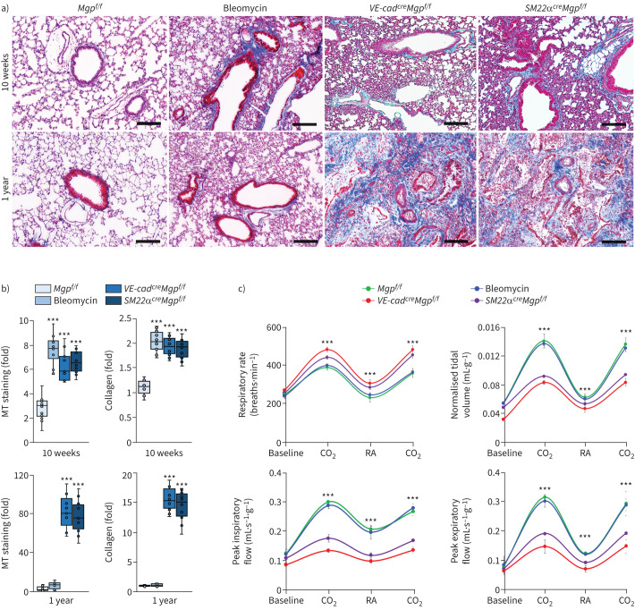
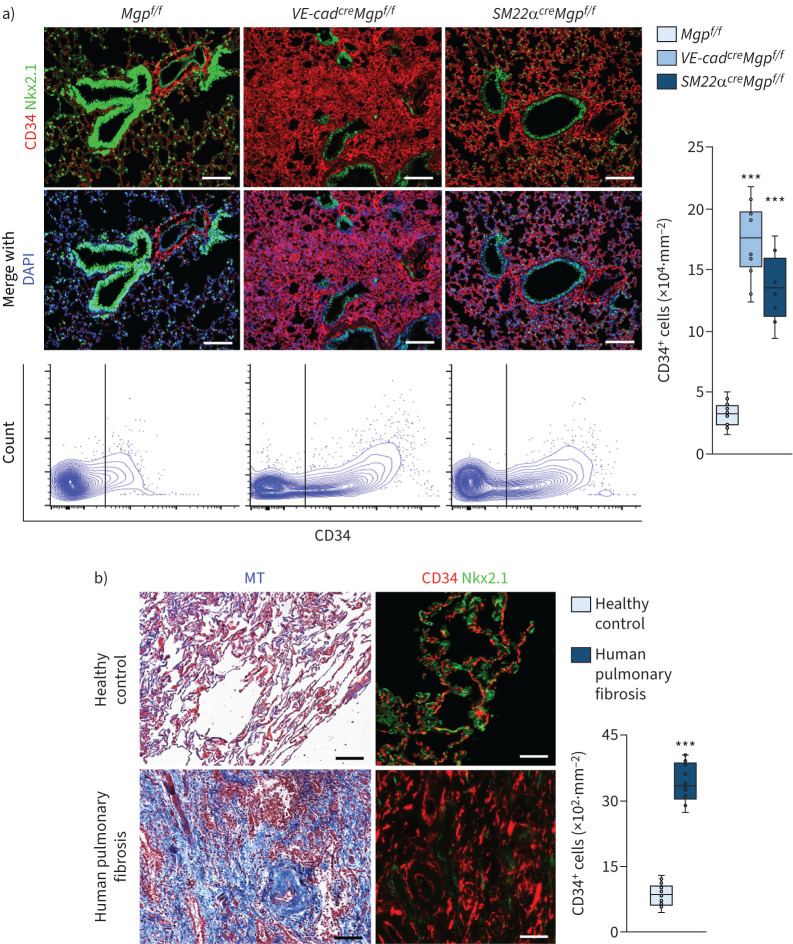
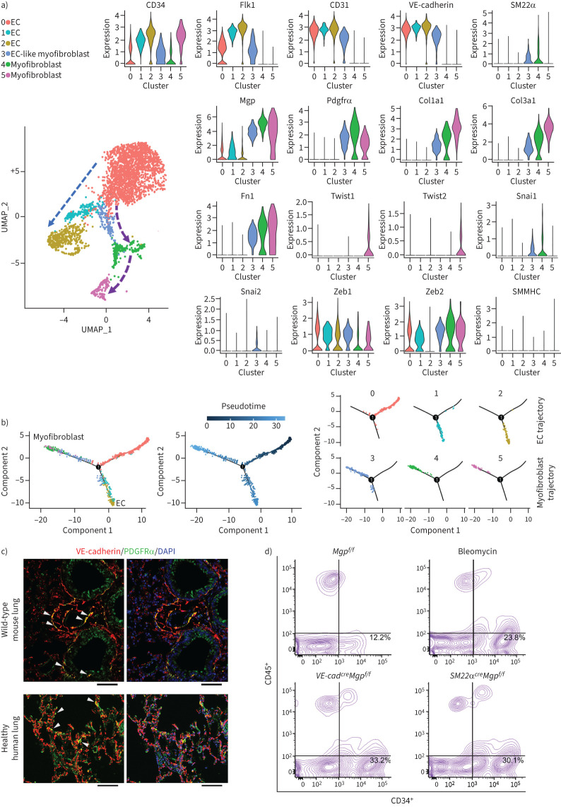
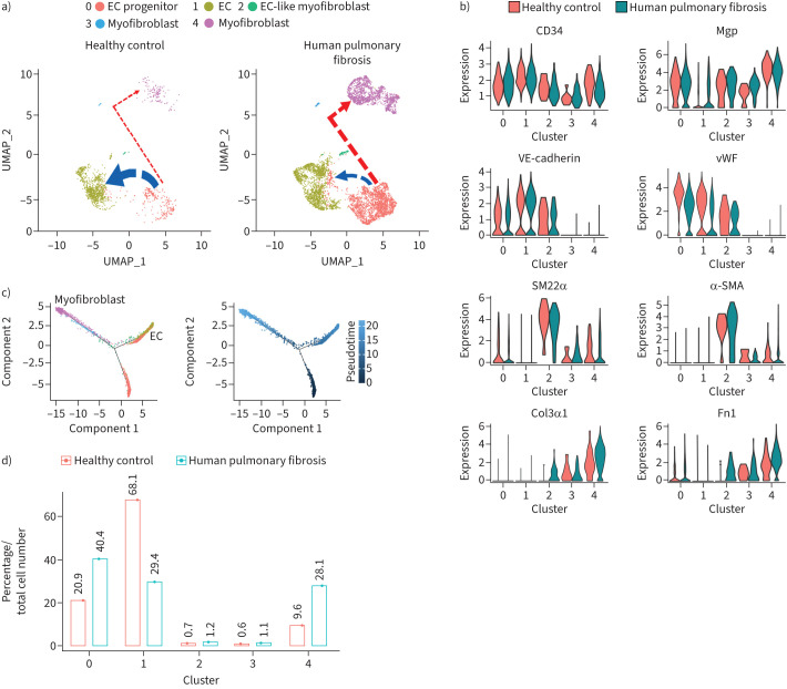
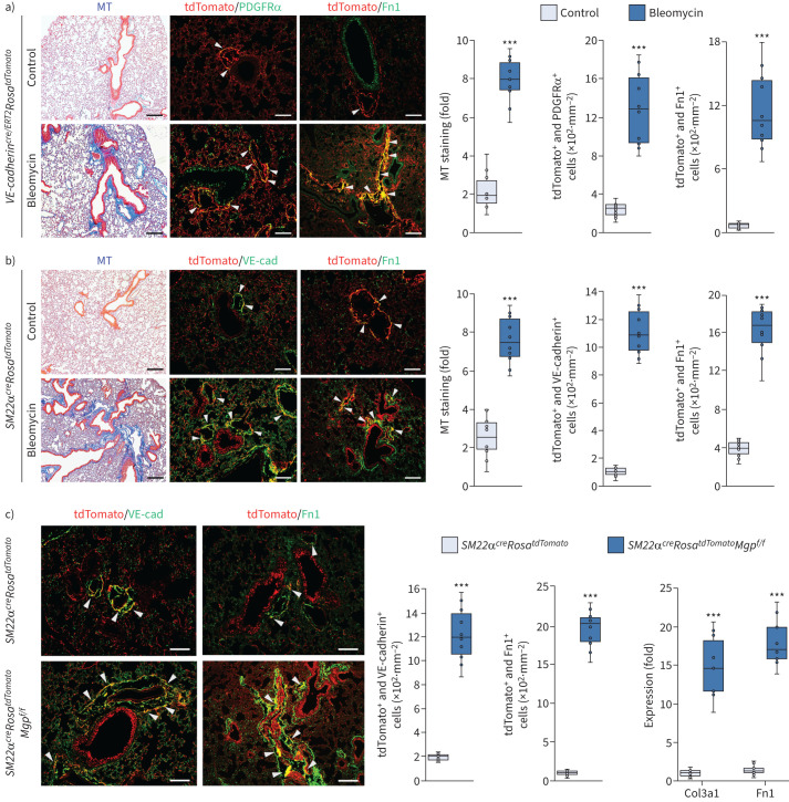
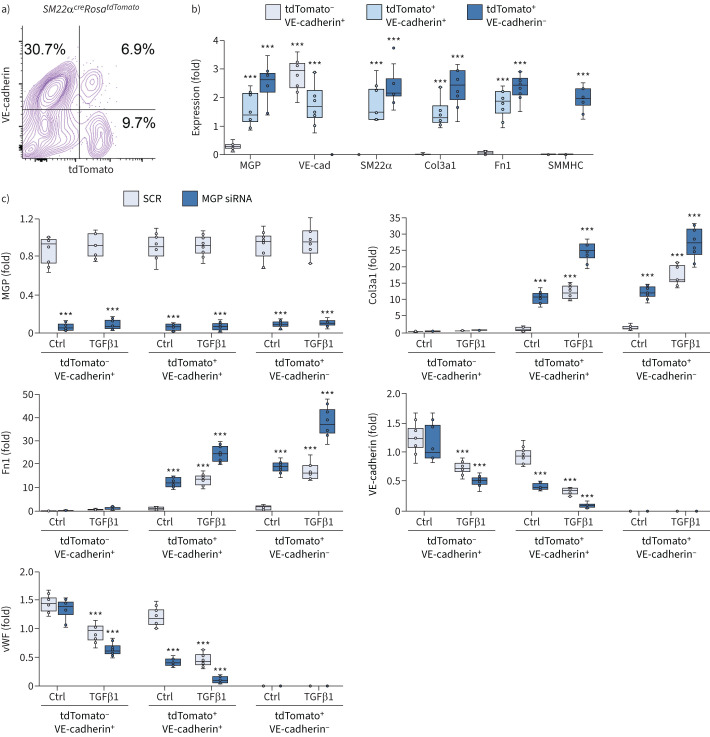
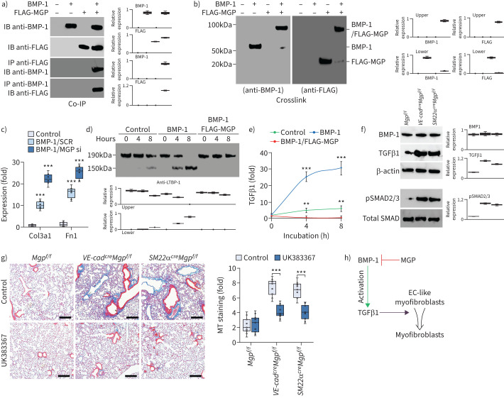
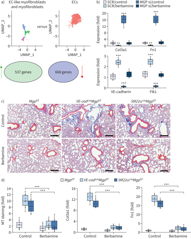
Pulmonary fibrosis is a common and severe fibrotic lung disease with high morbidity and mortality. Recent studies have reported a large number of unwanted myofibroblasts appearing in pulmonary fibrosis, and shown that the sustained activation of myofibroblasts is essential for unremitting interstitial fibrogenesis. However, the origin of these myofibroblasts remains poorly understood. Here, we create new mouse models of pulmonary fibrosis and identify a previously unknown population of endothelial cell (EC)-like myofibroblasts in normal lung tissue. We show that these EC-like myofibroblasts significantly contribute myofibroblasts to pulmonary fibrosis, which is confirmed by single-cell RNA sequencing of human pulmonary fibrosis. Using the transcriptional profiles, we identified a small molecule that redirects the differentiation of EC-like myofibroblasts and reduces pulmonary fibrosis in our mouse models. Our study reveals the mechanistic underpinnings of the differentiation of EC-like myofibroblasts in pulmonary fibrosis and may provide new strategies for therapeutic interventions.


Conflict of interest statement

Conflict of interest: The authors have no potential conflicts of interest to disclose.

Figures

FIGURE 1
FIGURE 1
Pulmonary fibrosis in VE-cadherincreMgpflox/flox and Sm22αcreMgpflox/flox mice. a) Masson's trichrome (MT) staining of pulmonary tissues (n=10). Scale bar=100 µm. b) Quantification of MT staining and collagen accumulation in pulmonary tissues (n=9). c) Mouse pulmonary tests (n=6). b) and c) were analysed for statistical significance by ANOVA with post hoc Tukey's test. The bounds of the boxes are upper and lower quartiles with data points; the line in the box is the median. In b), error bars are maximal and minimal values; in c), error bars are mean±sd. Bleomycin: wild-type mice injected with bleomycin; VE-cad: VE-cadherin; Mgp: matrix Gla protein; f/f: flox/flox; CO2: hypercapnia phase with 7% carbon dioxide, 21% oxygen and balanced nitrogen; RA: room air. ***: p<0.0001.
FIGURE 2
FIGURE 2
Enlarged CD34+ cell population in pulmonary fibrosis. a) Immunostaining with quantification of CD34+ cells and fluorescence-activated cell sorting in pulmonary tissues of VE-cadherincreMgpflox/flox and Sm22αcreMgpflox/flox mice (n=8). Mgpflox/flox was used as control. b) Masson's trichrome (MT) staining and immunostaining of human tissues with pulmonary fibrosis and quantification of CD34+ cells in human pulmonary fibrosis (n=6). Healthy lungs were used as control. Scale bars=100 µm. The data were analysed for statistical significance by unpaired two-tailed t-test; the bounds of the boxes are upper and lower quartiles with data points; the line in the box is the median; error bars are maximal and minimal values. VE-cad: VE-cadherin; Mgp: matrix Gla protein; f/f: flox/flox; DAPI: 4',6-diamidino-2-phenylindole. ***: p<0.0001.
FIGURE 3
FIGURE 3
Endogenous population of endothelial cell (EC)-like myofibroblasts in wild-type pulmonary tissues. a) Uniform manifold approximation and projection (UMAP) for the cell populations subclustered from the whole population of CD34+CD45 pulmonary cells and violin plots of the gene expression of the lineage markers in the cell clusters. b) Pseudotemporal trajectories of the cell clusters. c) Immunostaining of the lungs of wild-type mice and healthy humans (n=3). Scale bar=100 µm. d) Fluorescence-activated cell sorting (FACS) of whole lung cells isolated from bleomycin-injected mice, VE-cadherincreMgpflox/flox mice and Sm22αcreMgpflox/flox mice (n=6). Mgp: matrix Gla protein; PDGFR: platelet-derived growth factor receptor; Fn1: fibronectin 1; SMMHC: smooth muscle myosin heavy chain; DAPI: 4',6-diamidino-2-phenylindole.
FIGURE 4
FIGURE 4
Endothelial cell (EC)-like myofibroblasts are present in healthy human lungs and contribute to human pulmonary fibrosis. a) Uniform manifold approximation and projection (UMAP) for cell populations subclustered from the whole population of CD34+CD45 pulmonary cells of healthy human lungs and human pulmonary fibrosis. b) Violin plots of the gene expression of the lineage markers. c) Pseudotemporal trajectories of the cell clusters. d) Alterations in cell compositions of different populations in healthy human lungs and human pulmonary fibrosis. Mgp: matrix Gla protein; vWF: von Willebrand factor; SMA: smooth muscle actin; Fn1: fibronectin 1.
FIGURE 5
FIGURE 5
Cell lineage tracings reveal endothelial cell (EC)-like myofibroblasts contributing to pulmonary fibrosis. a) Masson's trichrome (MT) staining and immunostaining with cell quantification of the lungs of tamoxifen-injected VE-cadherincre/ERT2RosatdTomato mice after administration of bleomycin (n=10). b) MT staining and immunostaining with cell quantification of the lungs of Sm22acreRosatdTomato mice after administration of bleomycin (n=10). c) Immunostaining with cell quantification of the lungs of Sm22acreRosatdTomato and Sm22acreRosatdTomatoMgpFlox/Flox mice. The expression of Col3a1 and fibronectin (Fn)1 in tdTomato and VE-cadherin double-positive cells isolated from lungs of Sm22acreRosatdTomato and Sm22acreRosatdTomatoMgpFlox/Flox mice (n=10). Scale bars=100 µm. The data were analysed for statistical significance by unpaired two-tailed t-test. The bounds of the boxes are upper and lower quartiles with data points; the line in the box is the median; error bars are maximal and minimal values. PDGFR: platelet-derived growth factor receptor. ***: p<0.0001.
FIGURE 6
FIGURE 6
Transforming growth factor (TGF)β1 drives endothelial cell (EC)-like myofibroblasts towards myofibroblast differentiation. a) Fluorescence-activated cell sorting of whole lung cells of Sm22acreRosatdTomato mice. b) Gene expression of tdTomato-negative and VE-cadherin-positive (tdTomatoVE-cadherin+), tdTomato+VE-cadherin+ and tdTomato+VE-cadherin cells isolated from lungs of Sm22acreRosatdTomato mice (n=6). c) Gene expression of tdTomatoVE-cadherin+, tdTomato+VE-cadherin+ and tdTomato+VE-cadherin cells after matrix Gla protein (MGP) deletion with siRNA and treated with or without TGFβ1 (1 ng·mL−1) (n=6). Data were analysed for statistical significance by ANOVA with post hoc Tukey's test. The bounds of the boxes are upper and lower quartiles with data points; the line in the box is median; error bars are maximal and minimal values. Fn1: fibronectin 1; SMMHC: smooth muscle myosin heavy chain; vWF: von Willebrand factor. ***: p<0.0001.
FIGURE 7
FIGURE 7
Matrix Gla protein (MGP) inhibits bone morphogenic protein (BMP)-1, which regulates maturation of transforming growth factor (TGF)β1 and in turn controls the differentiation of endothelial cell (EC)-like myofibroblasts. a) Binding of BMP-1 to MGP shown by immunoprecipitation. BMP-1 (200 ng) and conditioned medium containing N-FLAG-MGP (100 μL; ∼200 ng) were combined as indicated in the top panel, and the presence of the respective protein was confirmed by immunoblotting (IB) (top two blots). Interactions between the proteins were analysed by immunoprecipitation (IP) followed by immunoblotting with antibodies as indicated and quantified by densitometry. b) Binding of BMP-1 to MGP shown by chemical crosslinking. BMP-1 (200 ng) and conditioned medium containing N-FLAG-MGP (100 μL; ∼200 ng) were combined as indicated in lane 2 and 3. Interactions were analysed using chemical crosslinking followed by immunoblotting with antibodies as indicated and quantified by densitometry. c) Expression of Col3a1 and Fn1 in tdTomato+VE-cadherin+ cells isolated from the lungs of SM22acreRosatdTomato mice and treated with BMP-1 (200 ng·mL−1) with transfection of MGP siRNA or scrambled siRNA (SCR) (n=6). d) Time-course immunoblotting with densitometry of latent TGFβ1 binding protein-1 (LTBP-1) in tdTomato+VE-cadherin+ cell lysates after treatment with BMP-1 (200 ng·mL−1) with or without conditioned medium containing N-FLAG-MGP (n=6). e) Levels of TGFβ1 in culture media of tdTomato+VE-cadherin+ cell after treatment with BMP-1 with or without conditioned medium containing N-FLAG-MGP (n=3). f) Immunoblotting with densitometry of lung tissues of VE-cadherincreMgpflox/flox and Sm22acreMgpflox/flox and control mice (n=3). g) Masson's trichrome (MT) staining with quantification of the lungs of VE-cadherincreMgpflox/flox and Sm22acreMgpflox/flox mice and control mice after treatment with UK388367 (n=8). Scale bars=100 µm. h) A schematic representation. c, e and g) were analysed for statistical significance by ANOVA with post hoc Tukey's test. The bounds of the boxes are upper and lower quartiles with data points; in c and g) error bars are maximal and minimal values; in e) error bars are mean±sd. **: p<0.001, ***: p<0.0001.
FIGURE 8
FIGURE 8
Berbamine ameliorates pulmonary fibrosis in mouse models. a) Uniform manifold approximation and projection (UMAP) and differential gene expression between endothelial cell (EC)-like myofibroblasts and ECs. b) Gene expression in tdTomato+VE-cadherin+ cells isolated from the lungs of SM22acreRosatdTomato mice and treated with berbamine (20 µM) with transfection of matrix Gla protein (MGP) siRNA or scrambled siRNA (SCR) (n=6). c) Masson's trichrome (MT) staining of pulmonary tissues from VE-cadherincreMgpflox/flox and Sm22acreMgpflox/flox mice after berbamine treatment (100 ng·g−1, daily) (n=12). Scale bars=100 µm. d) Quantification of MT staining and expression of Col3a1 and fibronectin 1 (Fn1) in pulmonary tissues from VE-cadherincreMgpflox/flox and Sm22acreMgpflox/flox mice after berbamine treatment (n=9 for quantification of MT and n=6 for gene expression). b) and d) were analysed for statistical significance by ANOVA with post hoc Tukey's test. The bounds of the boxes are upper and lower quartiles with data points; the line in the box is median; error bars are maximal and minimal values. **: p<0.001, ***: p<0.0001.
FIGURE 9
FIGURE 9
Berbamine induces FoxA2 to regulate transforming growth factor (TGF)β signalling and control endothelial cell (EC)-like myofibroblast differentiation. a) Expression of activin receptor-like kinase (ALK)5 in tdTomato+VE-cadherin+ cells isolated from the lungs of SM22acreRosatdTomato mice and treated with berbamine (20 µM). b) Gene expression in tdTomato+VE-cadherin+ cells transfected with matrix Gla protein (MGP) siRNA in combination with berbamine treatment with or without overexpression of ALK5 (n=5). c) Expression of FoxA2 in tdTomato+VE-cadherin+ cells isolated from the lungs of SM22acreRosatdTomato mice and treated with berbamine (20 µM). d) Immunoblotting with densitometry using tdTomato+VE-cadherin+ cell lysates treated with berbamine (Bbm) with or without overexpression of FoxA2 (cytomegalovirus promoter (CMV)-FoxA2) (n=3). e) Expression of endothelial markers in tdTomato+VE-cadherin+ cells treated with berbamine in combination with FoxA2 overexpression or FoxA2 knockdown (FoxA2 siRNA) (n=6). f) Immunoblotting with densitometry of the lungs of VE-cadherincreMgpflox/flox mice, Sm22acreMgpflox/flox mice and control mice after treatment of berbamine (Bbm) (n=6). g) Quantification of VE-cadherin-positive and platelet-derived growth factor receptor (PDGFR)α-positive (VE-cadherin+PDGFRα+) cells and VE-cadherin negative and PDGFRα positive (VE-cadherinPDGFRα+) cells after immunostaining of the lungs of bleomycin-injected wild-type mice, where gene expression was examined by real-time PCR (n=8). h) Immunostaining of ALK5 and FoxA2 of human pulmonary fibrosis (n=3). Scale bars=100 µm. a) and c) were analysed for statistical significance by unpaired two-tailed t-test. b), e) and g) were analysed for statistical significance by ANOVA with post hoc Tukey's test. The bounds of the boxes are upper and lower quartiles with data points; the line in the box is median; error bars are maximal and minimal values. SCR: scrambled siRNA; Ctr: control; Fn1: fibronectin 1. **: p<0.001, ***: p<0.0001.

Comment in

References

    1. Hutchinson J, Fogarty A, Hubbard R, et al. . Global incidence and mortality of idiopathic pulmonary fibrosis: a systematic review. Eur Respir J 2015; 46: 795–806. doi:10.1183/09031936.00185114 - DOI - PubMed
    1. Lederer DJ, Martinez FJ. Idiopathic pulmonary fibrosis. N Engl J Med 2018; 379: 797–798. doi:10.1056/NEJMc1807508 - DOI - PubMed
    1. Martinez FJ, Collard HR, Pardo A, et al. . Idiopathic pulmonary fibrosis. Nat Rev Dis Primers 2017; 3: 17074. doi:10.1038/nrdp.2017.74 - DOI - PubMed
    1. Raghu G, Collard HR, Egan JJ, et al. . An official ATS/ERS/JRS/ALAT statement: idiopathic pulmonary fibrosis: evidence-based guidelines for diagnosis and management. Am J Respir Crit Care Med 2011; 183: 788–824. doi:10.1164/rccm.2009-040GL - DOI - PMC - PubMed
    1. Raghu G, Weycker D, Edelsberg J, et al. . Incidence and prevalence of idiopathic pulmonary fibrosis. Am J Respir Crit Care Med 2006; 174: 810–816. doi:10.1164/rccm.200602-163OC - DOI - PubMed

Publication types