Review
. 2023 Apr;145(4):371-394.
doi: 10.1007/s00401-023-02540-w. Epub 2023 Feb 10.

Chronic traumatic encephalopathy (CTE): criteria for neuropathological diagnosis and relationship to repetitive head impacts

Affiliations
Review

Chronic traumatic encephalopathy (CTE): criteria for neuropathological diagnosis and relationship to repetitive head impacts

Ann C McKee et al. Acta Neuropathol. 2023 Apr.

Abstract

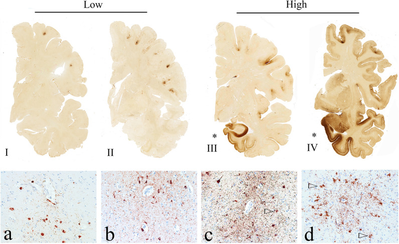
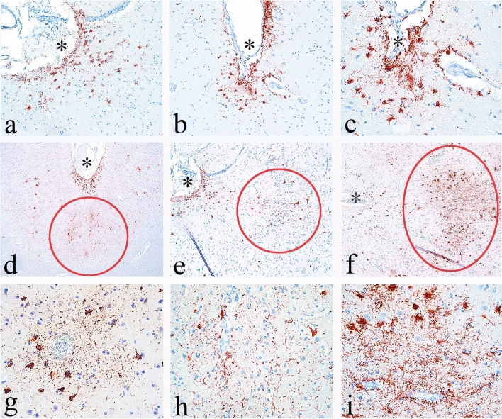
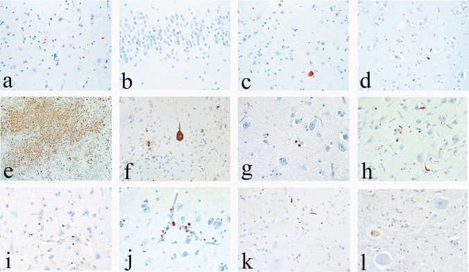
Over the last 17 years, there has been a remarkable increase in scientific research concerning chronic traumatic encephalopathy (CTE). Since the publication of NINDS-NIBIB criteria for the neuropathological diagnosis of CTE in 2016, and diagnostic refinements in 2021, hundreds of contact sport athletes and others have been diagnosed at postmortem examination with CTE. CTE has been reported in amateur and professional athletes, including a bull rider, boxers, wrestlers, and American, Canadian, and Australian rules football, rugby union, rugby league, soccer, and ice hockey players. The pathology of CTE is unique, characterized by a pathognomonic lesion consisting of a perivascular accumulation of neuronal phosphorylated tau (p-tau) variably alongside astrocytic aggregates at the depths of the cortical sulci, and a distinctive molecular structural configuration of p-tau fibrils that is unlike the changes observed with aging, Alzheimer's disease, or any other tauopathy. Computational 3-D and finite element models predict the perivascular and sulcal location of p-tau pathology as these brain regions undergo the greatest mechanical deformation during head impact injury. Presently, CTE can be definitively diagnosed only by postmortem neuropathological examination; the corresponding clinical condition is known as traumatic encephalopathy syndrome (TES). Over 97% of CTE cases published have been reported in individuals with known exposure to repetitive head impacts (RHI), including concussions and nonconcussive impacts, most often experienced through participation in contact sports. While some suggest there is uncertainty whether a causal relationship exists between RHI and CTE, the preponderance of the evidence suggests a high likelihood of a causal relationship, a conclusion that is strengthened by the absence of any evidence for plausible alternative hypotheses. There is a robust dose-response relationship between CTE and years of American football play, a relationship that remains consistent even when rigorously accounting for selection bias. Furthermore, a recent study suggests that selection bias underestimates the observed risk. Here, we present the advances in the neuropathological diagnosis of CTE culminating with the development of the NINDS-NIBIB criteria, the multiple international studies that have used these criteria to report CTE in hundreds of contact sports players and others, and the evidence for a robust dose-response relationship between RHI and CTE.

Keywords: CTE; Neurodegeneration; Repetitive head impacts; Tauopathy.


Conflict of interest statement

The authors have no relevant financial or non-financial interests to disclose.

Figures

Fig. 1
Fig. 1
The pathognomonic lesion of CTE and the staging schemes of pathological severity (adapted with permission from [83]). Representative images of p-tau pathology at Low and High chronic traumatic encephalopathy (CTE) pathological stage using the abbreviated staging scheme recommended by the second NINDS/NIBIB consensus panel (low–high) [11] and the McKee staging scheme (I–IV) [4, 79]. Low CTE is characterized by p-tau pathology restricted to focal cortical lesions. High CTE shows widespread p-tau pathology in the medial temporal lobe structures and diencephalon in addition to focal cortical lesions. McKee Stage I CTE is characterized by one or two isolated CTE lesions at the depths of the cortical sulci. In stage II, three or more cortical CTE lesions are found. In stage III CTE, there are multiple CTE lesions and diffuse NFTs in the medial temporal lobe. In stage IV CTE, CTE lesions and NFTs are widely distributed throughout the cerebral cortex, diencephalon, and brainstem. Top row: hemispheric 50-µm tissue sections immunostained with CP-13, directed against phosphoserine 202 of tau (courtesy of Peter Davies, Ph.D., Feinstein Institute for Medical Research; 1:200); positive p-tau immunostaining appears dark brown. Bottom row: 10-µm paraffin-embedded tissue sections immunostained for phosphorylated tau (AT8) (Pierce Endogen). Positive p-tau immunostaining appears dark red, hematoxylin counterstain. I A 26-year-old former college football player with stage I CTE (Low). Two perivascular p-tau CTE lesions are evident at the sulcal depths of the frontal cortex; there is no neurofibrillary degeneration in the medial temporal lobe. II A 49-year-old former NFL player with stage II CTE (Low). There are multiple perivascular p-tau CTE lesions at depths of sulci of the frontal cortex; there is no neurofibrillary degeneration in the amygdala or entorhinal cortex. III A 53-year-old former NFL player with stage III CTE (High). There are multiple CTE lesions in the frontal cortex and insula; there is diffuse neurofibrillary degeneration of hippocampus and entorhinal cortex (asterisk). IV A 62-year-old former NFL player with stage IV CTE (High). There are multiple CTE lesions in the cerebral cortex with widespread neurofibrillary degeneration. There is also extensive neurofibrillary degeneration of the amygdala and entorhinal cortex (asterisk). a Pathognomonic CTE lesion in stage I CTE. AT8 immunopositive neurofibrillary tangles, dot-like and threadlike neurites encircle a small blood vessel. b Pathognomonic CTE lesion in stage II CTE. A cluster of AT8 immunopositive neurofibrillary tangles and dense dot-like neurites surround several small blood vessels, c pathognomonic CTE lesion in stage III CTE. A large collection of AT8 immunopositive neurofibrillary tangles and dense dot-like neurites enclose several small blood vessels. With increasing age, AT8 immunoreactive astrocytes are increasingly evident within the pathognomonic lesion (open triangle). d Pathognomonic CTE lesion in stage IV CTE. A large accumulation of AT8 immunopositive neurofibrillary tangles, most of them ghost tangles, encompass several small blood vessels. With increasing age, AT8 immunoreactive astrocytes are increasingly prominent (open triangles) and there may be evidence of neuronal loss. a–d All magnification × 200. P-tau phosphorylated tau, CTE chronic traumatic encephalopathy, NFL National Football League
Fig. 2
Fig. 2
Differential diagnosis between mild CTE and ARTAG. 10-µm paraffin-embedded tissue sections immunostained for phosphorylated tau (AT8) (Pierce Endogen). Positive p-tau immunostaining appears dark red, hematoxylin counterstain. a–c p-tau-immunoreactive thorn-shaped astrocytes are present at the glial limitans at the depths of the sulcus, a form of ARTAG (a, b) magnification × 200, c magnification × 400). The depth of the sulcus is marked by an asterisk. d–f Clusters of p-tau neurons and dot-like neurites surrounding small blood vessels in deep cortical laminae at the depth of the sulci are representative of the diagnostic CTE lesion, all magnifications × 40. The diagnostic lesions are indicated by red circles and located deeper than subpial ARTAG, marked by an asterisks. g–i CTE lesions consist of p-tau-immunoreactive neurons, dot-like neurites, and variably astrocytes, surrounding small blood vessels, all magnifications × 200
Fig. 3
Fig. 3
Regional progression of semi-quantitative p-tau density in CTE by stage and age (n = 739). Top: among 739 cases of neuropathologically verified CTE cases in the UNITE brain bank, there is a hierarchical increase in p-tau pathology across CTE stages I–IV. In stage I CTE, p-tau pathology is most dense in CTE lesions in the dorsolateral frontal cortex and NFT are found in the locus coeruleus (LC). In CTE stage II, there are increasing densities of p-tau pathology in the cortex, especially dorsolateral frontal and temporal lobes, entorhinal cortex, amygdala, and LC. In CTE stage III, there are further increases in p-tau pathology in the same regions with expansion to CA1 hippocampus and substantia nigra. In stage IV, p-tau densities continue to increase with highest densities in dorsolateral frontal and superior temporal cortices, entorhinal cortex, amygdala, and LC. Bottom: across decade at death, p-tau pathology in CTE shows a similar pattern of progressive regional involvement with highest densities of p-tau pathology in the dorsolateral frontal and superior temporal cortices, entorhinal cortex, amygdala, and LC. The most common locations of pathognomonic CTE lesions differ slightly from the areas of highest p-tau density. In a preliminary analysis of a subset of 52 of the 739 cases, pathognomonic lesions were most often found in the dorsolateral (56%, often multiple) and superior frontal cortices (56%), followed by Rolandic (33%) and inferior parietal (33%), septal (17%), insula and superior temporal (each 12%), hippocampus and entorhinal (each 8%), inferior frontal (6%) and temporal pole (< 1%). While most pathognomonic lesions involved the neocortex, they were also found in hippocampus, amygdala and entorhinal cortex. Similarly, while most pathognomonic lesions were found at the depths of the neocortical sulci, occasionally they were present in areas with no sulci, e.g., along gyral banks, in hippocampal subfields, and in the amygdala. A superficial distribution of NFT is often most prominent in the temporal lobe. DLF dorsolateral frontal, IF inferior frontal, IP inferior parietal, ST superior temporal, CA1 CA1 hippocampus, CA2 CA2 hippocampus, CA4 CA4 hippocampus, EC entorhinal cortex, SN substantia nigra, LC locus coeruleus. In each region, dark green is the 1st percentile, light green is the 25th percentile, yellow is the 50th percentile, orange is the 75th percentile and dark red is the 100th percentile. The color scale is based on the distribution of all values, not by each individual stage. Values represent means of p-tau pathology among participants in each stage or age decade at death
Fig. 4
Fig. 4
The evolution of 3R/4R tau and astrocytic involvement in CTE pathology (adapted with permission from [21] and [17]). a Quantitative measurement of the relative ratios of 4R/3R p-tau present in the dorsolateral frontal cortex at the depth of the cortical sulcus across the stages of CTE. Values over 0 contain more 4R tau while values under 0 contain more 3R tau. Although 4R predominates at all stages of disease, there is relatively more 3R tau with increasing CTE stage. In cells containing 4R tau in CTE stage I and II, 95.8% ± 10.2% and 96.1% ± 4.7% were neurons, respectively. For CTE stage III and IV, 65.6% ± 28.7% and 69.7% ± 26.0% of cells containing 4R were neurons, respectively [21]. b Linear regression analysis of the 4R/3R ratio compared to the AT8 p-tau staining density in the dorsolateral frontal cortex. Data is log transformed. Each dot represents one case. As AT8 pathology accumulates, there is a decreasing ratio of 4R/3R p-tau [21]. c Quantitative measurement of the relative percentages of neurons and astrocytes that contain p-tau in the pathognomonic CTE lesion. Neurons contain both 3R and 4R while astrocytes only contain 4R ptau. Neurons were the primary cell type associated with the CTE lesion. Further ANCOVA analysis demonstrated that CTE stage was not significantly correlated with the percentage of 4R astrocytes around the lesion when including age at death as a covariate in the model (p = 0.031), suggesting age is a stronger driver of p-tau astrocytes than CTE stage. d Relative percentages of neurons and astrocytes in pathognomonic lesion that contain 4R tau stratified by decade of age at death. Data is presented as mean ± SD. There is a significant increase in astrocytic 4R p-tau with age at death, most prominent over age 60. e, f Percentage of neuronal, subpial astrocytic, and parenchymal astrocytic p-tau densities separated by CTE stage (e), and decade of age (f) further demonstrating the presence of p-tau positive astrocytes in the parenchyma are driven more by age than disease stage. Subpial astrocytes are also driven by age, first appearing at age > 40 years. Bar graphs represent mean ± standard error of the mean
Fig. 5
Fig. 5
TDP-43 pathology in CTE. Immunoreactivity for phosphorylated TDP-43 is common in CTE and consists of neuronal inclusions and neurites. Representative images of TDP-43 immunoreactive profiles in CTE cases: a CA1 hippocampus b dentate nucleus of the hippocampus c amygdala d frontal cortex. All magnifications × 400. TDP-43 immunoreactivity is also found in CTE cases with comorbid ALS. e–l A 49-year-old former NFL player with ALS and CTE stage IV. e A large CTE lesion at the depth of the dorsolateral frontal sulcus (AT8 immunostain, magnification × 40) f Betz cell in motor cortex with NFT (AT8-immunostain, magnification × 200) g TDP-43 immunoreactive inclusion and neurites in g amygdala, h dorsolateral frontal cortex i perivascular temporal cortex, j perivascular amygdala k lumbar spinal cord and l dentate nucleus of cerebellum. g–l All magnifications × 400

References

    1. Abdolmohammadi B, Uretsky M, Nair E, Saltiel N, Nicks R, Daneshvar D et al (2022) Duration of ice hockey play and chronic traumatic encephalopathy risk (P1–1.Virtual). Neurology 98:3026
    1. Ackermans NL, Varghese M, Williams TM, Grimaldi N, Selmanovic E, Alipour A, et al. Evidence of traumatic brain injury in headbutting bovids. Acta Neuropathol. 2022;144:5–26. doi: 10.1007/s00401-022-02427-2. - DOI - PMC - PubMed
    1. Adams JW, Alvarez VE, Mez J, Huber BR, Tripodis Y, Xia W, et al. Lewy body pathology and chronic traumatic encephalopathy associated with contact sports. J Neuropathol Exp Neurol. 2018;77:757–768. doi: 10.1093/jnen/nly065. - DOI - PMC - PubMed
    1. Alosco ML, Cherry JD, Huber BR, Tripodis Y, Baucom Z, Kowall NW, et al. Characterizing tau deposition in chronic traumatic encephalopathy (CTE): utility of the McKee CTE staging scheme. Acta Neuropathol. 2020;140:495–512. doi: 10.1007/s00401-020-02197-9. - DOI - PMC - PubMed
    1. Alosco ML, Stein TD, Tripodis Y, Chua AS, Kowall NW, Huber BR et al (2019) Association of white matter rarefaction, arteriolosclerosis, and tau with dementia in chronic traumatic encephalopathy. JAMA Neurol. 10.1001/jamaneurol.2019.2244 - PMC - PubMed

Publication types

LinkOut - more resources