. 2023 Apr;128(8):1592-1602.
doi: 10.1038/s41416-023-02192-3. Epub 2023 Feb 9.

An antibody-drug conjugate targeting GPR56 demonstrates efficacy in preclinical models of colorectal cancer

Affiliations

An antibody-drug conjugate targeting GPR56 demonstrates efficacy in preclinical models of colorectal cancer

Joan Jacob et al. Br J Cancer. 2023 Apr.

Abstract

Background: Long-term prognosis remains poor for colorectal cancer (CRC) patients with advanced disease due to treatment resistance. The identification of novel targets is essential for the development of new therapeutic approaches. GPR56, an adhesion GPCR, is highly expressed in CRC tumours and correlates with poor survival. Here, we describe the generation and preclinical evaluation of a novel ADC consisting of an anti-GPR56 antibody (10C7) conjugated with the DNA-damaging payload duocarmycin.

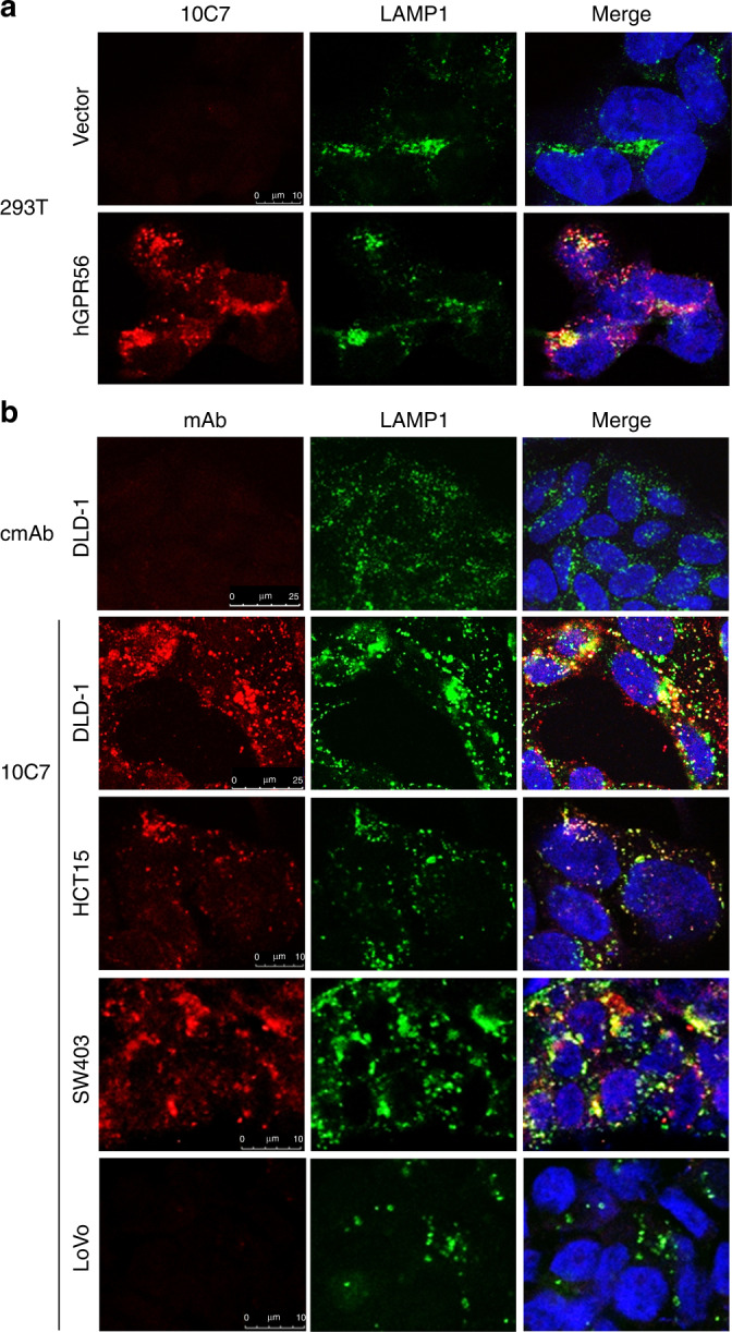
Methods: RNA-seq dataset analysis was performed to determine GPR56 expression in CRC subtypes. The specificity of binding, epitope mapping, and internalisation of 10C7 was examined. 10C7 was conjugated to payload and ADC cytotoxicity was assessed against a panel of CRC cell lines and tumour organoids. Antitumour efficacy was evaluated in xenograft models of CRC cell lines and patient-derived tumours.

Results: High GPR56 was shown to be associated with the microsatellite stable (MSS) subtype that accounts for 80-85% of CRC. GPR56 ADC selectively induced cytotoxicity in CRC cells and tumour organoids at low nanomolar potency in a GPR56-dependent manner and showed significant antitumour efficacy against GPR56-expressing xenograft models.

Conclusions: This study provides the rationale for the future development of a GPR56-targeted ADC approach to potentially treat a large fraction of MSS CRC patients.


Conflict of interest statement

The authors declare no competing interests.

Figures

Fig. 1
Fig. 1. GPR56 expression and correlations with microsatellite instability (MSI), chromosomal instability (CIN), CpG island methylator phenotype (CIMP) and mutational statuses in colorectal cancer.
a Box-and-whiskers plots showing GPR56 RNA-seq expression data of tumour (N = 383) and normal tissues (N = 50) from the TCGA colorectal adenocarcinoma (COADREAD) cohort. Values are RNA‐Seq by expectation–maximisation (RSEM). Distribution of b MSI/MSS, c CIN and d CIMP subtypes based on GPR56 expression analysis. GPR56 expression levels in tumours with WT and mutations in e KRAS, f TP53 and g BRAF. Values are read per kilobase of transcript per million (RPKM). h Western blot of GPR56 protein expression in a panel of colorectal cancer cell lines of different molecular subtypes. Statistical analysis performed using one-way analysis of variance (ANOVA) and Tukey’s multiple comparison test or unpaired two-tailed Student’s t test for two groups.
Fig. 2
Fig. 2. Mapping of the 10C7 mAb epitope within the GAIN domain of the hGPR56 ECD.
a Sequence alignment of amino acids 176–383 of the GAIN domains of human and mouse GPR56. Amino acids in red indicate where mutations were made to convert the human-to-mouse sequence for epitope mapping. Arrow indicates GPCR proteolysis site (GPS). * represents histidine critical for mAb binding. b Cell-based binding assay shows 10C7 mAb binds hGPR56-overexpressing 293T cells and not vector or mGPR56 cells. c Western blot of 10C7 detection of different hGPR56 mutants. d Cell-based binding assay and e confocal microscopy images show hGPR56 H360S mutation inhibits 10C7 binding. For ICC, 293T cells were treated with 10C7 (30 nmol/L) or myc-tag mAb for 30 min at 37 °C. f SRF-RE luciferase-reporter assay shows 10C7 activates WT, but not H360S mutant signalling in a concentration-dependent manner. Experiments were performed three times. Error Bars are SD.
Fig. 3
Fig. 3. GPR56 mAb internalises and traffics to the lysosomes in CRC cells.
Confocal images show the complex of 10C7 bound to GPR56 co-localises with lysosome marker, LAMP1, after 1 h at 37 °C in a hGPR56, but not vector 293T cells and b CRC cell lines DLD-1, HCT15, SW403 and LoVo. No binding of a non-targeting control mAb (cmAb) was detected nor was 10C7 in GPR56-negative LoVo cells. 293T and CRC cells were treated with 6.5  nmol/L and 30 nmol/L 10C7, respectively.
Fig. 4
Fig. 4. Generation and characterisation of a GPR56-targeted antibody–drug conjugate.
a Plot showing IC50 values of ADC payloads for different CRC cell lines. Payloads of monomethyl auristatin E (MMAE), mertansine (DM1), exatecan (DX8951), and duocarmycin SA (DMSA) were tested. b Schematic depiction of the GPR56 ADC consisting of 10C7 mAb conjugated to the DMSA payload via a cleavable linker (DAR = 3.54) attached to cysteine thiols. Linker comprises maleimidocaproyl (MC), valine-citrulline (VC), para-aminobenzyloxycarbonyl (PAB), dimethylethanolamine (DMEA), and polyethylene glycol (PEG). c Coomassie-stained SDS-PAGE of ~2 µg GPR56 mAb compared to ADC under reducing conditions. Image shows shift in the higher molecular weight (MW) of mAb after drug conjugation. Heavy chain (HC) and light chain (LC) are indicated. d Cell-based binding assay confirms no change in binding of ADC compared to parent 10C7 mAb on hGPR56 293T cells. e Cytotoxicity assay shows ADC selectively kills hGPR56 WT and not H360S mutant or vector 293T cells after 3 days. Experiments performed at least 3 times in triplicate. Error bars are SD.
Fig. 5
Fig. 5. GPR56 ADC potently and selectively kills GPR56-high CRC cell lines and patient-derived tumour organoids.
a Cytotoxicity assay shows ADC eliminates DLD-1 control (CTL) cells, but not GPR56 knockdown (KD) cells, in a concentration-dependent manner. b Western blot of GPR56 expression in CTL and GPR56 shRNA DLD-1 stable cell lines. Cytotoxicity of GPR56 ADC against c SW620 and d HCT15 cells treated with GPR56 mAb, GPR56 ADC, non-targeting control mAb (cmAb), or control ADC (cADC). e GPR56 ADC against multiple CRC cell lines with different levels of GPR56 expression. f Western blot of GPR56 expression in patient-derived tumour organoids (PDOs) of a liver metastasis of rectal adenocarcinoma (CRC-001). Different amounts of PDO protein lysate was assessed. hGPR56 293T and LS180 lysates represent positive controls for GPR56 expression. long exposure (L); short exposure (S). g Representative bright-field microscopy images and h cytotoxicity assay of PDOs treated with 30 nmol/L cADC, GPR56 ADC, or PBS vehicle. Experiments were performed two to three times in triplicates. Statistical analysis was performed using one-way ANOVA. Error Bars are SD.
Fig. 6
Fig. 6. GPR56 ADC induces antitumour activity CRC cell line and patient-derived xenograft models.
In vivo efficacy of GPR56 ADC was evaluated in a SW620 (n = 6, PBS vehicle; n = 4, GPR56 mAb and 1.5 mg/kg GPR56 ADC; n = 5, 5 mg/kg GPR56 ADC), b HT-29 (n = 5/cohort) and c SW403 (n = 6, vehicle; n = 5 cADC and GPR56 ADC) xenograft models. d Western blot of GPR56 expression in PDX models of CRC. SW620 lysate was used as a positive control. In vivo efficacy of GPR56 ADC in PDX models, e XST-GI-005 (n = 5/cohort) and f CRC-001 (n = 5/cohort). g Kaplan–Meier survival plot and log-rank test of GPR56 ADC compared to vehicle in CRC-001 model. h Bodyweight measurements of CRC-001 tumour-bearing NSG mice. Statistical analysis was performed using Student’s unpaired two-tailed t test for two groups or one-way ANOVA and Dunnett’s multiple comparison test. Error bars are SD.

References

    1. Society AC. Cancer Facts & Figures 2021. Atlanta, GA: American Cancer Society; 2021.
    1. Xie YH, Chen YX, Fang JY. Comprehensive review of targeted therapy for colorectal cancer. Signal Transduct Target Ther. 2020;5:22. doi: 10.1038/s41392-020-0116-z. - DOI - PMC - PubMed
    1. Le DT, Uram JN, Wang H, Bartlett BR, Kemberling H, Eyring AD, et al. PD-1 blockade in tumors with mismatch-repair deficiency. N Engl J Med. 2015;372:2509–20. doi: 10.1056/NEJMoa1500596. - DOI - PMC - PubMed
    1. Fu Z, Li S, Han S, Shi C, Zhang Y. Antibody drug conjugate: the “biological missile” for targeted cancer therapy. Signal Transduct Target Ther. 2022;7:93. doi: 10.1038/s41392-022-00947-7. - DOI - PMC - PubMed
    1. Leal M, Sapra P, Hurvitz SA, Senter P, Wahl A, Schutten M, et al. Antibody-drug conjugates: an emerging modality for the treatment of cancer. Ann N Y Acad Sci. 2014;1321:41–54. doi: 10.1111/nyas.12499. - DOI - PubMed

Publication types

MeSH terms

Substances