

Be aware of non-specific presentation of pulmonary embolism: a case report
- PMID: 36759780
- PMCID: PMC9912511
- DOI: 10.1186/s12872-023-03096-z
Be aware of non-specific presentation of pulmonary embolism: a case report
Abstract
Background: The early diagnosis of non-specific presentation of pulmonary embolism (PE) is difficult because the symptoms are non-specific and varied.
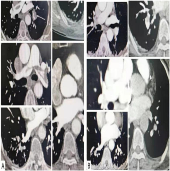
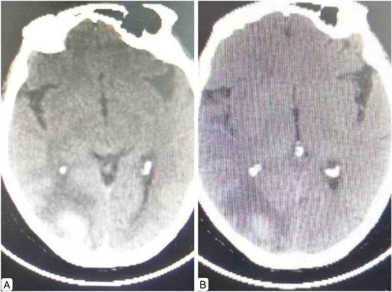
Case presentation: A 69-year-old female patient had syncope accompanied by gait disturbance, without obvious inducement. The patient was initially suspected to have cerebral infarction, but the symptoms did not improve and myocardial markers increased after two days of symptomatic treatment for myocardial infarction. Hence, PE was suspected and computed tomography pulmonary angiography (CTPA) examination confirmed the diagnosis. CTPA showed multiple emboli in pulmonary artery and its branches, so high-risk PE was diagnosed. Intravenous thrombolysis was administered, and pulmonary CTA showed a significant reduction of emboli in pulmonary artery and its left and right branches.
Conclusion: This case report highlights the importance of improving the clinical awareness about non-specific presentation of PE and avoiding misdiagnosis or missed diagnosis.
Keywords: Case report; Non-specific; Pulmonary embolism; Submassive; syncope.
© 2023. The Author(s).
Conflict of interest statement
The authors declare that they have no competing interests.
Figures
Similar articles
-
Unenhanced multidetector computed tomography findings in acute central pulmonary embolism.BMC Med Imaging. 2019 Aug 14;19(1):65. doi: 10.1186/s12880-019-0364-y. BMC Med Imaging. 2019. PMID: 31412797 Free PMC article.
-
Evaluation of pulmonary embolism in pediatric patients with nephrotic syndrome with dual energy CT pulmonary angiography.Acad Radiol. 2012 Mar;19(3):341-8. doi: 10.1016/j.acra.2011.11.002. Epub 2011 Dec 15. Acad Radiol. 2012. PMID: 22177283
-
Acute peripheral pulmonary embolism attributed to autoimmune haemolytic anaemia: a case report.BMC Cardiovasc Disord. 2020 Mar 4;20(1):106. doi: 10.1186/s12872-020-01401-8. BMC Cardiovasc Disord. 2020. PMID: 32131747 Free PMC article.
-
Acute Pulmonary Embolism and Chronic Thromboembolic Pulmonary Hypertension: Clinical and Serial CT Pulmonary Angiographic Features.J Korean Med Sci. 2022 Mar 14;37(10):e76. doi: 10.3346/jkms.2022.37.e76. J Korean Med Sci. 2022. PMID: 35289137 Free PMC article. Review.
-
Submassive Pulmonary Embolism.Am J Respir Crit Care Med. 2018 Sep 1;198(5):588-598. doi: 10.1164/rccm.201711-2302CI. Am J Respir Crit Care Med. 2018. PMID: 29672125 Review.
References
Publication types
MeSH terms
LinkOut - more resources
Full Text Sources
Medical
