Meta-Analysis
. 2023 Feb 9;21(1):103.
doi: 10.1186/s12967-023-03945-7.

ApoE4 associated with severe COVID-19 outcomes via downregulation of ACE2 and imbalanced RAS pathway

Affiliations
Meta-Analysis

ApoE4 associated with severe COVID-19 outcomes via downregulation of ACE2 and imbalanced RAS pathway

Feng Chen et al. J Transl Med. .

Abstract

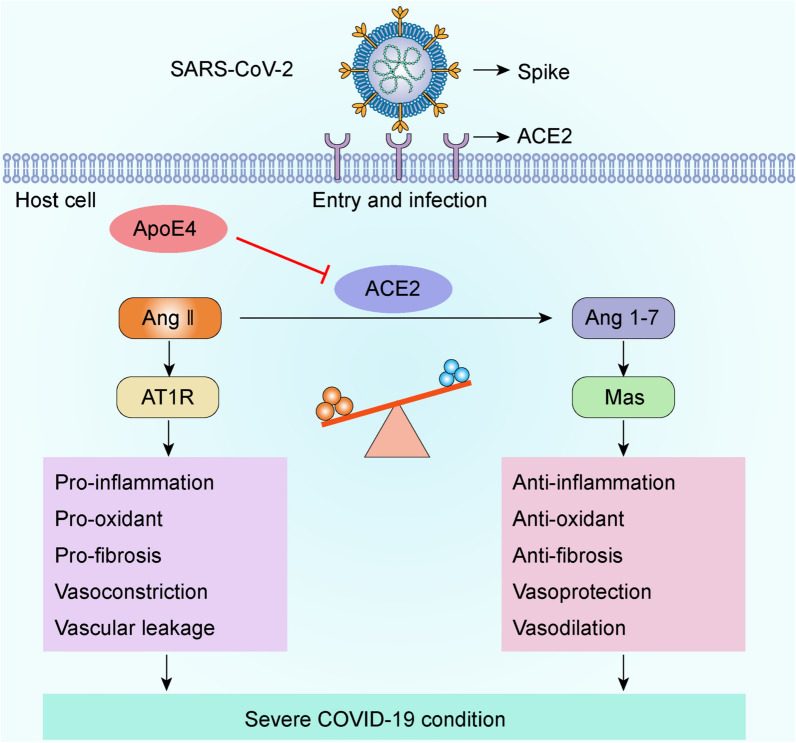
Background: Recent numerous epidemiology and clinical association studies reported that ApoE polymorphism might be associated with the risk and severity of coronavirus disease 2019 (COVID-19), and yielded inconsistent results. Severe acute respiratory syndrome coronavirus 2 (SARS-CoV-2) infection relies on its spike protein binding to angiotensin-converting enzyme 2 (ACE2) receptor expressed on host cell membranes.

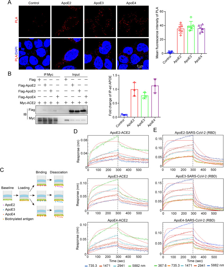
Methods: A meta-analysis was conducted to clarify the association between ApoE polymorphism and the risk and severity of COVID-19. Multiple protein interaction assays were utilized to investigate the potential molecular link between ApoE and the SARS-CoV-2 primary receptor ACE2, ApoE and spike protein. Immunoblotting and immunofluorescence staining methods were used to access the regulatory effect of different ApoE isoform on ACE2 protein expression.

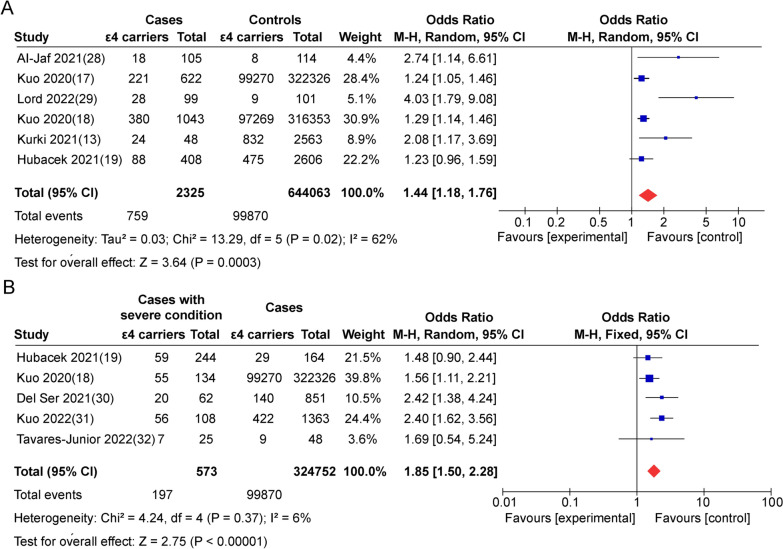
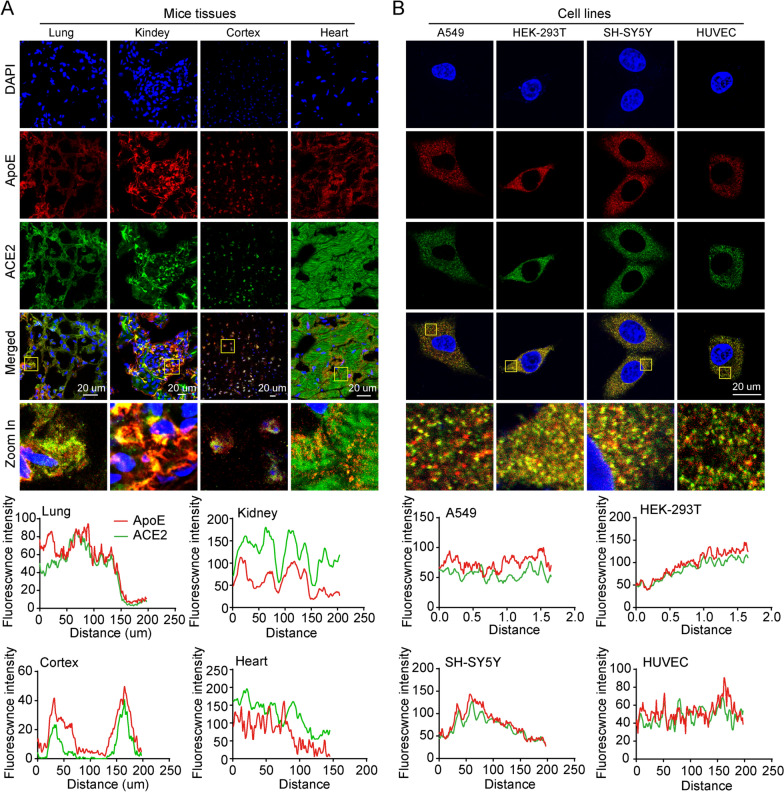
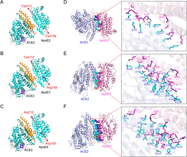
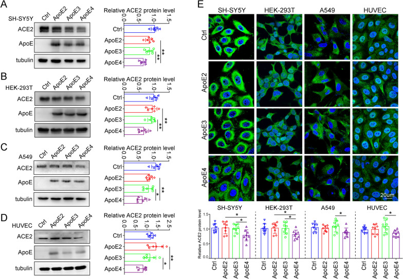
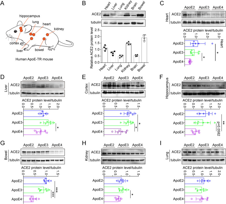
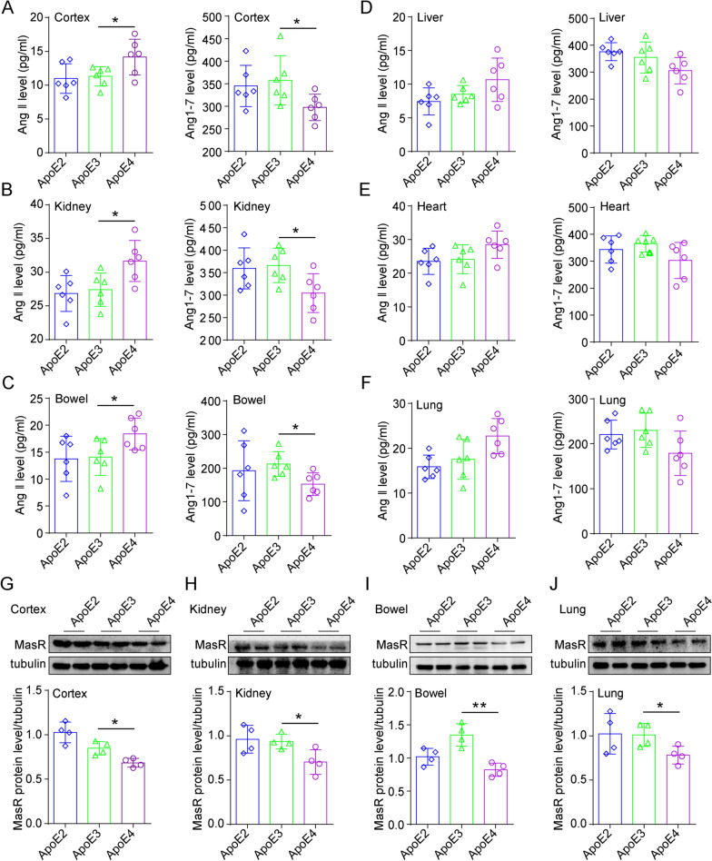
Results: ApoE gene polymorphism (ε4 carrier genotypes VS non-ε4 carrier genotypes) is associated with the increased risk (P = 0.0003, OR = 1.44, 95% CI 1.18-1.76) and progression (P < 0.00001, OR = 1.85, 95% CI 1.50-2.28) of COVID-19. ApoE interacts with both ACE2 and the spike protein but did not show isoform-dependent binding effects. ApoE4 significantly downregulates ACE2 protein expression in vitro and in vivo and subsequently decreases the conversion of Ang II to Ang 1-7.

Conclusions: ApoE4 increases SARS-CoV-2 infectivity in a manner that may not depend on differential interactions with the spike protein or ACE2. Instead, ApoE4 downregulates ACE2 protein expression and subsequently the dysregulation of renin-angiotensin system (RAS) may provide explanation by which ApoE4 exacerbates COVID-19 disease.

Keywords: ACE2; ApoE4; COVID-19; SARS-CoV-2; Spike.


Conflict of interest statement

The authors declare no potential conflicts of interest.

Figures

Fig. 1
Fig. 1
Flowchart summarising the search strategy and selection procedure following PRISMA guidelines
Fig. 2
Fig. 2
ApoE4 is associated with the increased risk and severity of COVID-19. Forest plot of the association of ApoE gene polymorphism (ε4 carrier genotypes versus non-ε4 carrier genotypes) with the risk (A) and severity (B) of COVID-19
Fig. 3
Fig. 3
ApoE colocalizes with ACE2 in vitro and in vivo. A Representative coexpression of ApoE (red) and ACE2 (green) in lung, kidney, brain cortex and heart sections of ApoE3-TR mice as shown by immunofluorescence staining. The nucleus (blue) was stained with DAPI. Scale bars = 20 μm. B Representative coexpression of ApoE (red) and ACE2 (green) in A549, HEK-293, SH-SY5Y and HUVECs as shown by immunofluorescence staining. The nucleus (blue) was stained with DAPI. Scale bars = 20 μm
Fig. 4
Fig. 4
Molecular docking and simulation analyses of the interaction between ApoE and ACE2. A-C Predicted binding mode of ACE2 with ApoE2, ApoE3 or ApoE4 (the orange region of the ACE2 protein represents the region that binds to the S1 unit of the spike protein, and amino acids 112 and 158 of the ApoE protein are represented by spheres). D-F The three-dimensional mode and structural representation of the interface residues of the ApoE and ACE2 complexes (dotted green lines indicate hydrogen bonds, and blue and purple lines represent amino acid residues in ACE2 and ApoE proteins, respectively)
Fig. 5
Fig. 5
ApoE interacts with ACE2 and the spike protein in an isoform-independent manner. A Representative images of individual immunofluorescence staining of ApoE and ACE2 interaction tested by Duolink PLA in HEK-293 T cells after 48 h of cotransfected by Myc-tagged ACE2 and 3 × Flag-tagged ApoE. The red particles (ApoE/ACE2 interaction) represent their interaction. DAPI as a nuclear marker. Scale bar: 5 μm. B Plasmids carrying Myc-tagged ACE2 and 3 × Flag-tagged ApoE (ApoE2, ApoE3 or ApoE4) were transiently cotransfected into HEK-293 T cells. After 48 h of transfection, the cell lysates were immunoprecipitated with anti-Myc antibody and subsequently immunoblotted with anti-Myc and anti-Flag antibodies. The data are presented as the mean ± SD, *P < 0.05, **P < 0.01, ***P < 0.001. C Schematic of the working principles of the BLI assay, which include loading, binding and dissociation. The binding affinities of ApoE and ACE2 were determined through BLI experiments. D The sensor surfaces were immersed in a solution of human ACE2 protein (20 µg/ml), and functionalized sensorgrams were captured upon incubation with human ApoE2, ApoE3, and ApoE4 at 735.3 (blue), 1471 (red), 2941 (light blue), and 5882 nM (green) (binding phase); then, the sensors were immersed in washing buffer (dissociation phase). E The binding affinities of ApoE and the SARS-CoV-2 RBD were determined through BLI experiments. The sensors were immersed in a solution of SARS-CoV-2 RBD (20 µg/ml), and functionalized sensorgrams were captured upon incubation with ApoE2, ApoE3, and ApoE4 at 367.6 (green), 735.3 (yellow), 1471 (light blue), 2941 (brown), and 5882 nM (blue); then, the sensors were immersed in washing buffer (dissociation phase)
Fig. 6
Fig. 6
ApoE4 downregulates ACE2 protein expression in vitro. A-D ACE2 protein levels in SH-SY5Y, HEK-293 T, A549 and HUVEC cells were examined by western blotting after transfection with 1 µg/ml plasmids expressing Flag, ApoE2-Flag, ApoE3-Flag or ApoE4-Flag for 48 h. The results were normalized to the expression of a-tubulin. The data are expressed as the mean ± SD. E Cells were stained with ACE2 (green) antibody and counterstained with DAPI (blue) after transfection with 1 µg/ml Flag, ApoE2-Flag, ApoE3-Flag or ApoE4-Flag plasmids for 48 h. The data are expressed as the mean ± SD. Statistical differences were evaluated by one-way ANOVA. Scale bars, 20 μm. *P < 0.05, **P < 0.01
Fig. 7
Fig. 7
ApoE4 downregulates ACE2 protein expression in vivo. A ApoE-TR mice were selected to evaluate the regulatory effect of ApoE4 on ACE2 protein expression. B ACE2 protein levels in the heart, liver, lung, kidney, brain, and bowel (equivalent amounts of total proteins) of ApoE3-TR mice were measured by western blotting, n = 4 mice per group. C-I ACE2 protein levels in the heart, liver, cortex, hippocampus, bowel, kidney, and lung of ApoE2-TR, ApoE3-TR and ApoE4-TR mice were measured by western blotting. The results were normalized to the expression of a-tubulin. n = 6 mice per group. The data are expressed as the mean ± SD. Statistical differences were evaluated by one-way ANOVA. *P < 0.05, **P < 0.01, ***P < 0.001
Fig. 8
Fig. 8
ApoE4 regulates Ang II and Ang 1–7 protein expression in vivo. A-F Ang II and Ang 1–7 proteins level in the cortex, kidney, bowel, liver, heart and lung (equivalent amounts of total proteins) of ApoE2-TR, ApoE3-TR, and ApoE4-TR mice were assessed by ELISA, n = 6 mice per group. G-J MasR protein level in the cortex, kidney, bowel and lung of ApoE2-TR, ApoE3-TR, and ApoE4-TR mice were measured by western blotting, n = 4 mice per group. The data are expressed as the mean ± SD. One-way ANOVA tests were used. *p < 0.05; **p < 0.01
Fig. 9
Fig. 9
ApoE4 downregulates ACE2 protein expression and consequently decreases the conversion of Ang II to Ang 1–7, which may contribute to adverse COVID-19 outcomes

Similar articles

Cited by

References

    1. Clausen TM, Sandoval DR, Spliid CB, Pihl J, Perrett HR, Painter CD, Narayanan A, Majowicz SA, Kwong EM, McVicar RN, et al. SARS-CoV-2 infection depends on cellular heparan sulfate and ACE2. Cell. 2020;183(4):1043–1057 e1015. - PMC - PubMed
    1. Hamming I, Timens W, Bulthuis ML, Lely AT, Navis G, van Goor H. Tissue distribution of ACE2 protein, the functional receptor for SARS coronavirus. A first step in understanding SARS pathogenesis. J Pathol. 2004;203(2):631–637. - PMC - PubMed
    1. Khazaal S, Harb J, Rima M, Annweiler C, Wu Y, Cao Z, Abi Khattar Z, Legros C, Kovacic H, Fajloun Z, et al. The pathophysiology of long COVID throughout the renin-angiotensin system. Molecules (Basel, Switzerland) 2022;27(9):2903. - PMC - PubMed
    1. Rysz S, Al-Saadi J, Sjöström A, Farm M, Campoccia Jalde F, Plattén M, Eriksson H, Klein M, Vargas-Paris R, Nyrén S, et al. COVID-19 pathophysiology may be driven by an imbalance in the renin-angiotensin-aldosterone system. Nat Commun. 2021;12(1):2417. - PMC - PubMed
    1. Severe Covid GG, Ellinghaus D, Degenhardt F, Bujanda L, Buti M, Albillos A, Invernizzi P, Fernandez J, Prati D, Baselli G, et al. Genomewide association study of severe Covid-19 with respiratory failure. N Engl J Med. 2020;383(16):1522–1534. - PMC - PubMed

Publication types

MeSH terms

Substances