

Translational molecular imaging and drug development in Parkinson's disease
- PMID: 36759912
- PMCID: PMC9912681
- DOI: 10.1186/s13024-023-00600-z
Translational molecular imaging and drug development in Parkinson's disease
Abstract
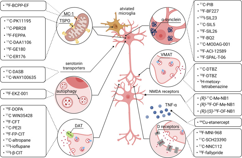
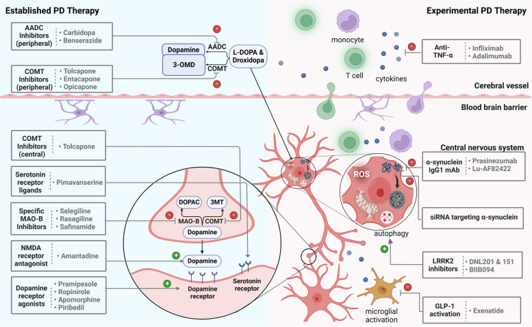
Parkinson's disease (PD) is a progressive neurodegenerative disorder that primarily affects elderly people and constitutes a major source of disability worldwide. Notably, the neuropathological hallmarks of PD include nigrostriatal loss and the formation of intracellular inclusion bodies containing misfolded α-synuclein protein aggregates. Cardinal motor symptoms, which include tremor, rigidity and bradykinesia, can effectively be managed with dopaminergic therapy for years following symptom onset. Nonetheless, patients ultimately develop symptoms that no longer fully respond to dopaminergic treatment. Attempts to discover disease-modifying agents have increasingly been supported by translational molecular imaging concepts, targeting the most prominent pathological hallmark of PD, α-synuclein accumulation, as well as other molecular pathways that contribute to the pathophysiology of PD. Indeed, molecular imaging modalities such as positron emission tomography (PET) and single-photon emission computed tomography (SPECT) can be leveraged to study parkinsonism not only in animal models but also in living patients. For instance, mitochondrial dysfunction can be assessed with probes that target the mitochondrial complex I (MC-I), while nigrostriatal degeneration is typically evaluated with probes designed to non-invasively quantify dopaminergic nerve loss. In addition to dopaminergic imaging, serotonin transporter and N-methyl-D-aspartate (NMDA) receptor probes are increasingly used as research tools to better understand the complexity of neurotransmitter dysregulation in PD. Non-invasive quantification of neuroinflammatory processes is mainly conducted by targeting the translocator protein 18 kDa (TSPO) on activated microglia using established imaging agents. Despite the overwhelming involvement of the brain and brainstem, the pathophysiology of PD is not restricted to the central nervous system (CNS). In fact, PD also affects various peripheral organs such as the heart and gastrointestinal tract - primarily via autonomic dysfunction. As such, research into peripheral biomarkers has taken advantage of cardiac autonomic denervation in PD, allowing the differential diagnosis between PD and multiple system atrophy with probes that visualize sympathetic nerve terminals in the myocardium. Further, α-synuclein has recently gained attention as a potential peripheral biomarker in PD. This review discusses breakthrough discoveries that have led to the contemporary molecular concepts of PD pathophysiology and how they can be harnessed to develop effective imaging probes and therapeutic agents. Further, we will shed light on potential future trends, thereby focusing on potential novel diagnostic tracers and disease-modifying therapeutic interventions.
Keywords: Dopamine; Drug development; Mitochondrial dysfunction; Neurodegeneration; Neuroinflammation; Parkinson’s disease; Translational molecular imaging; α-Synuclein.
© 2023. The Author(s).
Conflict of interest statement
The authors declare no competing interests.
Figures

References
-
- Bloem BR, Okun MS, Klein C. Parkinson’s disease. The Lancet. 2021;397:2284–303. - PubMed
-
- Ovallath S, Deepa P. The history of parkinsonism: descriptions in ancient Indian medical literature. Mov Disord. 2013;28:566–8. - PubMed
-
- Parkinson J. An Essay on the Shaking Palsy. 1817. J Neuropsychiatry Clin Neurosci. 2002;14:223–36. - PubMed
-
- Nussbaum RL, Ellis CE. Alzheimer’s disease and Parkinson’s disease. N Engl J Med. 2003;348:1356–64. - PubMed
Publication types
MeSH terms
Substances
Grants and funding
LinkOut - more resources
Full Text Sources
Medical
Research Materials
