

Accuracy verification of dental cone-beam computed tomography of mandibular incisor root canals and assessment of its morphology and aging-related changes
- PMID: 36760198
- PMCID: PMC10319490
- DOI: 10.5115/acb.22.247
Accuracy verification of dental cone-beam computed tomography of mandibular incisor root canals and assessment of its morphology and aging-related changes
Abstract
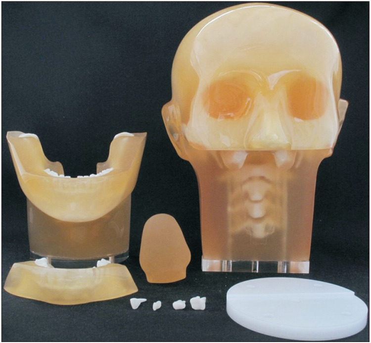
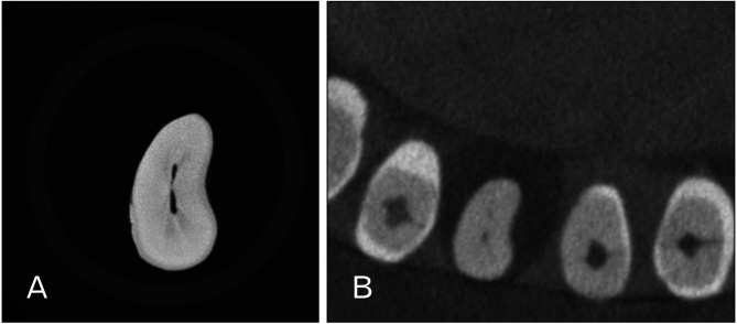
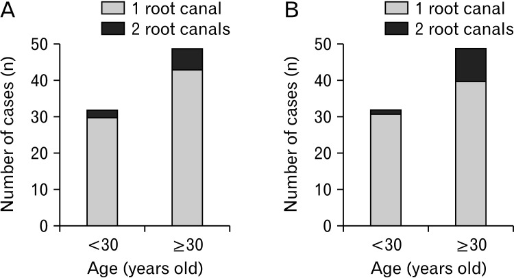
The root canal morphology undergoes aging-related changes, and relevant quantitative analyses have not yet been reported. We compared the cone beam computed tomography (CBCT) and micro-computed tomography (micro-CT) scans of extracted mandibular incisors to check the accuracy of morphological measurements. Thereafter, the root canal morphology and aging-related changes in the mandibular incisors of Japanese individuals were assessed using CBCT. Six extracted teeth were fixed in a phantom head and imaged using CBCT and micro-CT. The correlation between the findings of the two imaging modalities was examined. Further, CBCT reconstructed images of the mandibular incisors of 81 individuals were observed. Age-related changes of the root canals were compared between participants aged <30 years and those aged ≥30 years. The CBCT and micro-CT findings regarding the root canals of the extracted teeth coincided in 94.4% of the cases. Mandibular incisors exhibiting two root canals in either cross-section accounted for 9.9% of central incisors and 12.4% of lateral incisors. Mandibular central incisors with two root canals were observed in two (6.3%) individuals aged <30 years and six (12.2%) aged ≥30 years. Mandibular lateral incisors with two root canals were observed in one (3.1%) individual aged <30 years and nine (18.4%) aged ≥30 years. CBCT allows accurate evaluation of complex root canal morphologies and is useful for endodontic preoperative assessment. Mandibular incisors have more frequent occurrence of two root canals with aging.
Keywords: Accuracy verification; Aging; Cone-beam computed tomography; Incisors; X-ray microtomography.
Conflict of interest statement
No potential conflict of interest relevant to this article was reported.
Figures
Similar articles
-
Detection of various anatomic patterns of root canals in mandibular incisors using digital periapical radiography, 3 cone-beam computed tomographic scanners, and micro-computed tomographic imaging.J Endod. 2014 Jan;40(1):42-5. doi: 10.1016/j.joen.2013.09.039. Epub 2013 Oct 31. J Endod. 2014. PMID: 24331989
-
Ex vivo detection of mandibular incisors' root canal morphology using cone-beam computed tomography with 4 different voxel sizes and micro-computed tomography.BMC Oral Health. 2023 Sep 9;23(1):656. doi: 10.1186/s12903-023-03376-2. BMC Oral Health. 2023. PMID: 37689620 Free PMC article.
-
Cone-beam-computed-tomography of the symmetry of root canal anatomy in mandibular incisors.J Oral Sci. 2020;62(2):180-183. doi: 10.2334/josnusd.19-0113. J Oral Sci. 2020. PMID: 32224571
-
3-dimensional Analysis and Literature Review of the Root Canal Morphology and Physiological Foramen Geometry of 125 Mandibular Incisors by Means of Micro-Computed Tomography in a German Population.J Endod. 2020 Feb;46(2):184-191. doi: 10.1016/j.joen.2019.11.006. Epub 2019 Dec 27. J Endod. 2020. PMID: 31889585 Review.
-
Application of a new system for classifying root and canal anatomy in studies involving micro-computed tomography and cone beam computed tomography: Explanation and elaboration.Int Endod J. 2021 Jul;54(7):1056-1082. doi: 10.1111/iej.13486. Epub 2021 Apr 18. Int Endod J. 2021. PMID: 33527452 Review.
References
LinkOut - more resources
Full Text Sources