Review
. 2023 Feb 6;18(13):e1038-e1065.
doi: 10.4244/EIJ-D-22-00627.

Left atrial appendage occlusion

Affiliations
Review

Left atrial appendage occlusion

David R Holmes Jr et al. EuroIntervention. .

Abstract

Prevention of stroke represents a goal of primary importance in health systems due to its associated morbidity and mortality. As several patient groups with increased stroke rates have been identified, multiple approaches have been developed and implemented: oral anticoagulation (OAC) for patients with atrial fibrillation, surgical and percutaneous revascularisation in patients with carotid disease, device closure for patients with patent foramen ovale, and now, left atrial appendage occlusion (LAAO) for selected patients with non-valvular atrial fibrillation (NVAF). The latter group of patients are the focus of this review which evaluates the pathophysiology, selection of patients, procedural performance, outcomes of treatment both during and post-procedure, adjunctive therapy, complications, and longer-term outcomes.


Conflict of interest statement

D.R. Holmes is a member of the advisory board of Boston Scientific; and reports an institutional research grant from Boston Scientific. K. Korsholm has received speaker’s honoraria from Abbott and Boston Scientific; and has received unrestricted institutional education grants from Abbott and Boston Scientific. J. Rodés-Cabau has an institutional research grant from Boston Scientific. J. Saw received consultation fees from Abbott, Boston Scientific, Baylis, and Gore; and proctored for Abbott and Boston Scientific. S. Berti is a proctor for Abbott, Edwards Lifesciences, and Boston Scientific. M. Alkhouli received consultation fees from Boston Scientific, Abbott, Philips, and Johnson & Johnson.

Figures

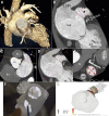
Central illustration
Central illustration. Imaging strategies for LAAO.
Aa-Ae) Procedural planning based on TOE images: multiple views with 2D technique, 3D full volume of LAA with component 2D images. MultiVue of 3D full volume shows the blue plane's alignment with the landing zone and landing zone orifice measurements. B) 3D volume rendering reconstruction of CT scan imaging of successful LAA occlusion with WATCHMAN FLX. C) Procedural planning with CT scan: 3D image volume rendering 3D MPR analysis (1), identification and measurements of the LAA ostium and landing zone (2), (3), (4), (5), (6) and CT-based software simulation of Amulet implantation (7). CT: computed tomography; LAA: left atrial appendage; MPR: multiplanar reconstruction; TOE: transoesophageal echocardiogram
Figure 1
Figure 1. Measurement of the left atrial appendage.
The left atrial appendage (LAA) is formed during the 4th week of embryogenic development. The relationship between the LAA, the left superior pulmonary vein (LSPV) and LA can be seen. A prominent feature is the presence of multiple pectinate muscles between which the atrial myocardium is very thin; thrombi may develop and become the source for an embolic stroke. Important measurements include length (L), width (W) and the ostial dimensions defined both echocardiographically (Oe) and anatomically (Oa). These dimensions and the angulation are important for device selection. (Reproduced with permission from31)
Figure 2
Figure 2. “The most lethal human attachment”.
LAA external view and TOE with thrombus − document specific anatomic characteristics including a thin-walled LAA with the crevasses identified produced by the pectinate muscles (A) and the thrombus which may be present (B). (Reproduced with permission from105). LAA: left atrial appendage; TOE: transoesophageal echocardiogram
Figure 3
Figure 3. Morphology classifications.
A) Modified caricatures of the 4 types: 1. Cactus, 2. Cauliflower, 3. Chicken-wing, and 4. Windsock. B) More detailed subtypes have been identified by CT. Four general shapes have been identified: chicken-wing (a, b), windsock (c,d), cauliflower (e) and cactus (f). In addition, there is a variability in the shape of the ostium ranging from oval (g) to triangular (h), foot-like (i,j), water drop (k), and round (l). The specific frequencies vary. Some specific features have also been associated with increased potential for ischaemic stroke/systemic embolic events. This morphologic information may be helpful in selecting access for LAAO or even for selection of a more optional device configuration. (A) reproduced from Adukauskaite A, et al. ECR 2019 Congress, C-2357, Innsbruck AT; (B) reproduced with permission from. CT: computed tomography; LAAO: left atrial appendage occlusion
Figure 4
Figure 4. Summary of clinical outcomes.
Data continue to accrue on the longer-term outcomes of the patients treated in the first 2 WATCHMAN 2.5 RCT of device versus warfarin and now the PRAGUE-17 with device versus DOAC. Several metrics have been used for comparison. As seen, there is no significant difference between LAAC and OAC in all stroke or systemic embolism. There is a marked reduction in haemorrhagic stroke (RR 0.22), and a significant reduction in CV and all-cause mortality. Non-procedure major bleeding was also significantly decreased. (Adapted with permission from22). CI: confidence interval; CV: cardiovascular; DOAC: direct OAC; LAAC: left atrial appendage closure; OAC: oral anticoagulants; RCT: randomised controlled trial; RR: risk ratio; SE: systemic embolism
Figure 5
Figure 5. Addressing leak and device-related thrombus.
Since the initial WATCHMAN 2.5 device (legacy device), a new device has been introduced and is now the only device available. As seen, there are structural differences in strut frames, with 18 frames in the new generation, and changes to the threaded metal insert. These changes have been associated with a marked improvement in closure rates at 12 months and a decrease in DRT at 12 months in the current FLX device. (Courtesy of Boston Scientific). DRT: device-related thrombus
Figure 6
Figure 6. Amulet device.
The Amulet device has a dual-seal technology with a lobe that fills the left atrial appendage cavity and a disc to seal the ostium.
Figure 7
Figure 7. Mechanism of action (45 days); Primary safety endpoint (12 months).
A) The mechanism of action was assessed as residual leak; differing significantly between the Amulet (63% –no residual leak) and the WATCHMAN 2.5 (46% – no residual leak). B) Safety was assessed using as procedural complications all-cause death or major bleeding at 12 months and was not significantly different. LAA: left atrial appendage
Figure 8
Figure 8. In-hospital adverse event rates in the NCDR-LAAO Registry.
Compared with the pivotal trial results of LAAO, in-hospital adverse rates in this clinically indicated (non-research protocol) document improved outcome. In particular, pericardial effusion and procedure-related stroke were markedly decreased. (Reproduced with permission from24). LAAO: left atrial appendage occlusion; TIA: transient ischaemic attack
Figure 9
Figure 9. TOE imaging documenting a large LAA thrombus.
At the time of preprocedural evaluation, the presence of LAA thrombus represents a contraindication because of the potential for embolisation during catheter placement. LAA: left atrial appendage; TOE: transoesophageal echocardiogram
Figure 10
Figure 10. 2D TOE planning for LAAO.
Four views are evaluated: A) 45°, B) 95°, C) Biplane view, 85°, 175°. In addition, 3D full volume of the LAA with the component 2D images are helpful. Finally, D) 3D full volume images showing alignment of the blue plane allows measurement of the landing zone orifice. E) The measurement box cannot be removed because it is the layout of the machine – the same image in 3D can be seen in the picture on the left – 3D MPR for accurate measurements of LAA diameters, an image without measurements would be incomplete. LAA: left atrial appendage; LAAO: left atrial appendage occlusion; MPR: multiplanar reconstruction; TOE: transoesophageal echocardiogram
Figure 11
Figure 11. CT has become widely used for preprocedural evaluation.
Different views as seen are useful to identify the relationships between the left upper pulmonary vein (LUPV), left atrium (LA), LAA, and mitral valve (MV). The circumflex coronary artery (arrow) is an important landmark. Lines drawn with that from the circumflex to the LUPV identify the anatomical orifice. A) Overall CT image documenting relationship between the pulmonary vein and LAA; B) Landmark can be used to identify the anatomical orifice of the LAA. It is also useful for measuring the ostium of the landing zone; C) Another view documents the relationship between the tip of the LAA and pulmonary artery. It also documents the position of the left atrium in relation to the aorta which is important for avoiding inadvertent puncturing of the aorta. The lines are useful for identifying the orifice of the LAA as well as planning optimal location of the implant; D) The route of the transseptal to the LAA can be identified. This is used to identify optimal position for transseptal puncture; E) A cross-section area of the LAA and its relationship with the circumflex coronary artery (arrow); F) Composite documenting the optimal transseptal location leading to the Watchman implant (see at the top of frame); G) The FEops can be used for preprocedural planning to identify optimal position of the Amulet device. AO: aorta; LAA: left atrial appendage; PA: pulmonary artery; RA: right atrium
Figure 12
Figure 12. MPR analysis of preprocedural CT images for AMPLATZER Amulet implantation.
Multiplanar reconstructions aid to determine the maximum diameter, minimum diameter, and circumference of the LAA landing zone (10-12 mm from the ostium and perpendicular to the LAA wall) as well as the length of the LAA. A) Length of LAA; B) and C) circumference of LAA. CT: computed tomography; LAA: left atrial appendage; MPR: multiplanar reconstruction
Figure 13
Figure 13. CT-based software simulation (FEops) of WATCHMAN FLX implantation.
Computational simulation is based on patient-specific morphology. The geometry has been reconstructed from the CT dataset. The software demonstrates potential opportunities to attempt the prediction of device interaction with LAA anatomy. CT: computed tomography; LAA: left atrial appendage
Figure 14
Figure 14. Step by step ICE-guided LAAC with an Amulet device.
Top panel: A) Angiography showing a chicken-wing LAA anatomy. B–D) Illustration of the ICE positions in the 3 standard views: B) left upper pulmonary vein (LUPV) view, C) mid-LA view, and D) supra-mitral view. Middle panel: E–H) LAA from the LUPV; Amulet lobe in the ball configuration; lobe in the triangular configuration and lobe deployed in the neck of the LAA. Lower panel: I–L) Amulet disc being deployed (American football configuration); disc fully deployed; colour Doppler without signs of peri-device leakage; the Amulet device seen in the supra-mitral view. (Reproduced with permission from45). ICE: intracardiac echocardiography; LA: left atrium; LAA: left atrial appendage; LAAC: left atrial appendage closure
Figure 15
Figure 15. LAA closure guided by 3D intracardiac echo.
A) The location of the ICE probe on fluoroscopy during deployment. B, C) Multiplanar reconstruction and 3D images of the LAA after closure with the WATCHMAN FLX device. (Reproduced with permission from53). ICE: intracardiac echocardiography; LAA: left atrial appendage; LAAO: left atrial appendage occlusion
Figure 16
Figure 16. How are we doing with LAAC?
As seen, the initial LAAO Registry documented that major complications are seen in 2.16% of patients. These rates have decreased since the early trials and registries. Implantation success has continued to improve. (Reproduced with permission from24). LAAC: left atrial appendage closure; LAAO: left atrial appendage occlusion; NCDR: National Cardiovascular Data Registry
Figure 17
Figure 17. Successful LAA occlusion by WATCHMAN FLX seen on a 3-month follow-up CT scan.
A) CT image shows the completely sealed LAA with a correct compression and complete exclusion. B) 3D volume rendering CT imaging of successful LAA occlusion. CT: computed tomography; LAA: left atrial appendage
Figure 18
Figure 18. Peri-device leaks on 3D volume rendering CT images.
LAA patency related to a superior leak (blue curved line) in a patient implanted with an AMPLATZER device. CT: computed tomography; LAA: left atrial appendage
Figure 19
Figure 19. Association of peri-device leak with thromboembolic events after LAAO.
Among >50,000 patients who underwent left atrial appendage occlusion in the United States (2016-2019), 1 in 4 patients had peri-device leak detected on follow-up imaging at 45 days. The majority of leaks were small (<5 mm in diameter). Small leaks, however, were associated with a modest increase in the composite endpoint of stroke, transient ischaemic attack, or systemic embolisation at 1-year follow-up. (Reprinted with permission from92). CI: confidence interval; HR: hazard ratio; LAAO: left atrial appendage occlusion
Figure 20
Figure 20. Device-related thrombus on left atrial apendage device.
Amplatzer amulet left atrial appendage occulsion (LAAO) device (asterisk) secured within the left atrial appendage. Prominent device-related thrombus (DRT) attached to the proximal disc with protrusion into the left atrium. Visualised in two views (A) and (B).
Figure 21
Figure 21. Hypoattenuated thickening in the WATCHMAN FLX and Amplatzer devices.
Analysis of hypoattenuated thickening (HAT) includes several considerations: Localisation: isolated to screw-hub cove (A) or on atrial surface (B,C,D). Extent: smooth continuation onto the left atrium (LA) wall (B), thickness of HAT with <3 mm described as flat. Morphology: homogeneity of the HAT surface (E). Sessile (F) or pedunculated (G). High-grade features: protruding sessile or (H) pedunculated HAT without LA wall continuity or inhomogeneous surface (highlighted with red). (Reproduced with permission from99).

References

    1. Wolf PA, Abbott RD, Kannel WB. Atrial fibrillation: a major contributor to stroke in the elderly. The Framingham Study. Arch Intern Med. 1987;147:1561–4. - PubMed
    1. Noseworthy PA, Kaufman ES, Chen LY, Chung MK, Elkind MSV, Joglar JA, Leal MA, McCabe PJ, Pokorney SD, Yao X American Heart Association Council on Clinical Cardiology Electrocardiography and Arrhythmias Committee; Council on Arteriosclerosis, Thrombosis and Vascular Biology; Council on Cardiovascular and Stroke Nursing; and Stroke Council. Subclinical and Device-Detected Atrial Fibrillation: Pondering the Knowledge Gap: A Scientific Statement From the American Heart Association. Circulation. 2019;140:e944–63. - PMC - PubMed
    1. January CT, Wann LS, Calkins H, Chen LY, Cigarroa JE, Cleveland JC, Ellinor PT, Ezekowitz MD, Field ME, Furie KL, Heidenreich PA, Murray KT, Shea JB, Tracy CM, Yancy CW. 2019 AHA/ACC/HRS Focused Update of the 2014 AHA/ACC/HRS Guideline for the Management of Patients With Atrial Fibrillation: A Report of the American College of Cardiology/American Heart Association Task Force on Clinical Practice Guidelines and the Heart Rhythm Society in Collaboration With the Society of Thoracic Surgeons. Circulation. 2019;140:e125–51. - PubMed
    1. Hindricks G, Potpara T, Dagres N, Arbelo E, Bax JJ, Blomström-Lundqvist C, Boriani G, Castella M, Dan GA, Dilaveris PE, Fauchier L, Filippatos G, Kalman JM, La Meir, Lane DA, Lebeau JP, Lettino M, Lip GYH, Pinto FJ, Thomas GN, Valgimigli M, Van Gelder, Van Putte, Watkins CL ESC Scientific Document Group. 2020 ESC Guidelines for the diagnosis and management of atrial fibrillation developed in collaboration with the European Association for Cardio-Thoracic Surgery (EACTS): The Task Force for the diagnosis and management of atrial fibrillation of the European Society of Cardiology (ESC) Developed with the special contribution of the European Heart Rhythm Association (EHRA) of the ESC. Eur Heart J. 2021;42:373–498. - PubMed
    1. Ruff CT, Giugliano RP, Braunwald E, Hoffman EB, Deenadayalu N, Ezekowitz MD, Camm AJ, Weitz JI, Lewis BS, Parkhomenko A, Yamashita T, Antman EM. Comparison of the efficacy and safety of new oral anticoagulants with warfarin in patients with atrial fibrillation: a meta-analysis of randomised trials. Lancet. 2014;383:955–62. - PubMed

MeSH terms