Review
. 2023 Jan 24:11:1094356.
doi: 10.3389/fcell.2023.1094356. eCollection 2023.

Mitochondrial calcium cycling in neuronal function and neurodegeneration

Affiliations
Review

Mitochondrial calcium cycling in neuronal function and neurodegeneration

Grant C Walters et al. Front Cell Dev Biol. .

Abstract

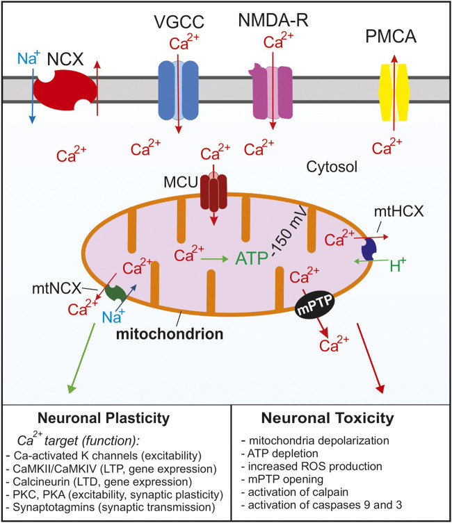
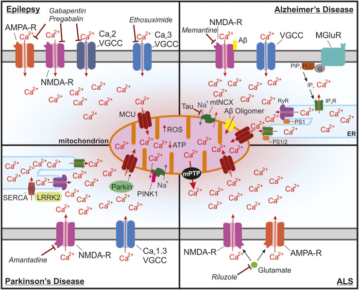
Mitochondria are essential for proper cellular function through their critical roles in ATP synthesis, reactive oxygen species production, calcium (Ca2+) buffering, and apoptotic signaling. In neurons, Ca2+ buffering is particularly important as it helps to shape Ca2+ signals and to regulate numerous Ca2+-dependent functions including neuronal excitability, synaptic transmission, gene expression, and neuronal toxicity. Over the past decade, identification of the mitochondrial Ca2+ uniporter (MCU) and other molecular components of mitochondrial Ca2+ transport has provided insight into the roles that mitochondrial Ca2+ regulation plays in neuronal function in health and disease. In this review, we discuss the many roles of mitochondrial Ca2+ uptake and release mechanisms in normal neuronal function and highlight new insights into the Ca2+-dependent mechanisms that drive mitochondrial dysfunction in neurologic diseases including epilepsy, Alzheimer's disease, Parkinson's disease, and amyotrophic lateral sclerosis. We also consider how targeting Ca2+ uptake and release mechanisms could facilitate the development of novel therapeutic strategies for neurological diseases.

Keywords: MCU; calcium; mitochondria; neurodegeneration; neuronal calcium homeostasis.


Conflict of interest statement

The authors declare that the research was conducted in the absence of any commercial or financial relationships that could be construed as a potential conflict of interest.

Figures

FIGURE 1
FIGURE 1
Summary of the Ca2+ signaling mechanisms in neurons that regulate cytosolic and mitochondrial Ca2+ concentrations. Ca2+ enters the cell through voltage gated Ca2+ channels (VGCC) following depolarization or through glutamate NMDA receptors. As the cytosolic Ca2+ concentration increases, the MCU complex opens and Ca2+ enters the mitochondria. Ca2+ is rapidly removed from the cytosol via Na+/Ca2+ exchangers (NCX) and plasma membrane Ca2+ ATPases (PMCA). As the cytosolic Ca2+ concentration decreases Ca2+ is slowly released from the mitochondria through Na+/Ca2+ (mtNCX) exchangers and H+/Ca2+ exchangers (mtHCX). Proper Ca2+ buffering is critical for normal neuronal functions and neuronal plasticity. Impaired buffering and Ca2+ overload leads to the opening of the mPTP and activation of apoptotic pathways and is implicated in many neurological diseases. For simplicity, the ER and other Ca2+ organelles are omitted from the model.
FIGURE 2
FIGURE 2
The structure of the greater mitochondrial Ca2+ uniporter (MCU) complex and its subunits that can regulate its permeability. (A) The assembled complex containing a tetramer of pore forming MCU and four subunits of the essential MCU regulator (EMRE), which are critical for the proper assembly of the transmembrane MCU to the mitochondrial Ca2+ uptake isoforms 1, 2, and 3 (MICU 1–3). The MCU complex also contains the MCU regulator 1 (MCUR1), which is thought to bind to EMRE. (B) The MCU paralog MCUb has been shown to replace subunits of pore forming MCU, which leads to reduced Ca2+ through the channel and as more MCUb subunits replace MCU the Ca2+ conductivity is reduced. (C) MICU1 and MICU two are critical for the Ca2+ sensing function of the channel and MICU2 has been shown to regulate Ca2+ sensitivity. When Ca2+ is low, MICU1 blocks the pore, but in the presence of high Ca2+ the conformational changes in MICU1 and MICU2 result in opening of the channel and allow Ca2+ to enter the mitochondria. (D) In neurons, MICU3 is more abundant and has been shown to replace MICU2 in dimers with MICU1. This replacement leads to increeased sensitivity to cytosolic Ca2+ of the MCU complex and results in MCU channel opening to lower concentrations of Ca2+ than compared to MICU2.
FIGURE 3
FIGURE 3
Mitochondrial Ca2+ overload leading to activation of the mPTP is a common pathway in many neurological diseases. (A) In epilepsy, seizures lead to buildup of cytosolic and mitochondrial Ca2+ through NMDA receptors, Ca2+ permeable AMPA receptors and Cav2 and Cav3 voltage-gated Ca2+ channels (VGCCs) in neurons that are in a state of hyperexcitability. Long seizures or repeated seizures can lead to Ca2+ overload in the mitochondria and trigger activation of the mPTP, ultimately leading to neuronal toxicity. Commonly prescribed antiepileptic drugs target these channels including: ethosuximide, which is a Cav3 VGCC inhibitor, as well as gabapentin and pregabalin, which act by binding to the with α2δ-1 subunit of voltage-gated Ca2+ channels, which could disrupt plasma membrane expression and function of Cav2 VGCCs, NMDA receptors, and Ca2+ permeable AMPA receptors. (B) In Alzheimer’s disease, Ca2+ overload in the mitochondria occurs through multiple mechanisms. Ca2+ entry into the cytosol can be enhanced by binding of Aβ to NMDA receptors. Internal release of Ca2+ from the ER through activation of metabotropic glutamate receptors (mGluR) also increases the accumulation of Ca2+ into the mitochondria. Enhanced Ca2+ extrusion from the ER has also been shown to occur through interactions between presenilin 1/2 (PS1/2) and inositol trisphosphate receptors (IP3R) and ryanodine receptors (RyR). This interaction seems to occur at mitochondria associated membranes (MAMs), which due to the proximity to the mitochondria, drives Ca2+ uptake into the mitochondria. Aβ oligomers also can form a pore in the mitochondria that can lead to increases in Ca2+ in the mitochondria. Ca2+ extrusion through the mitochondrial Na+/Ca2+ exchanger (mtNCX) has been shown to be impaired by tau accumulation leading to excessive Ca2+ accumulation in the mitochondria. Memantine, one of the FDA approved drugs for AD, inhibits NMDA receptors, which could block excessive Ca2+ uptake and to reduce glutamate induced excitotoxicity. (C) In Parkinson’s disease (PD), substantia nigra dopaminergic neurons are at risk of Ca2+ overload due to the presence of voltage-gated Cav1.3 channels, which are critical for the pacemaking activity of these neurons. One common form of autosomal recessive familial PD occurs through mutations in PARK2, the gene for Parkin. Parkin is responsible for regulating mitochondrial Ca2+ uptake isoform 1 (MICU1) degradation and may lead to disrupted MCU assembly altering mitochondrial Ca2+ uptake. Another form of autosomal recessive PD is caused by mutations in PINK1, which can impair mtNCX function and increase Ca2+ accumulation into the mitochondria. In astrocytes, ER Ca2+ release at MAMs may be enhanced in PD due to mutations in leucine-rich repeat kinase 2 (LRRK2), which enhance Ca2+ uptake into the ER through activation of sarcoplasmic reticulum Ca2+ -ATPase (SERCA). The NMDA receptor antagonist amantadine is used to prevent dyskinesia in PD patients. (D) In amyotrophic lateral sclerosis (ALS), motor neurons of the spinal cord and brain begin to degenerate. These neurons have been shown to have increased Ca2+ uptake through glutamate NMDA and Ca2+ permeable AMPA receptors, which enhances their susceptibility to mitochondrial Ca2+ overload. Excitotoxic glutamate, which signals through NMDA and GluA2-lacking Ca2+ permeable AMPA receptors, is the target of Riluzole, one of the few FDA approved drugs for ALS.

References

    1. Akundi R. S., Huang Z., Eason J., Pandya J. D., Zhi L., Cass W. A., et al. (2011). Increased mitochondrial calcium sensitivity and abnormal expression of innate immunity genes precede dopaminergic defects in Pink1-deficient mice. PLoS One 6 (1), e16038. 10.1371/journal.pone.0016038 - DOI - PMC - PubMed
    1. Alavian K. N., Beutner G., Lazrove E., Sacchetti S., Park H.-A., Licznerski P., et al. (2014). An uncoupling channel within the c-subunit ring of the F1FO ATP synthase is the mitochondrial permeability transition pore. Proc. Natl. Acad. Sci. 111(29), 10580–10585. 10.1073/pnas.1401591111 - DOI - PMC - PubMed
    1. Alzheimer's and Dementia (2020a). Alzheimer's disease facts and figures. Alzheimer's Dementia 16 (3), 391–460. 10.1002/alz.12068 - DOI
    1. Angelova P. R., Abramov A. Y. (2018). Role of mitochondrial ROS in the brain: From physiology to neurodegeneration. FEBS Lett. 592 (5), 692–702. 10.1002/1873-3468.12964 - DOI - PubMed
    1. Appel S. H., Beers D., Siklos L., Engelhardt J. I., Mosier D. R. (2001). Calcium: The darth vader of ALS. Amyotroph. Lateral Scler. Other Mot. Neuron Disord. 2 (1), 47–54. 10.1080/146608201300079418 - DOI - PubMed