Case Reports
. 2023 Jan 25:10:1100425.
doi: 10.3389/fcvm.2023.1100425. eCollection 2023.

Case report: Mechanical-electric feedback and atrial fibrillation-Revelation from the treatment of a rare atrial fibrillation caused by annular constrictive pericarditis

Affiliations
Case Reports

Case report: Mechanical-electric feedback and atrial fibrillation-Revelation from the treatment of a rare atrial fibrillation caused by annular constrictive pericarditis

Dong Yi et al. Front Cardiovasc Med. .

Abstract

Atrial fibrillation (AF) is one of the most common arrhythmias encountered in clinical practice. The pathophysiological mechanisms responsible for its development are complex, vary amongst individuals, and associated with predisposing factors. Here, we report a case of AF caused by annular constrictive pericarditis (ACP), which is extremely rare due to its unusual anatomical form. In our patient, AF was refractory to multiple antiarrhythmic medications; however, spontaneous conversion to sinus rhythm occurred when the ring encircling the right and left ventricular (RV and LV) cavities along the atrioventricular (AV) groove was severed. This suggests that atrial stretch due to atrial enlargement and increased left atrial (LA) pressure may contribute to the initiation and maintenance of AF. This report highlights the importance of the careful investigation of rare predisposing factors for AF using non-invasive diagnostic approaches and mechanical-electric feedback (MEF) as a pathophysiological mechanism for AF initiation and maintenance.

Keywords: annular constrictive pericarditis; atrial fibrillation; atrioventricular groove; mechanical-electric feedback; predisposing factors.


Conflict of interest statement

The authors declare that the research was conducted in the absence of any commercial or financial relationships that could be construed as a potential conflict of interest.

Figures

FIGURE 1
FIGURE 1
Physical examination revealed pitting edema of the lower extremities and abdominal distension (A). Chest radiology showed a pleural effusion (B). Electrocardiography (ECG) at admission indicated the diagnosis of atrial fibrillation (AF) [heart rate (HR), 134 bpm] with low voltage in all leads (C). Echocardiography showed left (D) and RA enlargement (E), and a normal LV chamber. Cardiac computed tomographic angiography of the pulmonary arteries excluded pulmonary embolism (F). LV, left ventricle; RV, right ventricle; LA, left atrium; RA, right atrium; AAO, ascending aortic artery; MPA, main pulmonary artery; DAO, descending aortic artery.
FIGURE 2
FIGURE 2
Tissue Doppler imaging (TDI) using echocardiography revealed that e’ = 18.4 m/s (medial) > 14.4 m/s (lateral) in the mitral valve (A,B). Significant respiratory variation of mitral or tricuspid peak E velocity was indeterminable due to atrial fibrillation (AF) (C). Paradoxical septal motion during respiration was not observed (D). The inferior vena cava (IVC) was dilated without inspiratory collapse (E). Pulmonary artery pressure was elevated according to the velocity of tricuspid regurgitation (F).
FIGURE 3
FIGURE 3
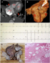
Cardiac computed tomographic angiogram of four-chamber view (A) and volume rendering views (B) showed calcified annular constrictive pericarditis (ACP) trapping both ventricles (red arrows). Spontaneous conversion from atrial fibrillation (AF) to sinus rhythm as the calcified pericardial ring was severed (C). Complete pericardiectomy was performed and revealed a thickened pericardium with calcification embedded in the atrioventricular (AV) groove (D). Pathological examination of the pericardial tissue indicated fibrotic tissue with calcification (E).

Similar articles

References

    1. Andrade J, Khairy P, Dobrev D, Nattel S. The clinical profile and pathophysiology of atrial fibrillation: relationships among clinical features, epidemiology, and mechanisms. Circ Res. (2014) 114:1453–68. 10.1161/CIRCRESAHA.114.303211 - DOI - PubMed
    1. Feinberg W, Blackshear J, Laupacis A, Kronmal R, Hart R. Prevalence, age distribution, and gender of patients with atrial fibrillation. Analysis and implications. Arch Intern Med. (1995) 155:469–73. 10.1001/archinte.155.5.469 - DOI - PubMed
    1. Peters N, Schilling R, Kangaratnam P, Markides V. Atrial fibrillation: strategies to control, combat, and cure. Lancet. (2002) 359:593–603. 10.1016/S0140-6736(02)07748-6 - DOI - PubMed
    1. Chugh S, Havmoeller R, Narayanan K, Narayanan K, Singh D, Rienstra M, et al. Worldwide epidemiology of atrial fibrillation: a global burden of disease 2010 study. Circulation. (2014) 129:837–47. 10.1161/CIRCULATIONAHA.113.005119 - DOI - PMC - PubMed
    1. Kannel W, Wolf P, Benjamin E, Levy D. Prevalence, incidence, prognosis, and predisposing conditions for atrial fibrillation: population-based estimates. Am J Cardiol. (1998) 82:2N–9N. 10.1016/S0002-9149(98)00583-9 - DOI - PubMed

Publication types

LinkOut - more resources