LncRNA MALAT1 mediates osteogenic differentiation in osteoporosis by regulating the miR-485-5p/WNT7B axis
- PMID: 36760811
- PMCID: PMC9904362
- DOI: 10.3389/fendo.2022.922560
LncRNA MALAT1 mediates osteogenic differentiation in osteoporosis by regulating the miR-485-5p/WNT7B axis
Abstract
Introduction: Accumulating evidence demonstrates that long non-coding RNAs (lncRNAs) are associated with the development of osteoporosis.
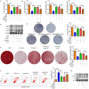
Methods: This study aimed to investigate the effects of MALAT1 on osteogenic differentiation and cell apoptosis in osteoporosis. MALAT1 level, detected by RT-qPCR, was downregulated in hindlimb unloading (HU) mice and simulated microgravity (MG)-treated MC3T3-E1 cells. Moreover, osteogenic differentiation-related factor (Bmp4, Col1a1, and Spp1) levels were measured by RT-qPCR and Western blot. ALP activity was detected, and ALP staining was performed. Cell apoptosis was assessed by flow cytometry.
Results: The results revealed that MALAT1 upregulated the expression of Bmp4, Col1a1, and Spp1, and enhanced ALP activity. Knockdown of MALAT1 suppressed their expression and ALP activity, suggesting that MALAT1 promoted osteogenic differentiation. Additionally, MALAT1 inhibited apoptosis, increased Bax and caspase-3 levels, and decreased Bcl-2 level. However, knockdown of MALAT1 had opposite results. In MG cells, MALAT1 facilitated osteogenic differentiation and suppressed apoptosis. Furthermore, miR-485-5p was identified as a target of MALAT1, and WNT7B was verified as a target of miR-485-5p. Overexpression of miR-485-5p rescued the promotion of osteogenic differentiation and the inhibition of apoptosis induced by MALAT1. Knockdown of WNT7B abolished the facilitation of osteogenic differentiation and the suppression of apoptosis induced by downregulation of miR-485-5p.
Discussion: In conclusion, MALAT1 promoted osteogenic differentiation and inhibited cell apoptosis through the miR-485-5p/WNT7B axis, which suggested that MALAT1 is a potential target to alleviate osteoporosis.
Keywords: MALAT1; MiR-485-5p; WNT7B; osteogenesis differentiation; osteoporosis.
Copyright © 2023 Zhou, Xu, Wang, Song and Yin.
Conflict of interest statement
The authors declare that the research was conducted in the absence of any commercial or financial relationships that could be construed as a potential conflict of interest.
Figures


Similar articles
-
lncRNA MALAT1 promotes osteogenic differentiation through the miR-217/AKT3 axis: A possible strategy to alleviate osteoporosis.J Gene Med. 2022 Jun;24(6):e3409. doi: 10.1002/jgm.3409. Epub 2022 May 6. J Gene Med. 2022. PMID: 35030644
-
LncRNA MALAT1 promotes osteogenic differentiation through the miR-93-5p/SMAD5 axis.Oral Dis. 2024 May;30(4):2398-2409. doi: 10.1111/odi.14705. Epub 2023 Aug 3. Oral Dis. 2024. PMID: 37533355
-
LncRNA metastasis-associated lung adenocarcinoma transcript-1 promotes osteogenic differentiation of bone marrow stem cells and inhibits osteoclastic differentiation of Mø in osteoporosis via the miR-124-3p/IGF2BP1/Wnt/β-catenin axis.J Tissue Eng Regen Med. 2022 Mar;16(3):311-329. doi: 10.1002/term.3279. Epub 2022 Jan 11. J Tissue Eng Regen Med. 2022. PMID: 34962086
-
LncRNA MALAT1 promotes osteoarthritis by modulating miR-150-5p/AKT3 axis.Cell Biosci. 2019 Jul 1;9:54. doi: 10.1186/s13578-019-0302-2. eCollection 2019. Cell Biosci. 2019. PMID: 31304004 Free PMC article. Review.
-
A potential therapeutic drug for osteoporosis: prospect for osteogenic LncRNAs.Front Endocrinol (Lausanne). 2023 Aug 3;14:1219433. doi: 10.3389/fendo.2023.1219433. eCollection 2023. Front Endocrinol (Lausanne). 2023. PMID: 37600711 Free PMC article. Review.
Cited by
-
Noncoding RNAs: the crucial role of programmed cell death in osteoporosis.Front Cell Dev Biol. 2024 May 10;12:1409662. doi: 10.3389/fcell.2024.1409662. eCollection 2024. Front Cell Dev Biol. 2024. PMID: 38799506 Free PMC article. Review.
-
CircZfp644-205 inhibits osteoblast differentiation and induces apoptosis of pre-osteoblasts via sponging miR-455-3p and promoting SMAD2 expression.Eur J Med Res. 2024 Jun 8;29(1):315. doi: 10.1186/s40001-024-01903-7. Eur J Med Res. 2024. PMID: 38849933 Free PMC article.
-
LINC00941 affects the proliferation, apoptosis and differentiation of osteoblasts by regulating the miR-335-5p/KAT7 axis.J Orthop Surg Res. 2025 Jan 21;20(1):75. doi: 10.1186/s13018-025-05469-w. J Orthop Surg Res. 2025. PMID: 39838460 Free PMC article.
-
A New Tissue Engineering Strategy to Promote Tendon-bone Healing: Regulation of Osteogenic and Chondrogenic Differentiation of Tendon-derived Stem Cells.Orthop Surg. 2024 Oct;16(10):2311-2325. doi: 10.1111/os.14152. Epub 2024 Jul 23. Orthop Surg. 2024. PMID: 39043618 Free PMC article. Review.
-
Long noncoding RNA MALAT1 mediates fibrous topography-driven pathologic calcification through trans-differentiation of myoblasts.Mater Today Bio. 2024 Aug 8;28:101182. doi: 10.1016/j.mtbio.2024.101182. eCollection 2024 Oct. Mater Today Bio. 2024. PMID: 39205874 Free PMC article.
References
Publication types
MeSH terms
Substances
LinkOut - more resources
Full Text Sources
Medical
Research Materials
Miscellaneous

