. 2023 Jan 9;25(2):92.
doi: 10.3892/etm.2023.11790. eCollection 2023 Feb.

Inhibition of human oral squamous cell carcinoma proliferation and migration by prodrug-activating suicide gene therapies

Affiliations

Inhibition of human oral squamous cell carcinoma proliferation and migration by prodrug-activating suicide gene therapies

Naining Xu et al. Exp Ther Med. .

Erratum in

Abstract

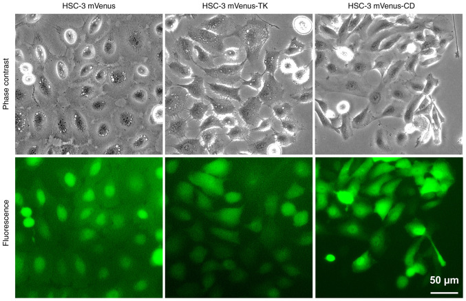
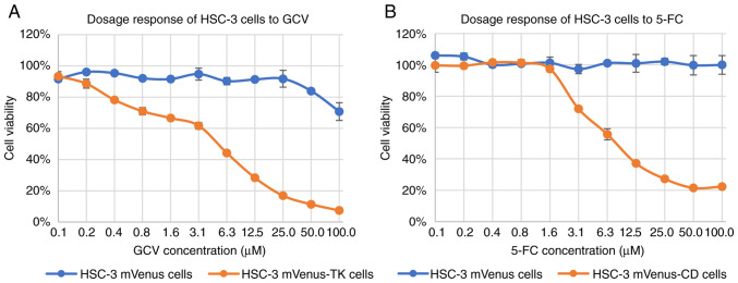
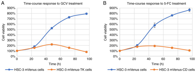
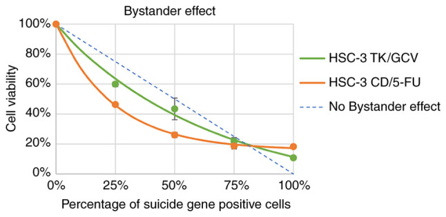
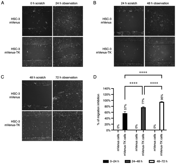
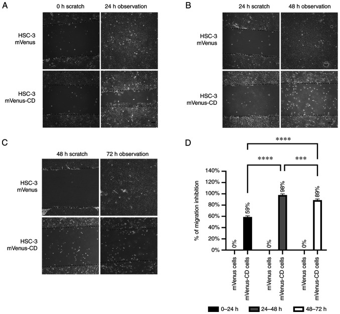
Head and neck squamous cell carcinoma (HNSCC), which originates from mucosal epithelium in the oral cavity, pharynx and larynx, is the sixth most common malignancy in the world. The prognosis of HNSCC is not satisfactory due to metastasis, resulting in 5-year survival rates ranging from 65.9 to 67.2%. Previously, we developed a method to evaluate the effect prodrug-activating suicide gene (PA-SG) therapy on the proliferation of HNSCC. The present study investigated PA-SG therapy on metastatic HNSCC by wound-healing assay and our previously established method. HSC-3 cells with stable expression of suicide genes thymidine kinase (TK) or cytosine deaminase (CD) were treated with prodrugs ganciclovir (GCV) or 5-fluorocytosine (5-FC), respectively. Both GCV and 5-FC inhibited HSC-3 proliferation while the bystander effect of CD/5-FC was greater compared with that of TK/GCV. GCV showed a greater anti-migration effect compared with that of 5-FC. To the best of our knowledge, the present study is the first to evaluate the anti-migratory and anti-proliferative effects of PA-SG therapies on metastatic HNSCC. This may also serve as a general method to quantify other types of PA-SC therapy. The present results demonstrated that PA-SG therapy is a promising treatment for anti-metastatic HNSCC therapy development.

Keywords: cytosine deaminase and 5-fluorocytosine; head and neck squamous cell carcinoma; metastasis; prodrug; suicide gene therapy; thymidine kinase and ganciclovir.


Conflict of interest statement

The authors declare that they have no competing interests.

Figures

Figure 1
Figure 1
HSC-3 stable cell lines expressing mVenus, mVenus-CD or mVenus-TK. Representative microscopy images of HSC-3 cells stably expressing mVenus, mVenus-CD or mVenus-TK. Scale bar, 50 µm. CD, cytosine deaminase; TK, thymidine kinase.
Figure 2
Figure 2
Dose-response of HSC-3 cells to GCV or 5-FC treatment. (A) Dose-response curve of HSC-3 cells treated with GCV. HSC-3 mVenus or HSC-3 mVenus-TK cells were treated with 2-fold dilutions of GCV ranging from 0.1 to 100.0 µM for three days. (B) Dose-response curve of HSC-3 cells treated with 5-FC. HSC-3 mVenus or HSC-3 mVenus-CD cells were treated with 2-fold dilutions of 5-FC ranging from 0.01 to 100.0 µM for three days. Cell viability was measured and normalized to untreated cells. Data are presented as mean ± SD of three experiments. CD, cytosine deaminase; TK, thymidine kinase; GCV, ganciclovir; 5-FC, 5-fluorocytosine; SD, standard deviation.
Figure 3
Figure 3
Time-course response of HSC-3 cells to GCV or 5-FC . (A) Time-course response of HSC-3 mVenus and HSC-3 mVenus-TK cells to 25 µM of GCV. (B) Time-course response of HSC-3 mVenus cells and HSC-3 mVenus-CD cells to 100 µM 5-FC. Cell viability was measured via MTT assay every 24 h. Cell viability was normalized to the MTT reading at 0 h. Data are presented as the mean ± SD of three experiments. CD, cytosine deaminase; TK, thymidine kinase; GCV, ganciclovir; 5-FC, 5-fluorocytosine; SD, standard deviation.
Figure 4
Figure 4
Bystander effects of GCV and 5-FC on HSC-3 cells. HSC-3 mVenus-TK or HSC-3 mVenus-CD cells were mixed with HSC-3 mVenus cells to a final percentage of suicide gene positive of 0, 25, 50, 75 and 100%. The cell mixtures were treated with 25 µM GCV or 100 µM 5-FC for 72 h. Cell viability was measured via MTT and normalized to the MTT reading of HSC-3 mVenus cells. Cell viability was plotted against the percentage of suicide gene-positive cells. Data are represented as the mean ± standard deviation of three experiments. CD, cytosine deaminase; TK, thymidine kinase; GCV, ganciclovir; 5-FC, 5-fluorocytosine.
Figure 5
Figure 5
Inhibition of HSC-3 mVenus-TK cell migration following treatment with GCV. (A) GCV treatment inhibited HSC-3 mVenus-TK but not HSC-3 mVenus cell migration during the 0-24 h period. Following generation of the wound, scratch images were captured and cells were treated with 25 µM GCV. The healing images were captured after a further 24 h GCV treatment (scale bar, 100 µm). (B) GCV treatment inhibited HSC-3 mVenus-TK cell migration during the 24-48 h period. HSC-3 mVenus and HSC-3 mVenus-TK cells were treated with 25 µM GCV for 24 h and wounds were generated. After taking the scratch images, cells were treated with 25 µM GCV for a further 24 h, then the healing images were captured (scale bar, 100 µm). (C) GCV treatment inhibited HSC-3 mVenus-TK cell migration during the 48-72 h period. The wounds were generated after cells were treated with 25 µM GCV for 48 h. The cells were treated for a further 24 h, then healing images were captured. Scale bar, 100 µm. (D) Quantification of inhibition of cell migration for treatment durations. The percentage of migration inhibition is the ratio of the area of healing images to the average strip area of the scratch images. Data are presented as the mean ± SD of >3 healing images. Data were analysed using one-way ANOVA followed by Tukey's post hoc test for multiple comparisons. ****P<0.0001. CD, cytosine deaminase; TK, thymidine kinase; GCV, ganciclovir; SD, standard deviation.
Figure 6
Figure 6
Inhibition of HSC-3 mVenus-CD cell migration by 5-FU. (A) 5-FU treatment inhibited HSC-3 mVenus-CD but not HSC-3 mVenus cell migration during the 0-24 h period. Following wound generation, scratch images were captured and cells were treated with 100 µM 5-FC. The healing images were captured after 5-FC treatment for 24 h. Scale bar, 100 µm. (B) 5-FU treatment inhibited HSC-3 mVenus-CD cell migration during the 24-48 h period. HSC-3 mVenus and HSC-3 mVenus-CD cells were treated with 100 µM 5-FC for 24 h and wounds were generated. After taking the scratch images, cells were continuously treated with 100 µM 5-FC for another 24 h. The healing images were captured at 48 h. Scale bar, 100 µm. (C) 5-FU treatment inhibited HSC-3 mVenus-CD cell migration during the 48-72 h period. The wounds were generated after cells were treated with 100 µM 5-FU for 48 h. The healing images were captured at 72 h. Scale bar, 100 µm. (D) Quantification of inhibition of cell migration of the three periods. The percentage of migration inhibition is the ratio of the area of healing images to mean area of the scratch images. Data are presented as the mean ± SD of >3 healing images. Data were analysed using one-way ANOVA followed by Tukey's post hoc test for multiple comparisons. ***P<0.001 and ****P<0.0001. CD, cytosine deaminase; 5-FC, 5-fluorocytosine; SD, standard deviation.

Similar articles

References

    1. Sung H, Ferlay J, Siegel RL, Laversanne M, Soerjomataram I, Jemal A, Bray F. Global cancer statistics 2020: GLOBOCAN estimates of incidence and mortality worldwide for 36 cancers in 185 countries. CA Cancer J Clin. 2021;71:209–249. doi: 10.3322/caac.21660. - DOI - PubMed
    1. Cramer JD, Burtness B, Le QT, Ferris RL. The changing therapeutic landscape of head and neck cancer. Nat Rev Clin Oncol. 2019;16:669–683. doi: 10.1038/s41571-019-0227-z. - DOI - PubMed
    1. Pulte D, Brenner H. Changes in survival in head and neck cancers in the late 20th and early 21st century: A period analysis. Oncologist. 2010;15:994–1001. doi: 10.1634/theoncologist.2009-0289. - DOI - PMC - PubMed
    1. Gonzalez-Garcia R, Naval-Gias L, Roman-Romero L, Sastre-Perez J, Rodriguez-Campo FJ. Local recurrences and second primary tumors from squamous cell carcinoma of the oral cavity: A retrospective analytic study of 500 patients. Head Neck. 2009;31:1168–1180. doi: 10.1002/hed.21088. - DOI - PubMed
    1. Pisani P, Airoldi M, Allais A, Valletti PA, Battista M, Benazzo M, Briatore R, Cacciola S, Cocuzza S, Colombo A, et al. Metastatic disease in head & neck oncology. Acta Otorhinolaryngol Ital. 2020;40:S1–S86. doi: 10.14639/0392-100X-suppl.1-40-2020. - DOI - PMC - PubMed