

Clinical and pathological studies of eight cases of lipidized fibrous histiocytoma
- PMID: 36761009
- PMCID: PMC9905647
- DOI: 10.3892/etm.2023.11792
Clinical and pathological studies of eight cases of lipidized fibrous histiocytoma
Abstract
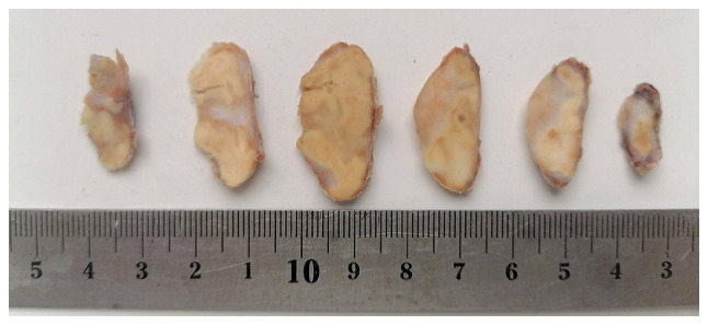
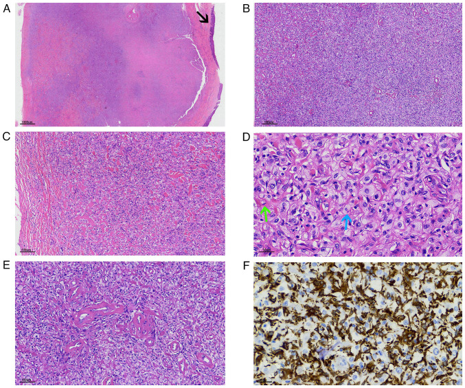
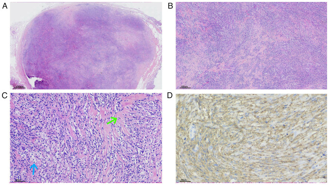
Lipidized fibrous histiocytoma (FH) is a rare type of FH. The present study aimed to describe the clinical and pathological features of lipidized FH. A total of eight patients diagnosed with lipidized FH were retrospectively reviewed in the present study. The cohort included three male and five female patients (male to female ratio, 1.7:1) with a mean age of 48 years (range, 38-62 years). In total, four tumors were located on the buttock, three on the lower leg and one on the forearm. Histological, lipidized FH showed a wide spectrum. Some cases included prominent stromal hyalinization and hyalinized vessels with scant lipid-laden histiocytes. Other cases exhibited the prominent lipid-laden histiocytes and scant stromal hyalinization. Overall, lipidized FH must be differentiated from other benign and malignant tumors, taking into account the therapeutic and prognostic differences between these different entities.
Keywords: dermatofibroma; fibrous histiocytoma; lipidized fibrous histiocytoma.
Copyright: © Liu et al.
Conflict of interest statement
The authors declare that they have no competing interests.
Figures

Similar articles
-
A Rare Case of Lipidized Fibrous Histiocytoma of the Oral Cavity.Cureus. 2025 Apr 14;17(4):e82265. doi: 10.7759/cureus.82265. eCollection 2025 Apr. Cureus. 2025. PMID: 40376352 Free PMC article.
-
Lipidized fibrous histiocytoma: clinicopathologic analysis of 22 cases.Am J Dermatopathol. 2000 Apr;22(2):126-34. doi: 10.1097/00000372-200004000-00007. Am J Dermatopathol. 2000. PMID: 10770432
-
A pruritic pedunculated pink nodule on the leg.Dermatol Online J. 2023 Apr 15;29(2). doi: 10.5070/D329260776. Dermatol Online J. 2023. PMID: 37220292
-
Conjunctival and Corneal Fibrous Histiocytoma: Brief review.Semin Ophthalmol. 2021 Oct 3;36(7):569-572. doi: 10.1080/08820538.2021.1900286. Epub 2021 Mar 25. Semin Ophthalmol. 2021. PMID: 33764259 Review.
-
[Fibrohistiocytic skin tumors].J Dtsch Dermatol Ges. 2006 Jul;4(7):544-55. doi: 10.1111/j.1610-0387.2006.06021.x. J Dtsch Dermatol Ges. 2006. PMID: 16827912 Review. German.
Cited by
-
Lipidized fibrous histiocytoma of the tongue.J Dent Sci. 2024 Oct;19(4):2461-2463. doi: 10.1016/j.jds.2024.06.008. Epub 2024 Jun 15. J Dent Sci. 2024. PMID: 39347091 Free PMC article. No abstract available.
-
A case of a giant lipidized dermatofibroma.JAAD Case Rep. 2025 Mar 22;61:116-118. doi: 10.1016/j.jdcr.2025.02.041. eCollection 2025 Jul. JAAD Case Rep. 2025. PMID: 40538790 Free PMC article. No abstract available.
References
LinkOut - more resources
Full Text Sources
Miscellaneous