Selective CD28 blockade impacts T cell differentiation during homeostatic reconstitution following lymphodepletion
- PMID: 36761170
- PMCID: PMC9904166
- DOI: 10.3389/fimmu.2022.1081163
Selective CD28 blockade impacts T cell differentiation during homeostatic reconstitution following lymphodepletion
Abstract
Introduction: Costimulation blockade targeting the CD28 pathway provides improved long-term renal allograft survival compared to calcineurin inhibitors but may be limited as CTLA-4-Ig (abatacept, belatacept) blocks both CD28 costimulation and CTLA-4 coinhibition. Directly targeting CD28 while leaving CTLA-4 intact may provide a mechanistic advantage. Fc-silent non-crosslinking CD28 antagonizing domain antibodies (dAb) are currently in clinical trials for renal transplantation. Given the current standard of care in renal transplantation at most US centers, it is likely that lymphodepletion via thymoglobulin induction therapy could be used in patients treated with CD28 antagonists. Thus, we investigated the impact of T cell depletion (TCD) on T cell phenotype following homeostatic reconstitution in a murine model of skin transplantation treated with anti-CD28dAb.
Methods: Skin from BALB/cJ donors was grafted onto C56BL/6 recipients which were treated with or without 0.2mg anti-CD4 and 10μg anti-CD8 one day prior to transplant and with or without 100μg anti-CD28dAb on days 0, 2, 4, 6, and weekly thereafter. Mice were euthanized six weeks post-transplant and lymphoid cells were analyzed by flow cytometry.
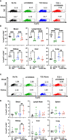
Results: Anti-CD28dAb reversed lymphopenia-induced differentiation of memory CD4+ T cells in the spleen and lymph node compared to TCD alone. Mice treated with TCD+anti-CD28dAb exhibited significantly improved skin graft survival compared to anti-CD28dAb alone, which was also improved compared to no treatment. In addition, the expression of CD69 was reduced on CD4+ and CD8+ T cells in the spleen and lymph node from mice that received TCD+anti-CD28dAb compared to TCD alone. While a reduced frequency of CD4+FoxP3+ T cells was observed in anti-CD28dAb treated mice relative to untreated controls, this was balanced by an increased frequency of CD8+Foxp3+ T cells that was observed in the blood and kidney of mice given TCD+anti-CD28dAb compared to TCD alone.
Discussion: These data demonstrate that CD28 signaling impacts the differentiation of both CD4+ and CD8+ T cells during homeostatic reconstitution following lymphodepletion, resulting in a shift towards fewer activated memory T cells and more CD8+FoxP3+ T cells, a profile that may underpin the observed prolongation in allograft survival.
Keywords: T cell depletion; alloimmunity; costimulation blockade; homeostatic reconstitution; lymphodepletion; transplantation.
Copyright © 2023 Habib, Liu, Crepeau, Wagener and Ford.
Conflict of interest statement
MF has received honoraria from Veloxis Pharmaceuticals. The remaining authors declare that the research was conducted in the absence of any commercial or financial relationships that could be constructed as a potential conflict of interest.
Figures


Similar articles
-
Impact of selective CD28 blockade on virus-specific immunity to a murine Epstein-Barr virus homolog.Am J Transplant. 2019 Aug;19(8):2199-2209. doi: 10.1111/ajt.15321. Epub 2019 Mar 29. Am J Transplant. 2019. PMID: 30801917 Free PMC article.
-
Aggressive skin allograft rejection in CD28-/- mice independent of the CD40/CD40L costimulatory pathway.Transpl Immunol. 2001 Oct;9(1):13-7. doi: 10.1016/s0966-3274(01)00043-0. Transpl Immunol. 2001. PMID: 11680567
-
Selective CD28 blockade attenuates CTLA-4-dependent CD8+ memory T cell effector function and prolongs graft survival.JCI Insight. 2018 Jan 11;3(1):e96378. doi: 10.1172/jci.insight.96378. eCollection 2018 Jan 11. JCI Insight. 2018. PMID: 29321374 Free PMC article.
-
Challenges and opportunities in targeting the CD28/CTLA-4 pathway in transplantation and autoimmunity.Expert Opin Biol Ther. 2017 Aug;17(8):1001-1012. doi: 10.1080/14712598.2017.1333595. Epub 2017 May 30. Expert Opin Biol Ther. 2017. PMID: 28525959 Free PMC article. Review.
-
Selective Costimulation Blockade With Antagonist Anti-CD28 Therapeutics in Transplantation.Transplantation. 2019 Sep;103(9):1783-1789. doi: 10.1097/TP.0000000000002740. Transplantation. 2019. PMID: 30951014 Review.
References
Publication types
MeSH terms
Substances
Grants and funding
LinkOut - more resources
Full Text Sources
Medical
Research Materials

