

Optic nerve sheath fenestration for visual impairment in cerebral venous diseases
- PMID: 36761350
- PMCID: PMC9902767
- DOI: 10.3389/fneur.2023.1065315
Optic nerve sheath fenestration for visual impairment in cerebral venous diseases
Abstract
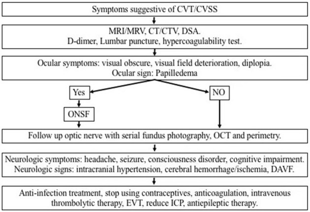
Objective: Visual impairment is the most common clinical feature of cerebral venous sinus occlusion or cerebral venous thrombosis-induced intracranial hypertension, which can result in optic atrophy, leading to irreversible vision loss, visual field defections, and finally, permanent blindness. Papilledema is a typical early pathophysiological alteration in visual impairment. Optic nerve sheath fenestration (ONSF) has become increasingly accepted as an option to prevent or halt progressive visual loss owing to its low risk and complications. The objective of this study is to review the latest research progress on ONSF for the treatment of visual impairment related to cerebral venous diseases.
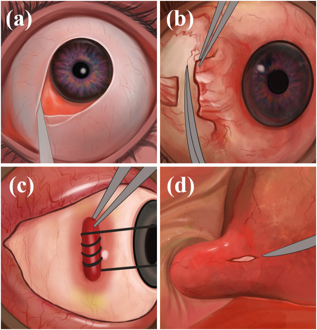
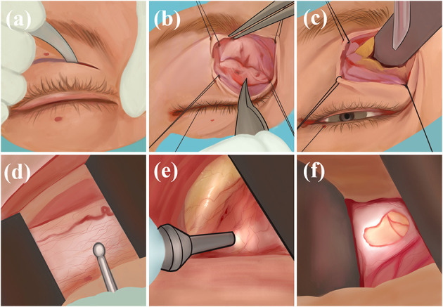
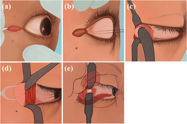
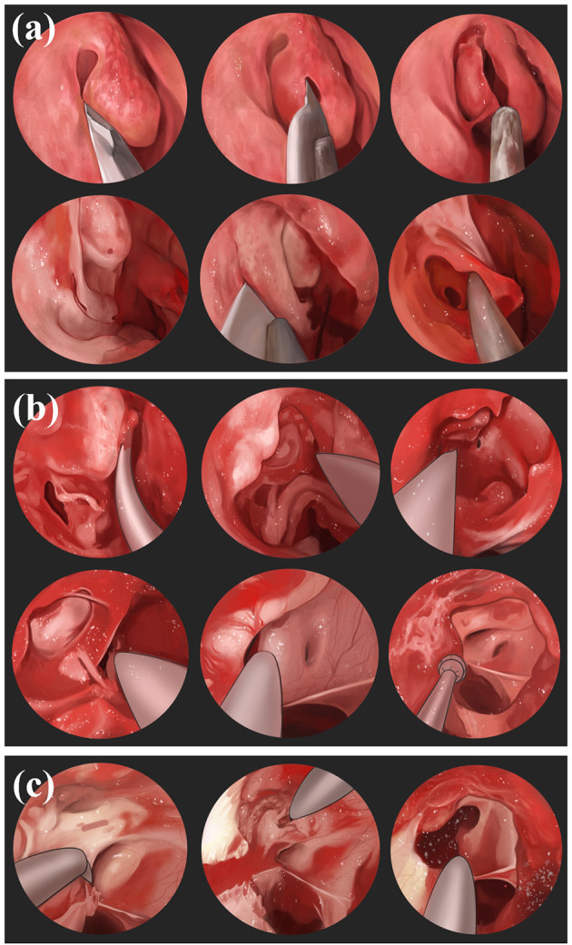
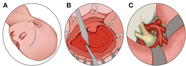
Methods: Study were searched following PRISMA guidelines based on three electronic databases (Pubmed, Embase and Medline-Ovid). We used the following keywords and variations as keywords to identify studies: "optic nerve sheath fenestration, papilledema, cerebral venous diseases, cerebral venous stenosis, cerebral venous thrombosis, idiopathic intracranial hypertension". The publication date of studies was restricted between 1,872.1.1 and 2,021.12.31. The application of ONSF in papilledema due to cerebral venous diseases is reviewed. Additionally, the common surgical approaches as well as advantages and disadvantages are also described graphically.
Results: With the improvement of specific details of the ONSF procedure and surgical instruments, complications of ONSF have reduced and its safety has been significantly improved, although the number of clinically investigated cases in the literature remains low.
Conclusion: We recommend that ONSF should be considered as an imperative alternative to reduce or delay the visual morbidity of cerebral venous diseases, although there is yet no consensus on the optimal surgical timing.
Keywords: cerebral venous diseases; cerebral venous stenosis; cerebral venous thrombosis; optic nerve sheath fenestration; papilledema.
Copyright © 2023 Xue, Zhou, Gao, Ji and Zhang.
Conflict of interest statement
The authors declare that the research was conducted in the absence of any commercial or financial relationships that could be construed as a potential conflict of interest.
Figures
Similar articles
-
Optic Nerve Sheath Fenestration for Progressive Visual Loss in Cerebral Venous Sinus Thrombosis: A Long-Term Retrospective Observational Study.Neurol Ther. 2023 Apr;12(2):441-457. doi: 10.1007/s40120-022-00434-9. Epub 2023 Jan 7. Neurol Ther. 2023. PMID: 36609961 Free PMC article.
-
Update on the application of optic nerve sheath fenestration.Restor Neurol Neurosci. 2017;35(3):275-286. doi: 10.3233/rnn-160693. Restor Neurol Neurosci. 2017. PMID: 28339414 Review.
-
Endoscopic Optic Nerve Sheath Fenestration for Treatment of Papilledema Secondary to Intracranial Venous Hypertension: Report of Two Cases.J Med Assoc Thai. 2016 Jun;99 Suppl 3:S141-6. J Med Assoc Thai. 2016. PMID: 29901363
-
Transient vision loss after optic nerve sheath fenestration.Orbit. 2020 Jun;39(3):217-220. doi: 10.1080/01676830.2019.1668433. Epub 2019 Sep 20. Orbit. 2020. PMID: 31537140
-
A 15-year review of secondary and tertiary optic nerve sheath fenestration for idiopathic intracranial hypertension.Orbit. 2018 Aug;37(4):266-272. doi: 10.1080/01676830.2017.1423337. Epub 2018 Jan 9. Orbit. 2018. PMID: 29313398 Review.
Cited by
-
Progress and recognition of idiopathic intracranial hypertension: A narrative review.CNS Neurosci Ther. 2024 Aug;30(8):e14895. doi: 10.1111/cns.14895. CNS Neurosci Ther. 2024. PMID: 39097911 Free PMC article. Review.
-
Visual Impairment in Cerebral Venous Thrombosis: The Various Shades of Gray.Ann Indian Acad Neurol. 2023 Sep-Oct;26(5):629-630. doi: 10.4103/aian.aian_449_23. Epub 2023 Oct 13. Ann Indian Acad Neurol. 2023. PMID: 38022438 Free PMC article. No abstract available.
-
Effectiveness of optic nerve sheath fenestration in preserving vision in idiopathic intracranial hypertension: an updated meta-analysis and systematic review.Acta Neurochir (Wien). 2024 Nov 25;166(1):476. doi: 10.1007/s00701-024-06345-y. Acta Neurochir (Wien). 2024. PMID: 39585430 Free PMC article.
References
-
- Ferro JM, Canhão P, Stam J, Bousser M-G, Barinagarrementeria F, ISCVT Investigators. Prognosis of cerebral vein and dural sinus thrombosis: results of the International Study on Cerebral Vein and Dural Sinus Thrombosis (ISCVT). Stroke. (2004) 35:664–70. 10.1161/01.STR.0000117571.76197.26 - DOI - PubMed
LinkOut - more resources
Full Text Sources