. 2023 Jan 24:14:1072142.
doi: 10.3389/fimmu.2023.1072142. eCollection 2023.

Silent neonatal influenza A virus infection primes systemic antimicrobial immunity

Affiliations

Silent neonatal influenza A virus infection primes systemic antimicrobial immunity

Anna Sophie Heinemann et al. Front Immunol. .

Abstract

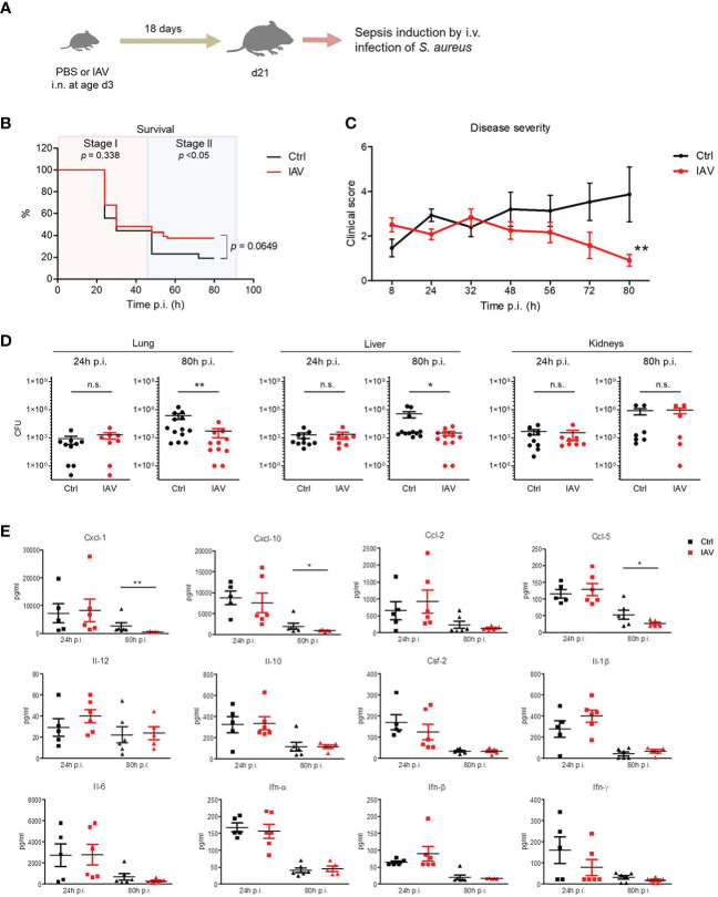
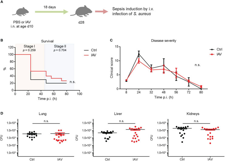
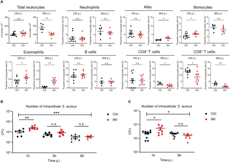
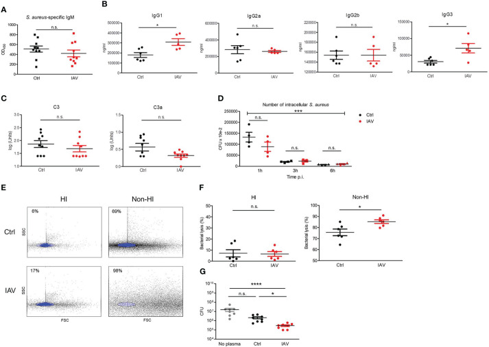
Infections with influenza A viruses (IAV) cause seasonal epidemics and global pandemics. The majority of these infections remain asymptomatic, especially among children below five years of age. Importantly, this is a time, when immunological imprinting takes place. Whether early-life infections with IAV affect the development of antimicrobial immunity is unknown. Using a preclinical mouse model, we demonstrate here that silent neonatal influenza infections have a remote beneficial impact on the later control of systemic juvenile-onset and adult-onset infections with an unrelated pathogen, Staphylococcus aureus, due to improved pathogen clearance and clinical resolution. Strategic vaccination with a live attenuated IAV vaccine elicited a similar protection phenotype. Mechanistically, the IAV priming effect primarily targets antimicrobial functions of the developing innate immune system including increased antimicrobial plasma activity and enhanced phagocyte functions and antigen-presenting properties at mucosal sites. Our results suggest a long-term benefit from an exposure to IAV during the neonatal phase, which might be exploited by strategic vaccination against influenza early in life to enforce the host's resistance to later bacterial infections.

Keywords: Staphylococcus aureus; antimicrobial immunity; influenza A virus; influenza vaccination; innate immunity training; neonate; sepsis.


Conflict of interest statement

The authors declare that the research was conducted in the absence of any commercial or financial relationships that could be construed as a potential conflict of interest.

Figures

Figure 1
Figure 1
Control of S. aureus infections in juvenile mice is improved after silent neonatal exposure to IAV. Newborn mice were treated intranasally (i.n.) at d3 of life with PBS (control, Ctrl) or 5 PFU of the H1N1 virus (IAV). After 18 days, sepsis was induced in these mice by intravenous (i.v.) application of S. aureus. (A) Experimental setup. (B) Survival was observed for 80 h post-infection (p.i.) with S. aureus and plotted as percentages over time (Mantel-Cox test). (C) Clinical scores at indicated time points p.i., plotted as means ± SEM. **p < 0.01 (post hoc 2-way ANOVA Bonferroni test) (Ctrl n = 52 from 7 litters, IAV n = 56 from 7 litters). (D) Bacterial burden was determined in independent experiments in indicated organs at 24 h and 80 h p.i. (each group n = 6-13 from 2-7 litters, respectively). Plotted are means ± SEM, *p < 0.05, **p < 0.005 (MWU test). (E) Plasma cytokine levels at 24 h and 80 h p.i. (each group n = 5-6, each from 2 independent experiments). Plotted are means ± SEM, *p < 0.05, **p < 0.005 (post hoc Kruskal-Wallis Dunn’s multiple comparison test). n.s., not significant.
Figure 2
Figure 2
Exposure to IAV beyond the neonatal period has no impact on the later defense of S. aureus. Mice were treated i.n. at d10 of life with PBS (control, Ctrl) or 5 PFU of IAV. After 18 days, sepsis was induced in these mice by i.v. application of S. aureus. (A) Experimental setup. (B) Survival was observed for 80 h p.i. and plotted as percentages over time (Mantel-Cox test). (C) Clinical scores assigned at indicated time points p.i., plotted as means ± SEM (post hoc 2-way ANOVA Bonferroni test) (Ctrl n = 10, IAV n = 15, each from 2 litters). (D) Bacterial burden was determined in independent experiments in indicated organs 80 h p.i. (each group n = 12-17 from 7 litters, respectively). Plotted are means ± SEM. n.s., not significant (MWU test).
Figure 3
Figure 3
Silent neonatal IAV infections train phagocyte functions against S. aureus infections. Experimental setup as illustrated in Figure 1A . (A) After 24 h and 80 h of infection of d21 mice with S. aureus, the total number of leukocytes and the proportion of indicated leukocytes harvested from the lungs were determined by flow cytometry in the Ctrl and IAV pretreatment group (each group n = 5-10 from 2 litters, respectively). Plotted are means ± SEM, *p < 0.05 (MWU test). (B, C) Bacterial phagocytosis and killing capacity of blood-derived macrophages (B) and BMDMs (C) obtained from d21 mice after neonatal i.n. pretreatment with PBS or 5 PFU of IAV (each group n = 7-10 from 2 litters, respectively). Plotted are the numbers of colony-forming units (CFU) of intracellular S. aureus at 1 h, 3 h and 6 h (in blood-derived macrophages) respective 1 h and 6 h (in BMDMs) after ex vivo infection of macrophages at MOI 10. Bars represent means ± SEM. Significant differences were determined by ANOVA across settings (***p < 0.0001, *p < 0.05) and by post hoc ANOVA Tukey’s multiple comparison tests between pretreatment groups at resp. time points p.i. (**p < 0.005, *p < 0.05). n.s., not significant.
Figure 4
Figure 4
Silent neonatal exposure to IAV enhances humoral antimicrobial functions. Plasma was collected from d21 mice pretreated i.n. on d3 with PBS (Ctrl) or 5 PFU of IAV. (A) Levels of S. aureus-specific IgM as determined by ELISA. OD450, optical density measured at 450 nm (Ctrl n = 10, IAV n = 10). (B) Plasma concentration of indicated murine IgG subclasses (Ctrl n = 6, IAV n = 5). (C) Complement levels of C3 and C3a (Ctrl n = 9, IAV n = 9). Bars represent means ± SEM. *p < 0.05 (MWU tests). (D) Opsonizing plasma activity was tested by preincubating S. aureus with heat-inactivated (HI) plasma from d21 mice of the Ctrl or IAV group (each n = 4). Opsonized bacteria were used at MOI 10 for infection of macrophages obtained from untreated adult C57BL/6 mice. Plotted is the number of CFU of intracellular S. aureus at 1 h, 3 h and 6 h p.i. Indicated is the overall significance of S. aureus elimination over time (***p < 0.0001, ANOVA) and differences between the pretreatment groups at respective time points p.i. (post hoc ANOVA Tukey’s multaiple comparison tests). (E, F) Plasma-mediated bacterial lysis was determined flow cytometrically by analyzing the number of bacteria left after incubation of 1 × 10⁷ CFU of heat-inactivated S. aureus with HI plasma and non-HI plasma obtained from d21 mice of the Ctrl and IAV group. (E) Representative FACS scatter plots with the gate of intact bacteria highlighted in blue. Indicated are the percentages of bacterial lysis. (F) Bacterial lysis was plotted as percentage of bacterial loss after treatment with HI and not-HI plasma compared to not plasma treated bacteria (Ctrl n = 6, IAV n = 6). Bars represent means ± SEM, *p < 0.05 (MWU test). (G) CFU of S. aureus after 1h of growth without plasma and in the presence of plasma from d21 mice of the Ctrl and IAV group (each n = 8). Bars represent means ± SEM, *p < 0.05, ****p < 0.0001 (Kruskal-Wallis test with post hoc Dunn’s multiple comparison tests). n.s., not significant.
Figure 5
Figure 5
Neonatal exposure to IAV induces long-lasting changes in the phenotype of lung and gut resident leukocytes. Single-cell suspensions of (A, B) the lungs and (C, D) the colonic lamina propria from d21 mice pretreated i.n. with PBS (Ctrl, n = 11-12) or 5 PFU of IAV (n = 17) on d3 of life. (A, C) Numbers and (B, D) MHC-II expression (MFI, mean fluorescence intensity) of indicated leukocytes in lungs and colons. Bars represent means ± SEM. *p < 0.05, **p < 0.01, ***p < 0.005 (MWU test). n.s., not significant. AM, alveolar macrophage; DC, dendritic cell; IM, interstitial monocyte; LPMP, lamina propria macrophage; Tregs, regulatory T cells.
Figure 6
Figure 6
Neonatal influenza vaccination improves control of S. aureus infections beyond childhood. Newborn mice were treated i.n. at d3 of life with PBS (Ctrl) or a live-attenuated influenza vaccine (LAIV). After 18 days, sepsis was induced in these mice by i.v. application of S. aureus. (A) Experimental setup. (B) Survival was observed for 80 h p.i. and plotted as percentages over time. n.s., not significant (Mantel-Cox test). (C) Clinical scores assigned at indicated time points p.i., plotted as means ± SEM. *p < 0.05 (post hoc 2-way ANOVA Bonferroni test) (Ctrl n = 52 from 7 litters, LAIV n = 26 from 4 litters). (D) Bacterial burden was determined in independent experiments in indicated organs 80 h p.i. (each group n = 6-13 from 3 litters, respectively). Plotted are means ± SEM, *p < 0.05, **p < 0.005 (MWU test).
Figure 7
Figure 7
Model of innate antimicrobial priming by IAV exposure early in life. Graphical summary how a respiratory exposure of murine d3 neonates to IAV shapes their antimicrobial immunity in the long-term. While an early-life encounter of IAV does not influence the performance of the antimicrobial response during the acute early stage of a systemic S. aureus infection in adolescence (stage I), it improves pathogen clearance and survival during the second stage of sepsis (stage II). Control mice are depicted on the left side, mice challenged with IAV on d3 on the right side. The numbers of mouse icons are an approximation based on our experimental findings. Created with BioRender.

Similar articles

Cited by

  • Influenza Immunization in Very-Low-Birth-Weight Infants: Epidemiology and Long-Term Outcomes.
    Dammann MT, Kraft H, Stichtenoth G, Hanke K, Zemlin M, Soler Wenglein J, Ricklefs I, Herz A, Humberg A, Viemann D, Engels G, Kopp MV, Brinkmann F, Fortmann-Grote C, Göpel W, Herting E, Härtel C, Fortmann I, On Behalf Of The German Neonatal Network. Dammann MT, et al. Vaccines (Basel). 2025 Jan 7;13(1):42. doi: 10.3390/vaccines13010042. Vaccines (Basel). 2025. PMID: 39852821 Free PMC article.

References

    1. Iuliano AD, Roguski KM, Chang HH, Muscatello DJ, Palekar R, Tempia S, et al. . Estimates of global seasonal influenza-associated respiratory mortality: A modelling study. Lancet (2018) 391:1285–300. doi: 10.1016/S0140-6736(17)33293-2 - DOI - PMC - PubMed
    1. Hayward AC, Fragaszy EB, Bermingham A, Wang L, Copas A, Edmunds WJ, et al. . Comparative community burden and severity of seasonal and pandemic influenza: Results of the flu watch cohort study. Lancet Resp Med (2014) 2:445–54. doi: 10.1016/S2213-2600(14)70034-7 - DOI - PMC - PubMed
    1. Thompson MG, Levine MZ, Bino S, Hunt DR, Al-Sanouri TM, Simões EAF, et al. . Underdetection of laboratory-confirmed influenza-associated hospital admissions among infants: A multicentre, prospective study. Lancet Child Adolesc Health (2019) 3:781–94. doi: 10.1016/S2352-4642(19)30246-9 - DOI - PMC - PubMed
    1. Gluckman PD, Hanson MA, Cooper C, Thornburg KL. Effect of in utero and early-life conditions on adult health and disease. N Engl J Med (2008) 359:61–73. doi: 10.1056/NEJMra0708473 - DOI - PMC - PubMed
    1. Renz H, Adkins BD, Bartfeld S, Blumberg RS, Farber DL, Garssen J, et al. . The neonatal window of opportunity-early priming for life. J Allergy Clin Immunol (2018) 141:1212–4. doi: 10.1016/j.jaci.2017.11.019 - DOI - PMC - PubMed

Publication types