. 2023 Jan 24:14:1089576.
doi: 10.3389/fimmu.2023.1089576. eCollection 2023.

Sestrin2 provides cerebral protection through activation of Nrf2 signaling in microglia following subarachnoid hemorrhage

Affiliations

Sestrin2 provides cerebral protection through activation of Nrf2 signaling in microglia following subarachnoid hemorrhage

Youqing Yang et al. Front Immunol. .

Abstract

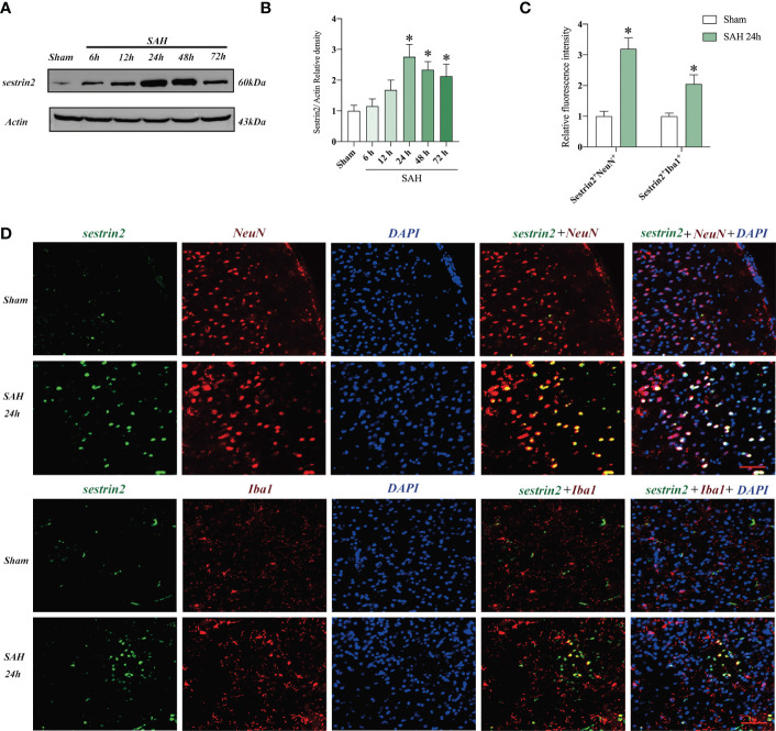
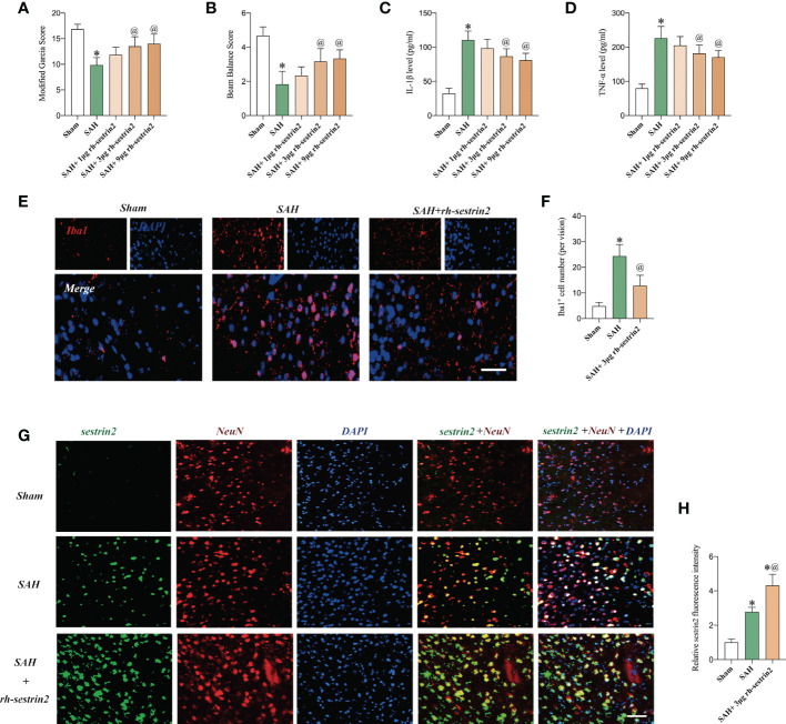
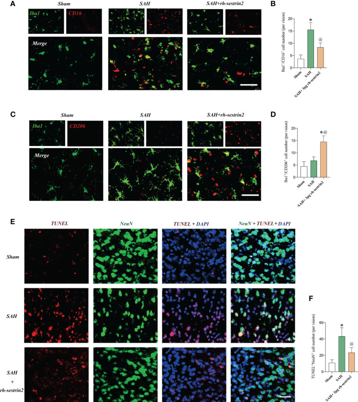
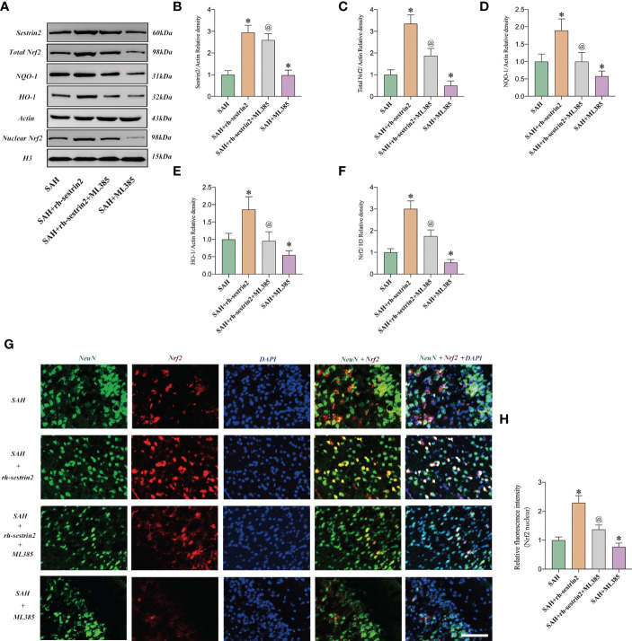
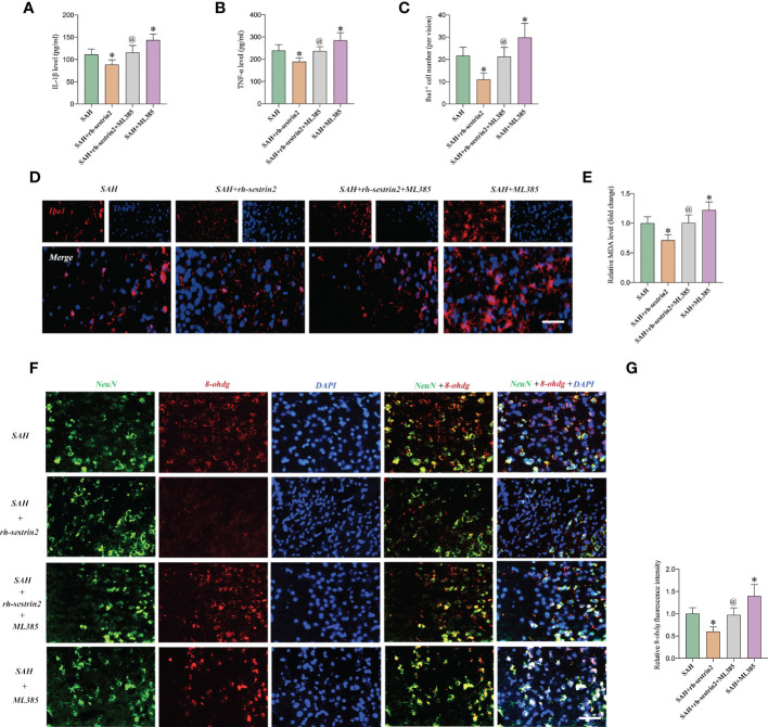
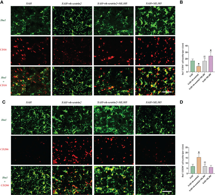
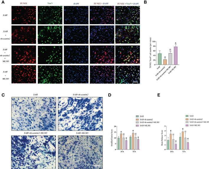
Subarachnoid hemorrhage (SAH) is a neurological emergency characterized by dysfunctional inflammatory response. However, no effective therapeutic options have been reported so far. Microglia polarization has been proposed to exert an essential role in modulating inflammatory response after SAH. Sestrin2 is a stress response protein. Growing evidence has reported that sestrin2 could inhibit M1 microglia and promote M2 microglia polarization. The current study investigated the effects of sestrin2 on microglia phenotype switching and the subsequent brain injury and sought to elucidate the underlying mechanism. We conducted an endovascular perforation SAH model in mice. It was found that sestrin2 was significantly increased after SAH and was mainly distributed in neurons and microglia. Exogenous recombinant human sestrin2 (rh-sestrin2) evidently alleviated inflammatory insults and oxidative stress, and improved neurofunction after SAH. Moreover, rh-sestrin2 increased M2-like microglia polarization and suppressed the number of M1-like microglia after SAH. The protection by rh-sestrin2 was correlated with the activation of Nrf2 signaling. Nrf2 inhibition by ML385 abated the cerebroprotective effects of rh-sestrin2 against SAH and further manifested M1 microglia polarization. In conclusion, promoting microglia polarization from the M1 to M2 phenotype and inducing Nrf2 signaling might be the major mechanism by which sestrin2 protects against SAH insults. Sestrin2 might be a new molecular target for treating SAH.

Keywords: Nrf2; microglial polarization; neuroinflammation; sestrin2; subarachnoid hemorrhage.


Conflict of interest statement

The authors declare that the research was conducted in the absence of any commercial or financial relationships that could be construed as a potential conflict of interest.

Figures

Figure 1
Figure 1
Expression and cellular distribution of sestrin2 in the early period after SAH. (A) Representative western blot bands and (B) quantitative analysis of sestrin2 in different groups (n = 6 per group). (C) Quantitative analysis of sestrin2 in neurons and microglia in sham and SAH groups (n = 6 per group). (D) Double immunofluorescence staining for sestrin2 in neurons and microglia in the brain cortex after SAH. * P < 0.05 versus sham group. Data indicated as mean ± SD. Scale bars = 50 μm.
Figure 2
Figure 2
Rh-sestrin2 improved neurological function and reduced neuroinflammatory insults after SAH. (A) Modified Garcia score and (B) beam balance score indifferent experimental groups (n = 6 per group). Quantitative analyses of (C) IL-1β and (D) TNF-α in different experimental groups (n = 6 per group). (E) Representative immunofluorescence staining for Iba1. (F) Quantitative analysis of Iba1 staining (n = 6 per group). (G) Representative images and (H)quantifications of sestrin2 staining in different groups (n = 6 per group). *P < 0.05 versus sham group. @P < 0.05 versus SAH group. Data indicated asmean ± SD. Scale bars = 50 mm.
Figure 3
Figure 3
Rh-sestrin2 inhibited M1 microglia, increased M2 microglia polarization, and reduced neuronal apoptosis after SAH. (A) Representative images and (B) quantifications of CD16+/Iba1+ staining in different groups (n = 6 per group). (C) Representative images and (D) quantifications of CD206+/Iba1+ staining in different groups (n = 6 per group). (E) Representative images and (F) quantifications of TUNEL staining in different groups (n = 6 per group). * P < 0.05 versus sham group. @ P < 0.05 versus SAH group. Data indicated as mean ± SD. Scale bars = 50 μm.
Figure 4
Figure 4
Rh-sestrin2 treatemtn induced Nrf2 signaling activation after SAH. (A) Representative western blot bands and quantitative analysis of (B) sestrin2, (C) total Nrf2, (D) NQO-1, (E) HO-1, and (F) nuclear Nrf2 in different groups (n = 6 per group). (G) Representative images and (H) quantifications of Nrf2 staining in different groups (n = 6 per group). * P < 0.05 versus sham group. @ P < 0.05 versus SAH group. Data indicated as mean ± SD. Scale bars = 50 μm.
Figure 5
Figure 5
ML385 reversed the anti-inflammatory and anti-oxidative effects of rh-sestrin2 against SAH. Quantitative analyses of (A) IL-1β and (B) TNF-α in different experimental groups (n = 6 per group). (C) Quantitative analysis and (D) representative images of Iba1 staining in different groups (n = 6 per group). (E) Quantitative analyses of MDA (n = 6 per group). (F) Representative images and (G) quantifications of 8-ohdg staining in different groups (n = 6 per group). * P < 0.05 versus SAH group. @ P < 0.05 versus SAH + rh- sestrin2 group. Data indicated as mean ± SD. Scale bars = 50 μm.
Figure 6
Figure 6
ML385 induced M1 microglia polarization and suppressed M2 microglia polarization after SAH. (A) Representative images and (B) quantifications of CD16+/Iba1+ staining in different groups (n = 6 per group). (C) Representative images and (D) quantifications of CD206+/Iba1+ staining in all groups (n = 6 per group). * P < 0.05 versus SAH group. @ P < 0.05 versus SAH + rh- sestrin2 group. Data indicated as mean ± SD. Scale bars = 50 μm.
Figure 7
Figure 7
ML385 aggravated neuronal apoptosis and exacerbated neurological deterioration after SAH. (A) Representative images and (B) quantifications of TUNEL staining in different groups (n = 6 per group). (C) Representative images of Nissl staining in all groups. Quantifications of (D) modified Garcia score and (E) beam balance score (n = 6 per group). * P < 0.05 versus SAH group. @ P < 0.05 versus SAH + rh- sestrin2 group. Data indicated as mean ± SD. Scale bars = 50 μm.

Similar articles

Cited by

References

    1. Claassen J, Park S. Spontaneous subarachnoid haemorrhage. Lancet (2022) 400:846–62. doi: 10.1016/S0140-6736(22)00938-2 - DOI - PMC - PubMed
    1. Geraghty JR, Davis JL, Testai FD. Neuroinflammation and microvascular dysfunction after experimental subarachnoid hemorrhage: Emerging components of early brain injury related to outcome. Neurocrit Care (2019) 31:373–89. doi: 10.1007/s12028-019-00710-x - DOI - PMC - PubMed
    1. Korja M, Kaprio J. Controversies in epidemiology of intracranial aneurysms and SAH. Nat Rev Neurol (2016) 12:50–5. doi: 10.1038/nrneurol.2015.228 - DOI - PubMed
    1. Macdonald RL, Schweizer TA. Spontaneous subarachnoid haemorrhage. Lancet (2017) 389:655–66. doi: 10.1016/S0140-6736(16)30668-7 - DOI - PubMed
    1. Rass V, Helbok R. Early brain injury after poor-grade subarachnoid hemorrhage. Curr Neurol Neurosci Rep (2019) 19:78. doi: 10.1007/s11910-019-0990-3 - DOI - PMC - PubMed

Publication types

LinkOut - more resources