. 2022 Sep 27:93:23-56.
doi: 10.3897/mycokeys.93.86580. eCollection 2022.

Four new species of Mycenasect.Calodontes (Agaricales, Mycenaceae) from northeast China

Affiliations

Four new species of Mycenasect.Calodontes (Agaricales, Mycenaceae) from northeast China

Zewei Liu et al. MycoKeys. .

Abstract

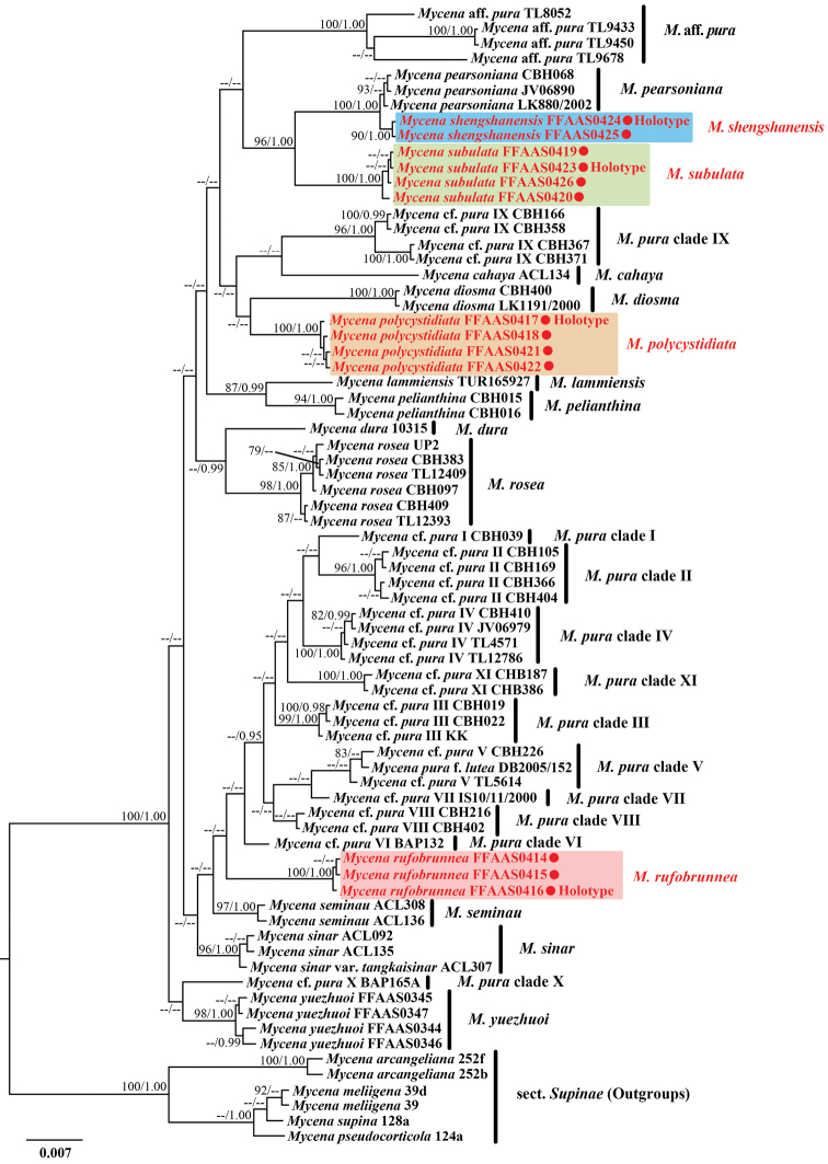
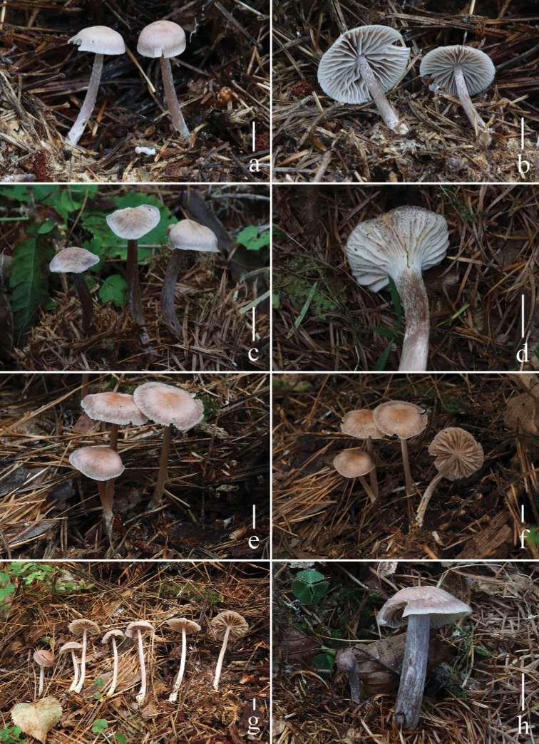
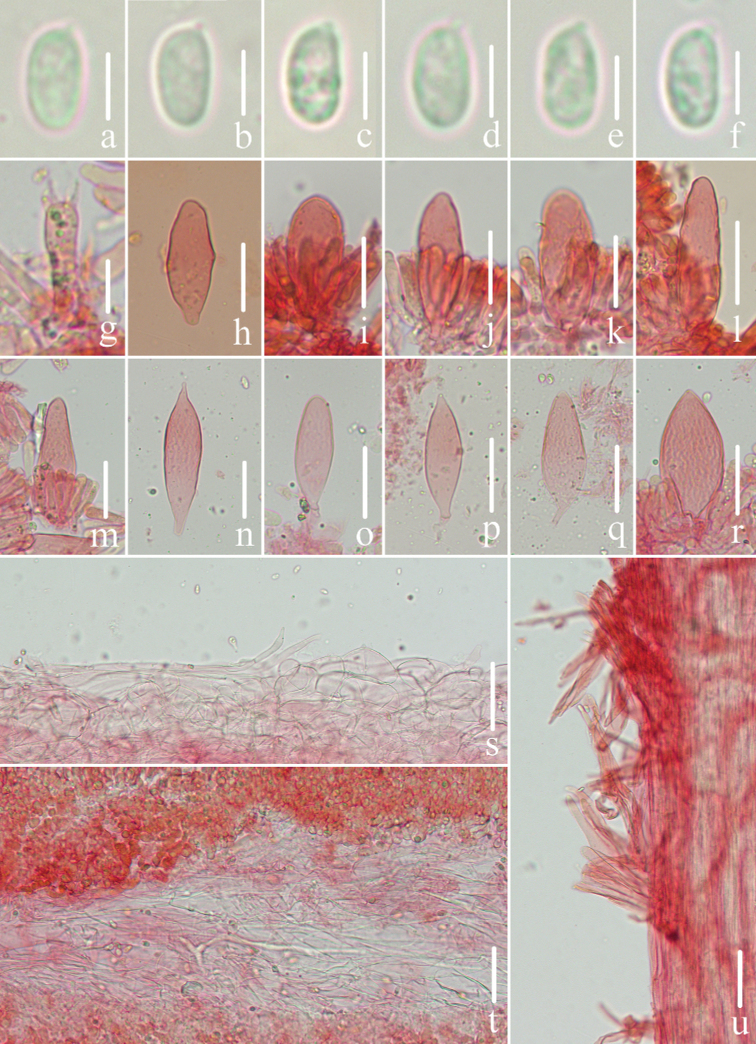
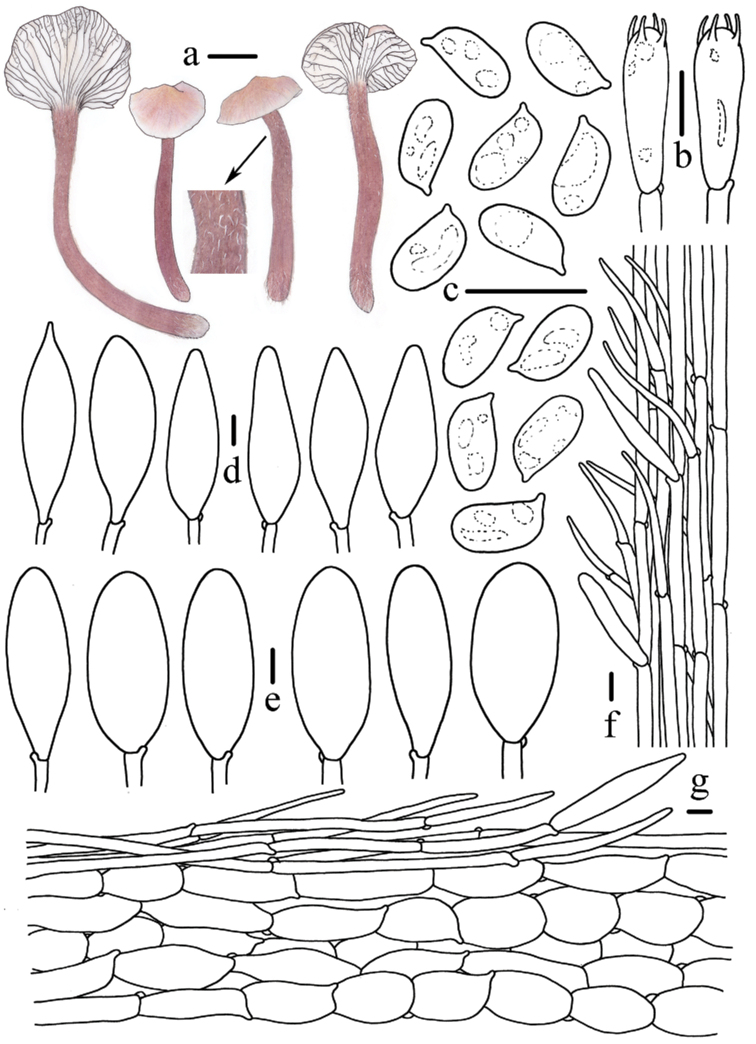
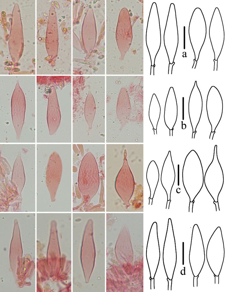
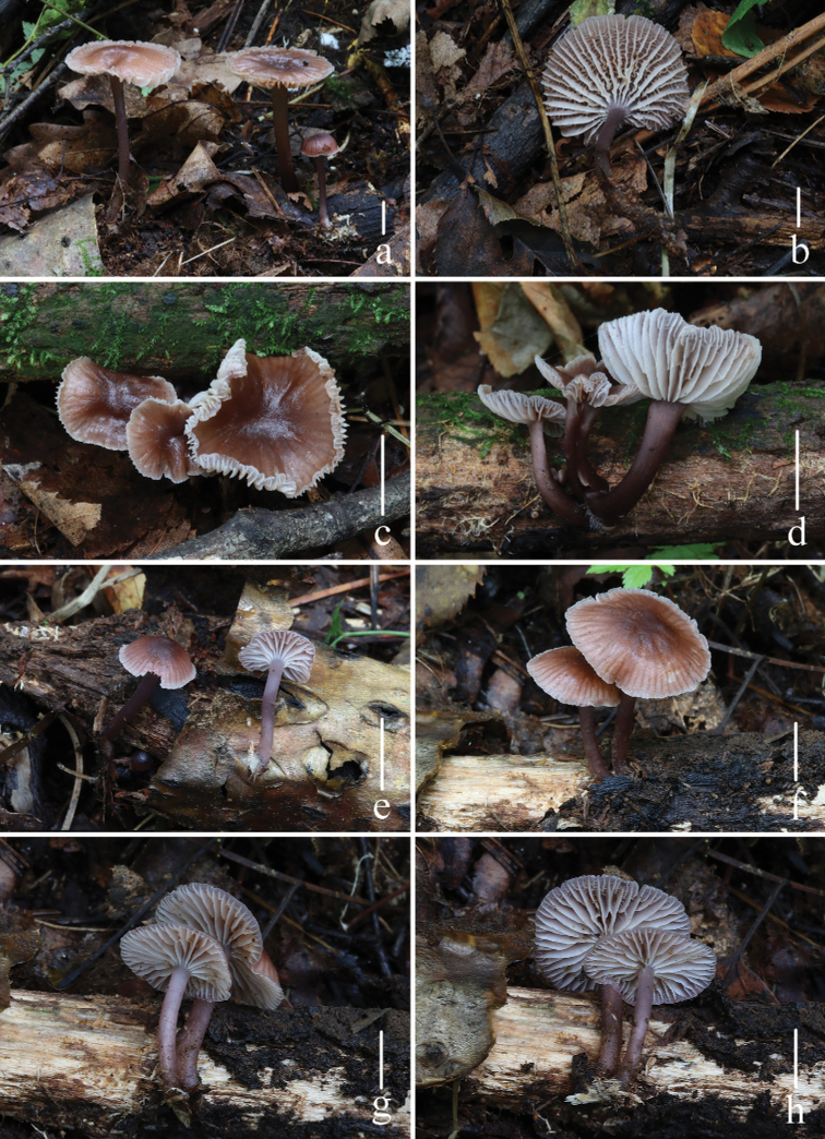
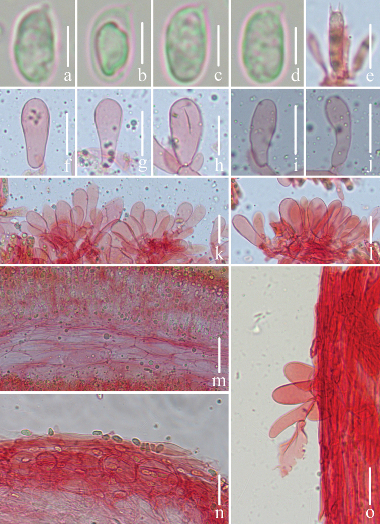
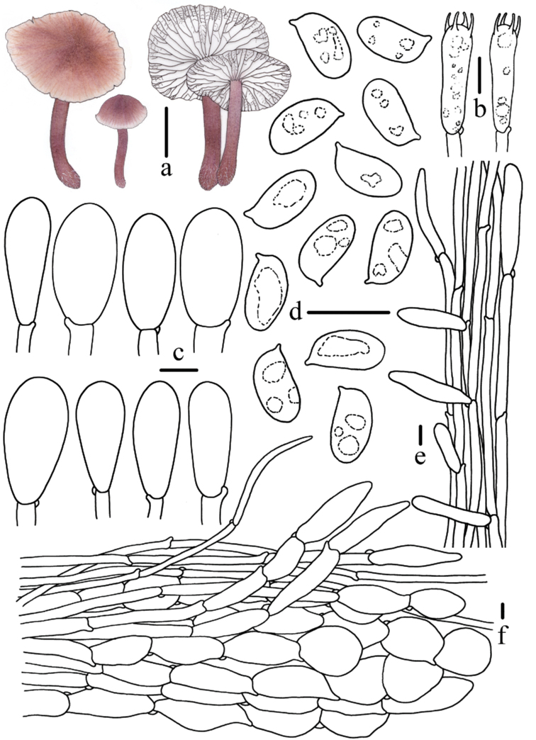
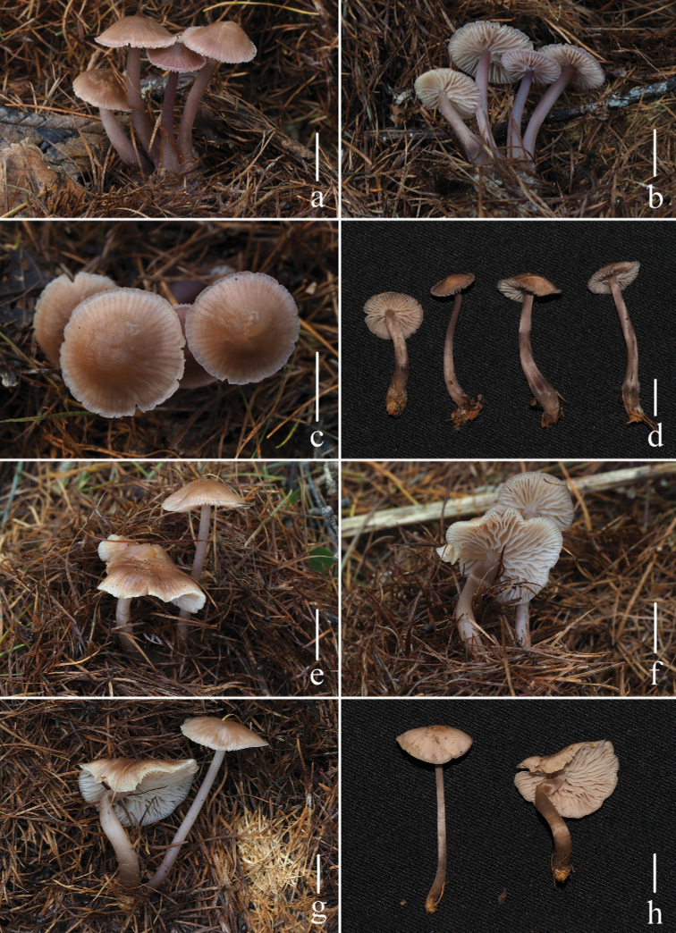
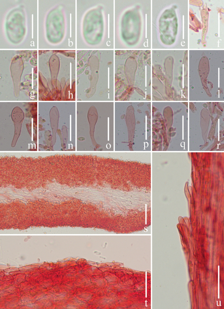
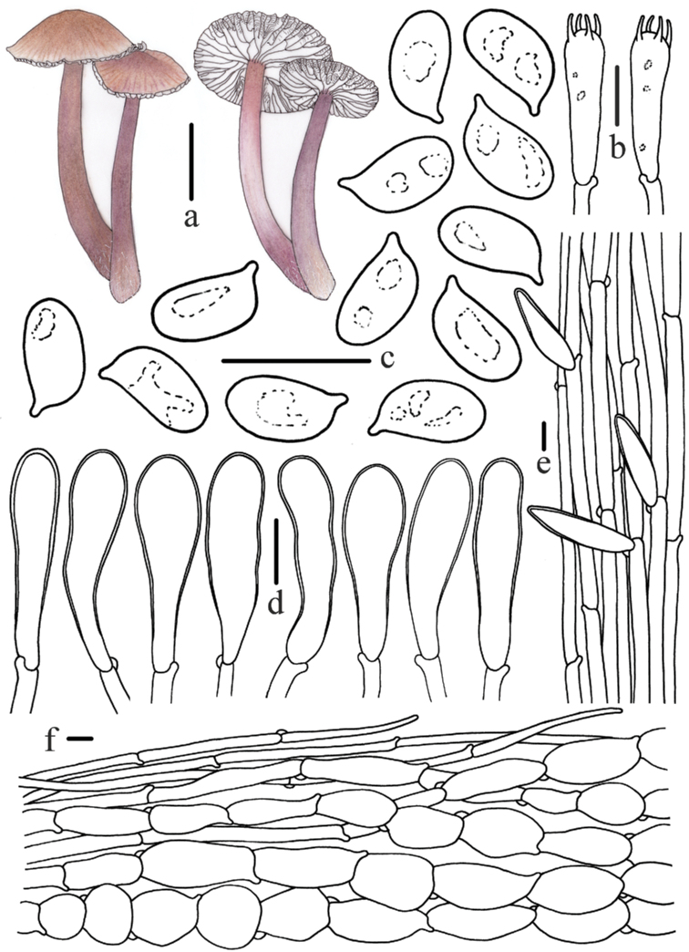
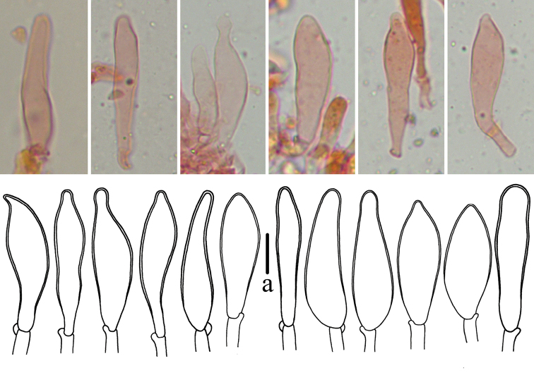
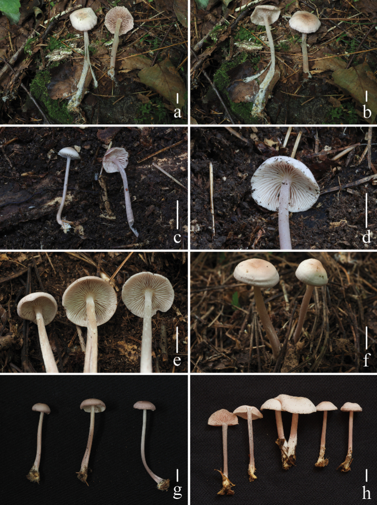
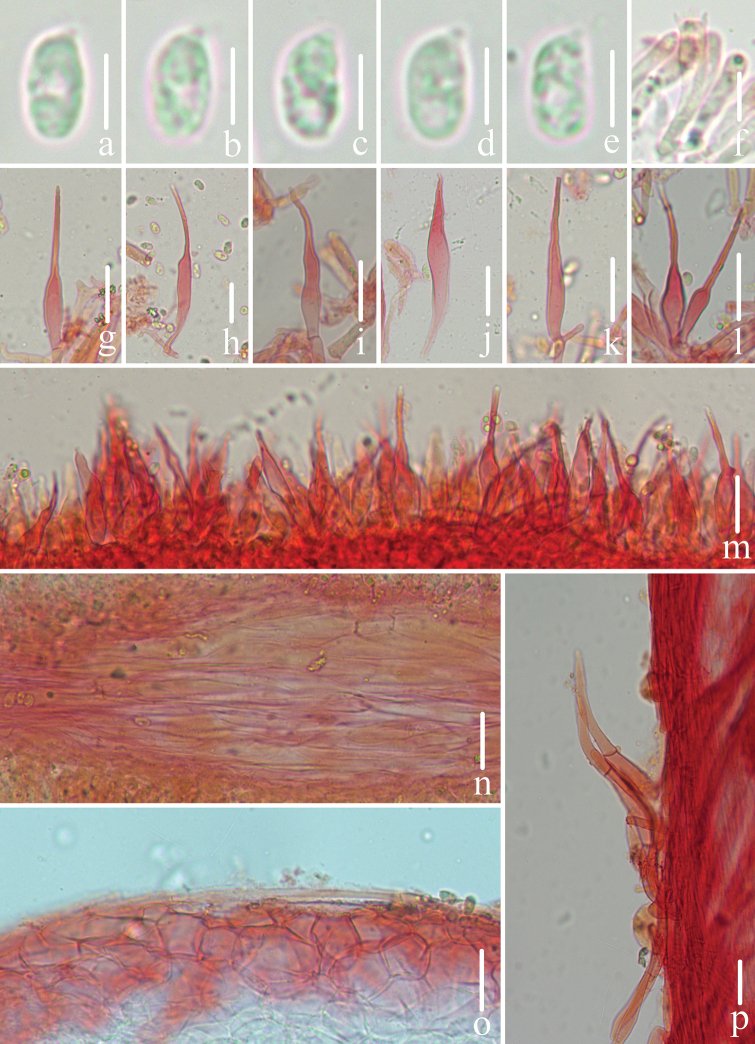
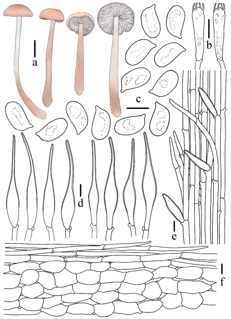
Species of Mycenasect.Calodontes are representative of the Mycena genus as a whole and are easily recognised by the pinkish, reddish, purplish to brownish pileus and larger basidiomata. Furthermore, the colour of the pileus in the species of sect. Calodontes often has a transition or changes in different stages and the combination of the colour of the pileus with cystidia and basidiospores can be used to recognise taxa within this section. To date, 19 species of Mycenasect.Calodontes have been reported worldwide. Including our recent description of M.yuezhuoi, five species of sect. Calodontes have been recorded in China. During examination of specimens collected in coniferous forests or mixed broadleaf-conifer forests in temperate regions of China, additional taxa assigned to sect. Calodontes were identified. Four new species are recognised, based mostly on characters of the pileus and cystidia. Phylogenetic analysis of sequence data from multiple DNA regions (ITS + rpb1 + tef1) supported the morphological evidence. Here, we propose M.polycystidiata, M.rufobrunnea, M.shengshanensis and M.subulata as new species in Mycenasect.Calodontes. Morphological descriptions, line drawings, habitat photos and comparisons with closely-related taxa are provided. A key to the 23 known species of sect. Calodontes is presented.

Keywords: coniferous forest; new taxa; phylogeny; saprobic; taxonomy.


Figures

Figure 1.
Figure 1.
Bayesian Inference analysis of Mycenasect.Calodontes with ITS, rpb1 and tef1 sequence data. Species in Mycenasect.Supinae served as outgroup. Bootstrap values (BS) from Maximum Likelihood ≥ 75 and Bayesian posterior probabilities (BPP) ≥ 0.95 are shown on each branch (BS/BPP). The new species are marked in red.
Figure 2.
Figure 2.
Basidiomata of Mycenapolycystidiata Z.W. Liu, Y.P. Ge, L. Zou & Q. Na a, bFFAAS0422c, dFFAAS0417, holotype e–gFFAAS00421hFFAAS0418 Scale bars: 10 mm (a–h). Photographs a–e, h by Qin Na f, g by Yupeng Ge.
Figure 3.
Figure 3.
Microscopic features of Mycenapolycystidiata (FFAAS0417, holotype) a–f basidiospores g basidia h–l cheilocystidia m–r pleurocystidia s pileipellis and hypodermium t lamellar trama u stipitipellis and caulocystidia. Scale bars: 5 μm (a–f); 10 μm (g); 30 μm (h–r); 40 μm (s–u).
Figure 4.
Figure 4.
Morphological features of Mycenapolycystidiata (FFAAS0417, holotype) a basidiomata b basidia c basidiospores d pleurocystidia e cheilocystidia f stipitipellis and caulocystidia g pileipellis and hypodermium. Scale bars: 10 mm (a); 10 μm (b–g). Drawings by Zewei Liu.
Figure 5.
Figure 5.
Pleurocystidia of MycenapolycystidiataaFFAAS0422bFFAAS0417, holotype cFFAAS0418dFFAAS0421. Scale bars: 25 μm (a–d).
Figure 6.
Figure 6.
Basidiomata of Mycenarufobrunnea Z.W. Liu, Y.P. Ge & Q. Na a, bFFAAS0414c, dFFAAS0415e–hFFAAS0416, holotype. Scale bars: 10 mm (a–h). Photographs a–h by Qin Na.
Figure 7.
Figure 7.
Microscopic features of Mycenarufobrunnea (FFAAS0416, holotype) a–d basidiospores e basidia f–l cheilocystidia m lamellar trama n pileipellis and hypodermiumo stipitipellis and caulocystidia. Scale bars: 5 μm (a–d); 20 μm (e–o).
Figure 8.
Figure 8.
Morphological features of Mycenarufobrunnea (FFAAS0416, holotype) a basidiomata b basidia c cheilocystidia d basidiospores e stipitipellis and caulocystidia f pileipellis and hypodermium. Scale bars: 10 mm (a); 10 μm (b–f). Drawings by Zewei Liu.
Figure 9.
Figure 9.
Basidiomata of Mycenashengshanensis Z.W. Liu, Y.P. Ge & Q. Na a–dFFAAS0424, holotype e–hFFAAS0425 Scale bars: 10 mm (a–h). Photographs a–c, e–g by Yupeng Ge d, h by Zewei Liu.
Figure 10.
Figure 10.
Microscopic features of Mycenashengshanensis (FFAAS0424, holotype) a–e basidiospores f basidia g–r cheilocystidia s lamellar trama t pileipellis and hypodermium u stipitipellis and caulocystidia. Scale bars: 5 μm (a–e); 20 μm (g–r); 40 μm (s–u).
Figure 11.
Figure 11.
Morphological features of Mycenashengshanensis (FFAAS0424, holotype) a basidiomata b basidia c basidiospores d cheilocystidia e stipitipellis and caulocystidia f pileipellis and hypodermium. Scale bars: 10 mm (a); 10 μm (b–f). Drawings by Zewei Liu.
Figure 12.
Figure 12.
Cheilocystidia of Mycenashengshanensis FFAAS0424, holotype. Scale bars: 10 μm (a).
Figure 13.
Figure 13.
Basidiomata of Mycenasubulata Z.W. Liu, Y.P. Ge & Q. Na a, bFFAAS0419c, dFFAAS0420e–gFFAAS0423, holotype hFFAAS0426 Scale bars: 10 mm (a–h). Photographs a, b, e, f by Yupeng Ge c, d by Qin Na g by Zewei Liu h by Shixin Wang.
Figure 14.
Figure 14.
Microscopic features of Mycenasubulata (FFAAS0423, holotype) a–e basidiospores f basidia g–m cheilocystidia n lamellar trama o pileipellis and hypodermium p stipitipellis and caulocystidia. Scale bars: 5 μm (a–e); 10 μm (f); 20 μm (g–p).
Figure 15.
Figure 15.
Morphological features of Mycenasubulata (FFAAS0423, holotype) a basidiomata b basidia c basidiospores d cheilocystidia e stipitipellis and caulocystidia f pileipellis and hypodermium. Scale bars: 10 mm (a); 5 μm (c); 10 μm (b, d–f). Drawings by Zewei Liu.

Similar articles

Cited by

References

    1. Aravindakshan DM, Manimohan P. (2015) Mycenas of Kerala. PhD Thesis, University of Calicut, Kerala, India. 10.13140/RG.2.1.2116.4003 - DOI
    1. Aronsen A, Læssøe T. (2016) The Genus Mycena s.l. Fungi of Northern Europe Vol. 5. Narayana Press, Gylling, Denmark, 373 pp.
    1. Berkeley MJ. (1836) Fungi. In: Smith JE (Eds) The English flora. Longman, Hurst, Rees, Orme, Brown, and Green, London, 5(2): 43. 10.5962/bhl.title.6340 - DOI
    1. Chew AL, Tan YS, Desjardin DE, Musa MY, Sabaratnam V. (2014) Four new bioluminescent taxa of Mycenasect.Calodontes from Peninsular Malaysia. Mycologia 106(5): 976–988. 10.3852/13-274 - DOI - PubMed
    1. Clémençon H, Emmett V, Emmett EE. (2004) Cytology and Plectology of the Hymenomycetes. Bibliotheca Mycologica, Vol. 199. J. Cramer, Berlin, Stuttgart, 488 pp.

LinkOut - more resources